. 2004 Mar 10;24(10):2542-50.
doi: 10.1523/JNEUROSCI.0239-03.2004.

Loss-of-function analysis of EphA receptors in retinotectal mapping

Affiliations

Loss-of-function analysis of EphA receptors in retinotectal mapping

David A Feldheim et al. J Neurosci. .

Abstract

EphA tyrosine kinases are thought to act as topographically specific receptors in the well-characterized projection map from the retina to the tectum. Here, we describe a loss-of-function analysis of EphA receptors in retinotectal mapping. Expressing patches of a cytoplasmically truncated EphA3 receptor in chick retina caused temporal axons to have reduced responsiveness to posterior tectal repellent activity in vitro and to shift more posteriorly within the map in vivo. A gene disruption of mouse EphA5, replacing the intracellular domain with beta-galactosidase, reduced in vitro responsiveness of temporal axons to posterior target membranes. It also caused map abnormalities in vivo, with temporal axons shifted posteriorly and nasal axons anteriorly, but with the entire target still filled by retinal axons. The anterior shift of nasal axons was not accompanied by increased responsiveness to tectal repellent activity, in contrast to the comparable anterior shift in ephrin-A knock-outs, helping to resolve a previous ambiguity in interpreting the ephrin gene knock-outs. The results show the functional requirement for endogenous EphA receptors in retinotectal mapping, show that the receptor intracellular domain is required for a forward signaling response to topographic cues, and provide new evidence for a role of axon competition in topographic mapping.


Figures
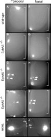
Figure 1.
Figure 1.
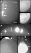
Mapping abnormalities in the SC of EphA5lacZ/lacZ mutant mice. Retinal axons labeled by focal DiI injection were visualized at P14 by florescence microscopy of SC whole mounts. The brackets indicate SC; anterior is at the top. A-D, Temporal injections. Temporal retinal injections in wild-type (A) or EphA5lacZ/+ heterozygous (B) mice show an arborization in the anterior portion of the SC. Similar injections in EphA5lacZ/lacZ mutants (C, D) show ectopic arborizations posterior to the normal location. F-I, Nasal injections. Nasal retinal injections in wild-type (F) or EphA5lacZ/+ heterozygous (G) mice show arborizations at the posterior extreme of the SC. EphA5lacZ/lacZ mutants show ectopic arborizations anterior to the normal site. In addition to ectopic spots, all labelings in the EphA5lacZ/lacZ mutants showed a prominent arborization at the normal location. The arrows indicate ectopic arborizations. E, J, Temporal or nasal retinal quadrants, respectively, showing typical labeling sites. Axons exit the retina at the optic disc (OD).
Figure 2.
Figure 2.
Analysis of the visual projections in EphA5lacZ/lacZ mice. A, Axons labeled approximately in the middle of the nasal-temporal axis by focal DiI injections in ventral retina were visualized at P14 by fluorescence microscopy of SC. EphA5lacZ/lacZ mutants show ectopic arborizations both anterior and posterior to the expected location. The arrows indicate likely ectopic arborizations. A bracket indicates the anterior and posterior boundaries of the SC. B, Parasagittal section through the SC shown in A. Ectopic arborizations branch to the correct layer of the SC. C, D, Eye fill with fluoresceinated cholera toxin β subunit to anterogradely label projections throughout the retina. Labeled axons fill the SC in both wild-type and EphA5lacZ/lacZ mutant animals in C and D, respectively. E, Horizontal section through the midbrain of an EphA5lacZ/lacZ mouse given an injection in the temporal retina. Axons can be seen at the extreme SC-IC border (arrow indicates path of axons along the lateral extreme of the SC). F, Whole-mount view of P3 mouse SC, after eye fill with DiI at P2. The arrow indicates axons passing through the SC to the posterior extreme of the IC.
Figure 3.
Figure 3.
Patterning of the retina and SC in EphA5lacZ/lacZ mice. A-C, Horizontal sections through the P0 eye from wild-type (A) or EphA5lacZ/lacZ (B, C) mice. A, EphA5 receptor RNA is in a temporal>nasal gradient in wild-type retina. B, EphA5 detected by β-galactosidase staining in EphA5lacZ/lacZ remains in a temporal>nasal gradient. C, EphA6 receptor RNA is also in a temporal>nasal gradient in EphA5lacZ/lacZ mutant retinas. D, E, Midbrains in EphA5lacZ/lacZ mice are patterned normally. D, Whole-mount β-galactosidase staining of EphA5lacZ/lacZ mutant midbrain is in an anterior>posterior gradient. E, Ligand expression detected by binding of an EphA3-AP probe remains in a posterior>anterior gradient in EphA5lacZ/lacZ mutants. N, Nasal; T, temporal; GCL, ganglion cell layer. The brackets indicate the SC and IC regions of the midbrain.
Figure 4.
Figure 4.
Mix-and-match stripe assays of retina and SC from EphA5lacZ/lacZ or wild-type mice. A, B, Repellent activity in the SC of EphA5lacZ/lacZ mice. Temporal axons from wild-type mice were tested with alternating anterior and posterior stripes of SC membranes from wild-type (A) or EphA5lacZ/lacZ (B) mice. C, D, Loss of responsiveness of temporal axons from EphA5lacZ/+ and EphA5lacZ/lacZ mutant mice. Temporal axons from EphA5lacZ/+ (C) or EphA5lacZ/lacZ (D) mice were grown on alternating anterior and posterior SC stripes from wild-type mice. E, Results of mix-and-match stripe assays using retina or SC from wild-type (WT) or EphA5lacZ/lacZ mice. Axon preference for anterior SC lanes was scored on a scale of 0 (no preference) to 4 (strong preference). The bars show means ± SEM. N, Nasal; T, temporal. Axons are shown in green; red fluorescent microspheres mark posterior SC stripes.
Figure 5.
Figure 5.
Cytoplasmically truncated EphA3 leads to mapping and guidance errors of temporal retinal axons. A, Pattern of retroviral vector infection in chick retina. Whole-mount in situ hybridization with an EphA3 RNA probe to detect EphA3▵C on chick retinas after injection of virus into the optic cup at E2. This is an example with a relatively extensive expression of exogenous EphA3 RNA; in other cases, patches were sparser. B, C, Truncated EphA3 leads to mapping and guidance errors of temporal retinal axons. Temporal retinal axons were labeled by DiI and visualized by fluorescence microscopy as whole-mount chick tectum. B, Wild-type temporal axons map to the anterior tectum. C, Temporal axons infected with RCAS-EphA3▵C project to abnormally posterior locations in the tectum. Arrows indicate ectopic arbors. Insets show schematic view of labeled axons with respect to the tectum.
Figure 6.
Figure 6.
Stripe assay of axons from RCAS-EphA3▵C-infected or normal chick retina. Explants from temporal retina were placed on membrane carpets consisting of alternating stripes of membranes derived from anterior or posterior tectum. Preference for anterior lanes was scored blind on a scale of 0 (no detectable preference) to 4 (strong preference). A, Temporal axons from normal chick retina prefer anterior stripes to posterior stripes (score, 4). B-D, Temporal axons from chick retina infected with RCAS-EphA3▵C virus tended to show a reduced preference. The results were somewhat variable, sometimes even within a single explant, perhaps reflecting patchy virus infection. B shows an extreme example with axons growing similarly over both anterior and posterior stripes (score, 0), whereas C (score, 2) and D (score, 3) show intermediate examples. Axons are shown in green; blue fluorescent microspheres mark posterior tectal stripes, and smaller microspheres were used in C and D.

References

    1. Birgbauer E, Cowan CA, Sretavan DW, Henkemeyer M (2000) Kinase independent function of EphB receptors in retinal axon pathfinding to the optic disc from dorsal but not ventral retina. Development 127: 1231-1241. - PubMed
    1. Brown A, Yates PA, Burrola P, Ortuno D, Vaidya A, Jessell TM, Pfaff SL, O'Leary DDM, Lemke G (2000) Topographic mapping from the retina to the midbrain is controlled by relative but not absolute levels of EphA receptor signaling. Cell 102: 77-88. - PubMed
    1. Bruckner K, Pasquale EB, Klein R (1997) Tyrosine phosphorylation of transmembrane ligands for Eph receptors. Science 275: 1640-1643. - PubMed
    1. Cheng H-J, Nakamoto M, Bergemann AD, Flanagan JG (1995) Complementary gradients in expression and binding of ELF-1 and Mek4 in development of the topographic retinotectal projection map. Cell 82: 371-381. - PubMed
    1. Ciossek T, Lerch MM, Ulrich A (1995) Cloning, characterization, and differential expresssion of MDK2 and MDK5, two novel receptor tyrosine kinases of the eck/eph family. Oncogene 11: 2085-2095. - PubMed

MeSH terms