The processing of visual shape in the cerebral cortex of human and nonhuman primates: a functional magnetic resonance imaging study
- PMID: 15014131
- PMCID: PMC6729498
- DOI: 10.1523/JNEUROSCI.3569-03.2004
The processing of visual shape in the cerebral cortex of human and nonhuman primates: a functional magnetic resonance imaging study
Abstract
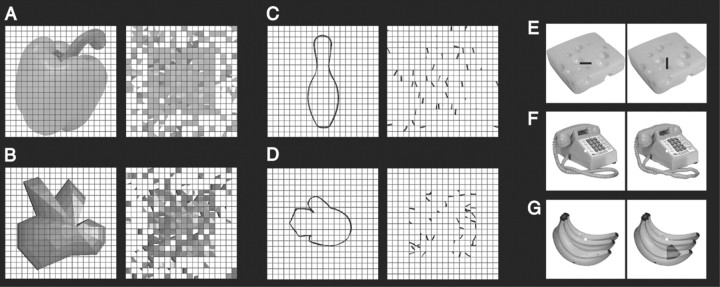
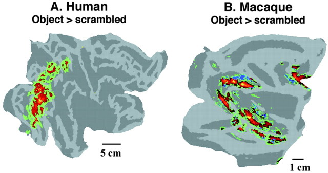
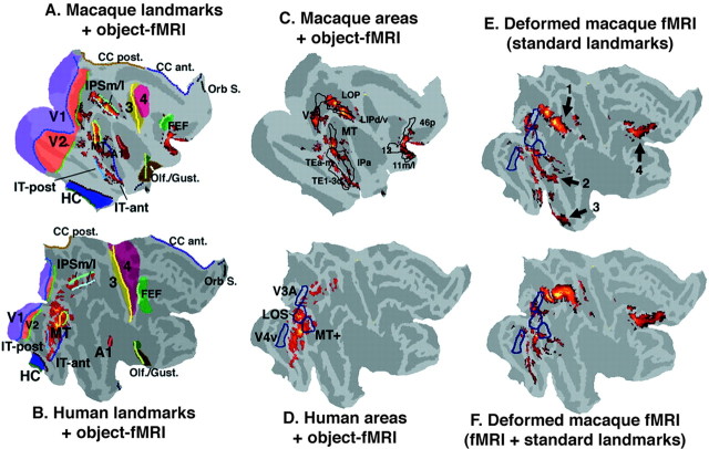
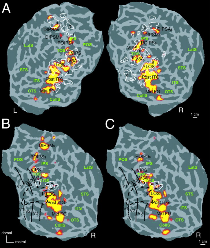
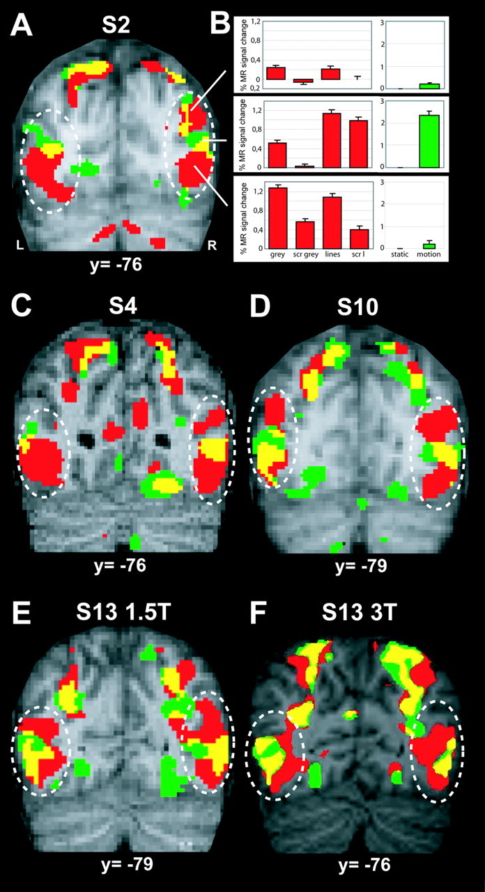
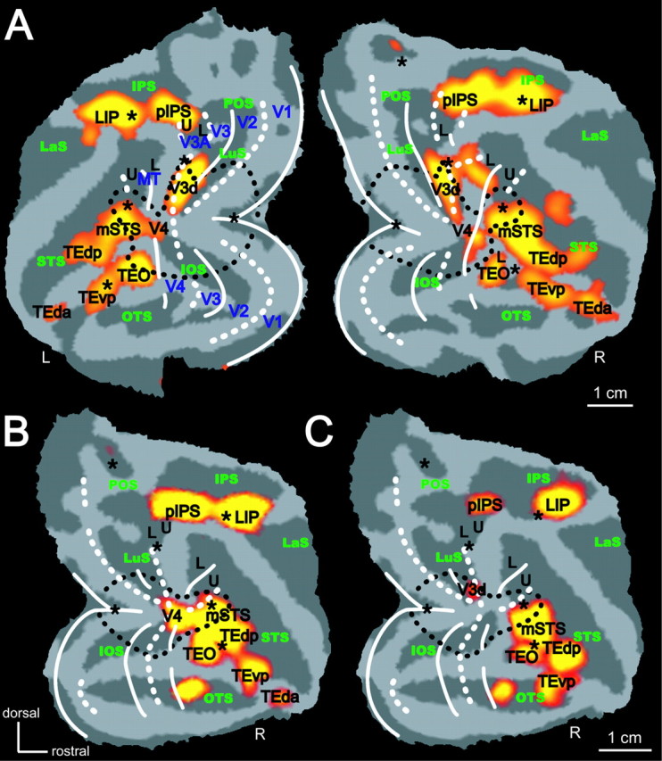
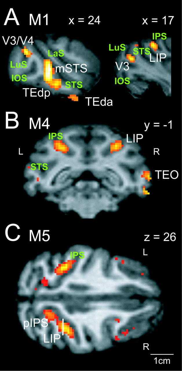
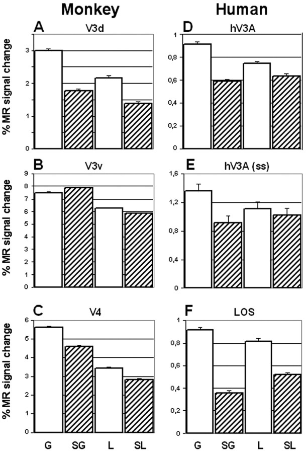
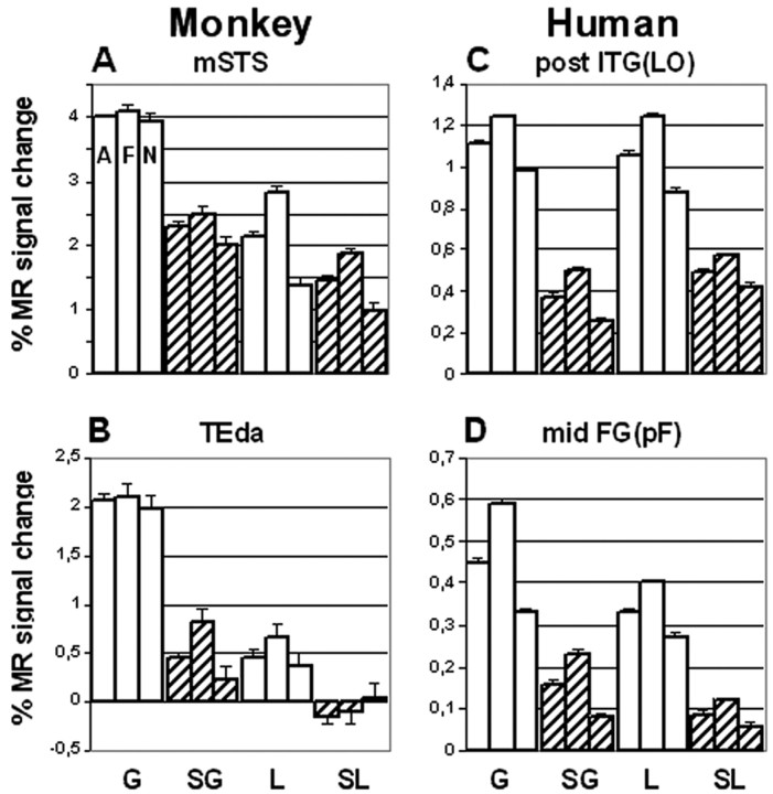
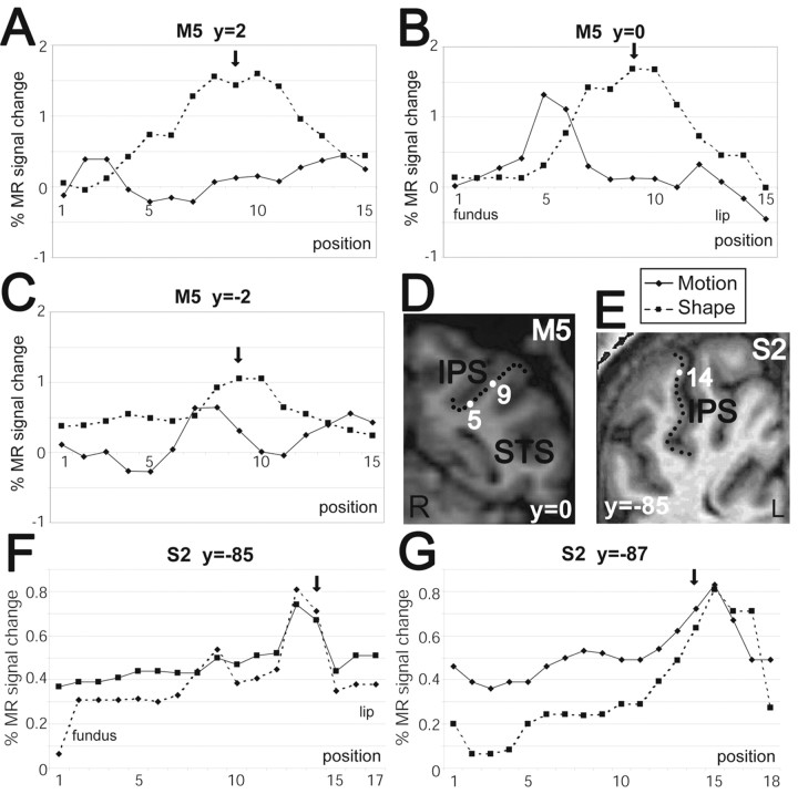
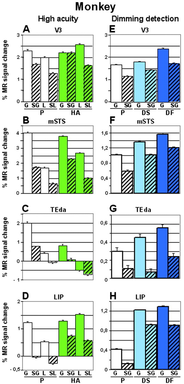
We compared neural substrates of two-dimensional shape processing in human and nonhuman primates using functional magnetic resonance (MR) imaging in awake subjects. The comparison of MR activity evoked by viewing intact and scrambled images of objects revealed shape-sensitive regions in occipital, temporal, and parietal cortex of both humans and macaques. Intraparietal cortex in monkeys was relatively more two-dimensional shape sensitive than that of humans. In both species, there was an interaction between scrambling and type of stimuli (grayscale images and drawings), but the effect of stimulus type was much stronger in monkeys than in humans. Shape- and motion-sensitive regions overlapped to some degree. However, this overlap was much more marked in humans than in monkeys. The shape-sensitive regions can be used to constrain the warping of monkey to human cortex and suggest a large expansion of lateral parietal and superior temporal cortex in humans compared with monkeys.
Figures


Similar articles
-
Color-Biased Regions of the Ventral Visual Pathway Lie between Face- and Place-Selective Regions in Humans, as in Macaques.J Neurosci. 2016 Feb 3;36(5):1682-97. doi: 10.1523/JNEUROSCI.3164-15.2016. J Neurosci. 2016. PMID: 26843649 Free PMC article.
-
Using functional magnetic resonance imaging to assess adaptation and size invariance of shape processing by humans and monkeys.J Neurosci. 2005 Apr 27;25(17):4294-306. doi: 10.1523/JNEUROSCI.0377-05.2005. J Neurosci. 2005. PMID: 15858056 Free PMC article.
-
Comparison of shape encoding in primate dorsal and ventral visual pathways.J Neurophysiol. 2007 Jan;97(1):307-19. doi: 10.1152/jn.00168.2006. Epub 2006 Oct 4. J Neurophysiol. 2007. PMID: 17021033
-
Stages of self-motion processing in primate posterior parietal cortex.Int Rev Neurobiol. 2000;44:173-98. doi: 10.1016/s0074-7742(08)60742-4. Int Rev Neurobiol. 2000. PMID: 10605646 Review. No abstract available.
-
The neural basis of biased competition in human visual cortex.Neuropsychologia. 2001;39(12):1263-76. doi: 10.1016/s0028-3932(01)00116-6. Neuropsychologia. 2001. PMID: 11566310 Review.
Cited by
-
A problem of overlap.Vis Neurosci. 2015 Jan;32:E001. doi: 10.1017/S0952523814000340. Vis Neurosci. 2015. PMID: 26239105 Free PMC article.
-
Impact of Visual Corticostriatal Loop Disruption on Neural Processing within the Parahippocampal Place Area.J Neurosci. 2016 Oct 5;36(40):10456-10471. doi: 10.1523/JNEUROSCI.0741-16.2016. J Neurosci. 2016. PMID: 27707978 Free PMC article.
-
The functional organization of the intraparietal sulcus in humans and monkeys.J Anat. 2005 Jul;207(1):3-17. doi: 10.1111/j.1469-7580.2005.00426.x. J Anat. 2005. PMID: 16011542 Free PMC article. Review.
-
Brain activity mediates the relation between emotional but not instrumental support and trait loneliness.Soc Cogn Affect Neurosci. 2018 Sep 11;13(9):995-1002. doi: 10.1093/scan/nsy067. Soc Cogn Affect Neurosci. 2018. PMID: 30137637 Free PMC article.
-
Impact of Huntington's Disease on Mental Rotation Performance in Motor Pre-Symptomatic Individuals.J Huntingtons Dis. 2019;8(3):339-356. doi: 10.3233/JHD-190348. J Huntingtons Dis. 2019. PMID: 31306138 Free PMC article.
References
-
- Avidan G, Harel M, Hendler T, Ben-Bashat D, Zohary E, Malach R (2002) Contrast sensitivity in human visual areas and its relationship to object recognition. J Neurophysiol 87: 3102-3116. - PubMed
-
- Belliveau JW, Kennedy Jr DN, McKinstry RC, Buchbinder BR, Weisskoff RM, Cohen MS, Vevea JM, Brady TJ, Rosen BR (1991) Functional mapping of the human visual cortex by magnetic resonance imaging. Science 254: 716-719. - PubMed
-
- Boussaoud D, Ungerleider LG, Desimone R (1990) Pathways for motion analysis: cortical connections of the medial superior temporal and fundus of the superior temporal visual areas in the macaque. J Comp Neurol 296: 462-495. - PubMed
Publication types
MeSH terms
Grants and funding
LinkOut - more resources
Full Text Sources