

The functional neuroanatomy of temporal discrimination
- PMID: 15014134
- PMCID: PMC6729480
- DOI: 10.1523/JNEUROSCI.4210-03.2004
The functional neuroanatomy of temporal discrimination
Abstract
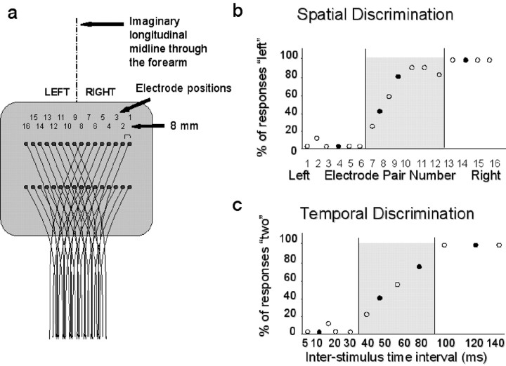
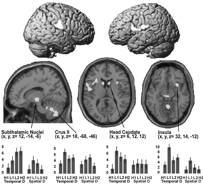
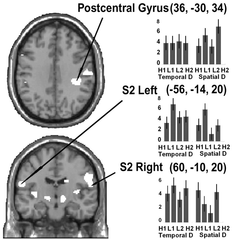
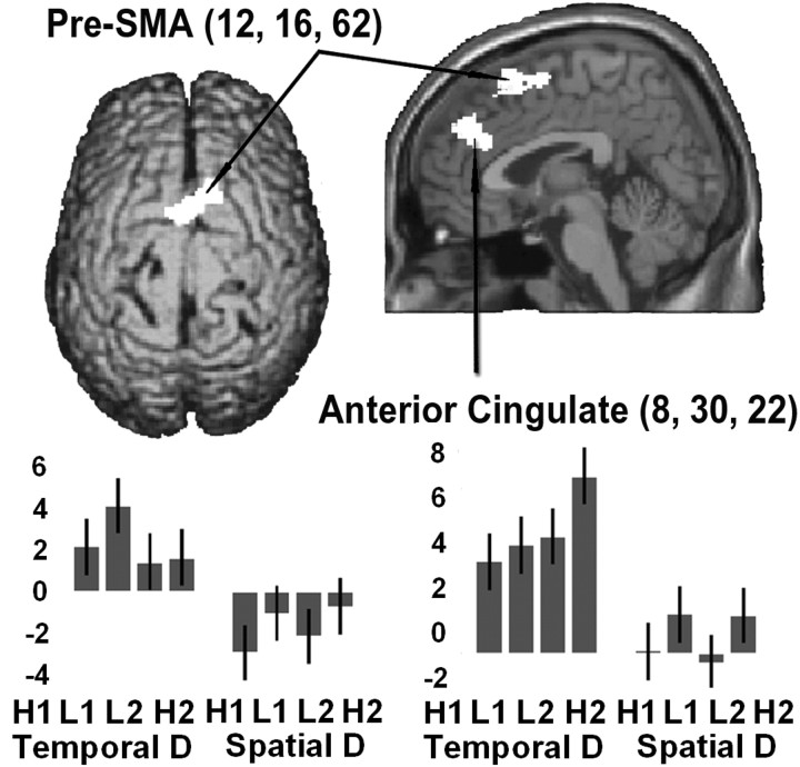
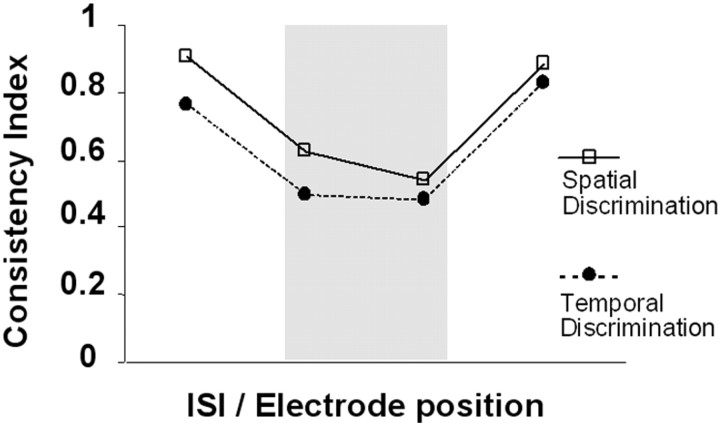
Two identical stimuli, such as a pair of electrical shocks to the skin, are readily perceived as two separate events in time provided the interval between them is sufficiently long. However, as they are presented progressively closer together, there comes a point when the two separate stimuli are perceived as a single stimulus. Damage to posterior parietal cortex, peri-supplementary motor area (peri-SMA), and basal ganglia can disturb this form of temporal discrimination. Our aim was to establish, in healthy subjects, the brain areas that are involved in this process. During functional magnetic resonance imaging scanning, paired electrical pulses, separated by variable inter-stimulus intervals (5-110 msec), were delivered to different sites on one forearm (8-64 mm from the midline). Subjects were required to simply detect the stimulus (control task) or to identify a stimulus property. For temporal discrimination (TD), subjects reported whether they felt one or two stimuli. For spatial discrimination, they reported whether the stimuli were located on the right or left side of the forearm. Subjects reported their choice by pressing a button with the opposite hand. Our results showed that discrimination, as opposed to simply detection, activated several brain areas. Most were common to both discrimination tasks. These included regions of prefrontal cortex, right postcentral gyrus and inferior parietal lobule, basal ganglia, and cerebellum. However, activation of pre-SMA and anterior cingulate was found to be specific to the TD task. This suggests that these two frontal regions may play a role in the temporal processing of somatosensory events.
Figures
Similar articles
-
The neural basis of temporal auditory discrimination.Neuroimage. 2006 Apr 1;30(2):512-20. doi: 10.1016/j.neuroimage.2005.09.053. Epub 2005 Nov 9. Neuroimage. 2006. PMID: 16289998
-
Basal ganglia and supplementary motor area subtend duration perception: an fMRI study.Neuroimage. 2003 Aug;19(4):1532-44. doi: 10.1016/s1053-8119(03)00159-9. Neuroimage. 2003. PMID: 12948709
-
A functional MRI study of motor dysfunction in Friedreich's ataxia.Brain Res. 2012 Aug 30;1471:138-54. doi: 10.1016/j.brainres.2012.06.035. Epub 2012 Jul 3. Brain Res. 2012. PMID: 22771856
-
The neurobiological basis of time estimation and temporal order.Rev Neurosci. 1999;10(2):151-73. doi: 10.1515/revneuro.1999.10.2.151. Rev Neurosci. 1999. PMID: 10658957 Review.
-
Neural underpinnings of temporal processing: a review of focal lesion, pharmacological, and functional imaging research.Rev Neurosci. 1999;10(2):91-116. doi: 10.1515/revneuro.1999.10.2.91. Rev Neurosci. 1999. PMID: 10658954 Review.
Cited by
-
Aristotle's illusion in Parkinson's disease: evidence for normal interdigit tactile perception.PLoS One. 2014 Feb 11;9(2):e88686. doi: 10.1371/journal.pone.0088686. eCollection 2014. PLoS One. 2014. PMID: 24523929 Free PMC article.
-
Characteristics of bilateral hand function in individuals with unilateral dystonia due to perinatal stroke: sensory and motor aspects.J Child Neurol. 2014 May;29(5):623-32. doi: 10.1177/0883073813512523. Epub 2014 Jan 5. J Child Neurol. 2014. PMID: 24396131 Free PMC article.
-
Revisiting the Cerebellum's Linguistic Role: Evidence for Cerebellar Involvement in Expressive Syntax.Cerebellum. 2025 Jul 10;24(5):126. doi: 10.1007/s12311-025-01879-y. Cerebellum. 2025. PMID: 40637927 Free PMC article.
-
Neural representations of beat and rhythm in motor and association regions.Cereb Cortex. 2024 Oct 3;34(10):bhae406. doi: 10.1093/cercor/bhae406. Cereb Cortex. 2024. PMID: 39390710 Free PMC article.
-
The neural substrate of predictive motor timing in spinocerebellar ataxia.Cerebellum. 2011 Jun;10(2):233-44. doi: 10.1007/s12311-010-0237-y. Cerebellum. 2011. PMID: 21110147
References
-
- Artieda J, Pastor MA, Lacruz F, Obeso JA (1992) Temporal discrimination is abnormal in Parkinson's disease. Brain 115: 199-210. - PubMed
-
- Bara-Jimenez W, Shelton P, Hallett M (2000) Spatial discrimination is abnormal in focal hand dystonia. Neurology 55: 1869-1873. - PubMed
-
- Bower JM (1997) Control of sensory data acquisition. Int Rev Neurobiol 41: 489-513. - PubMed
-
- Burton H, Abend NS, MacLeod AM, Sinclair RJ, Snyder AZ, Raichle ME (1999) Tactile attention tasks enhance activation in somatosensory regions of parietal cortex: a positron emission tomography study. Cereb Cortex 9: 662-674. - PubMed
-
- Drevets WC, Burton H, Videen TO, Snyder AZ, Simpson Jr JR, Raichle ME (1995) Blood flow changes in human somatosensory cortex during anticipated stimulation. Nature 373: 249-252. - PubMed
Publication types
MeSH terms
Grants and funding
LinkOut - more resources
Full Text Sources