. 2004 Mar 24;23(6):1289-300.
doi: 10.1038/sj.emboj.7600156. Epub 2004 Mar 11.

Mechanism controlling perpendicular alignment of the spindle to the axis of cell division in fission yeast

Affiliations

Mechanism controlling perpendicular alignment of the spindle to the axis of cell division in fission yeast

Yannick Gachet et al. EMBO J. .

Abstract

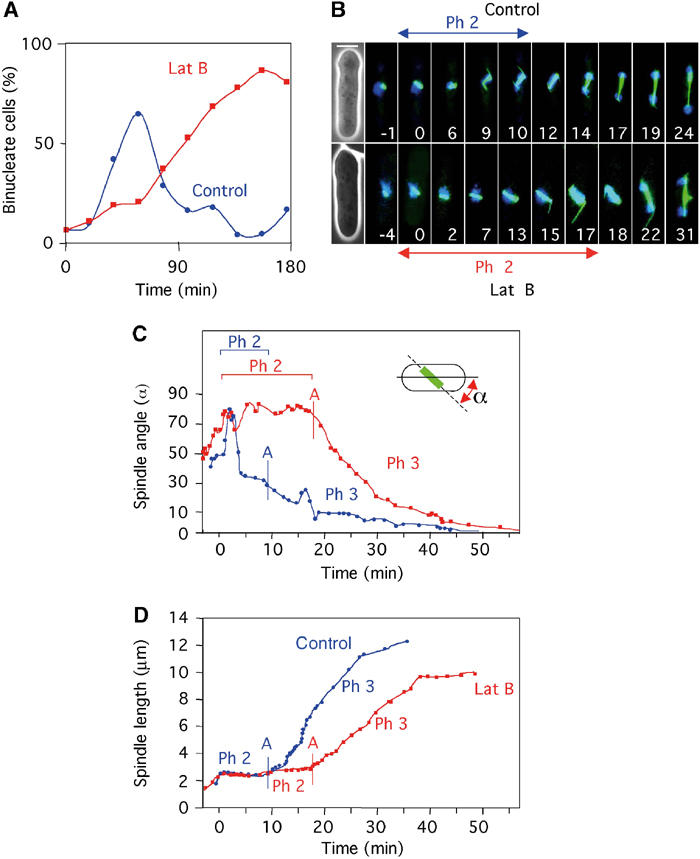
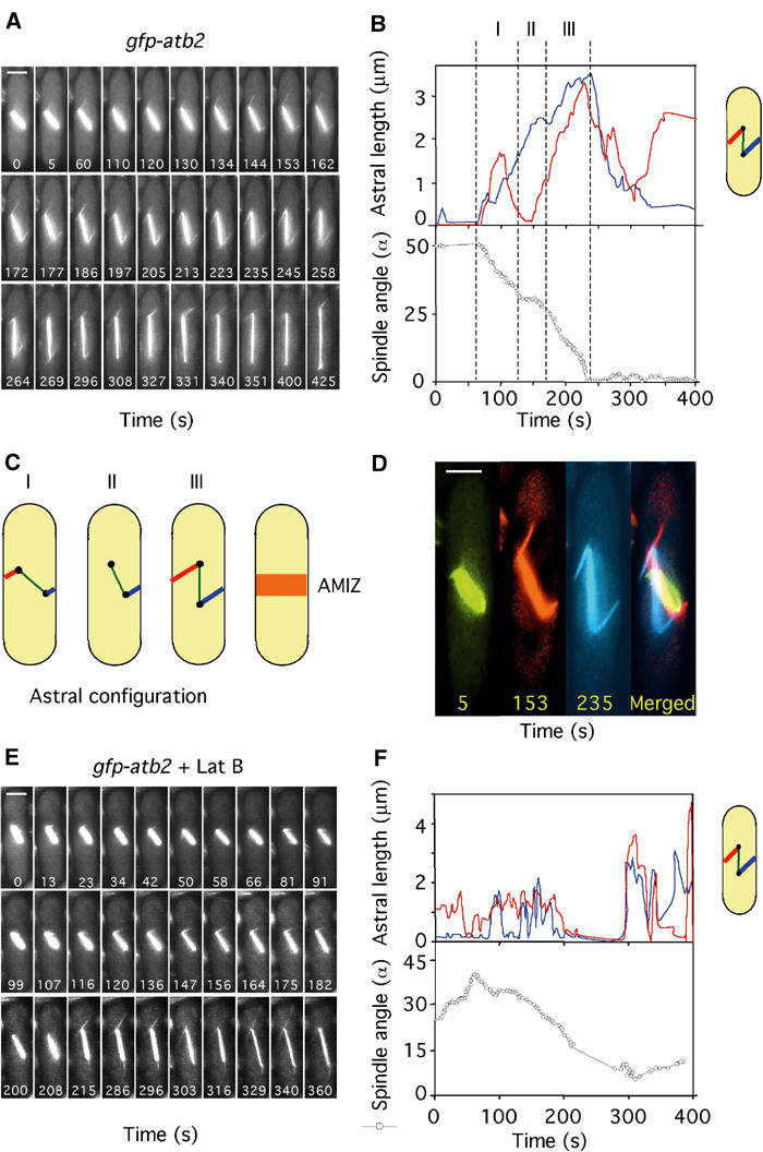
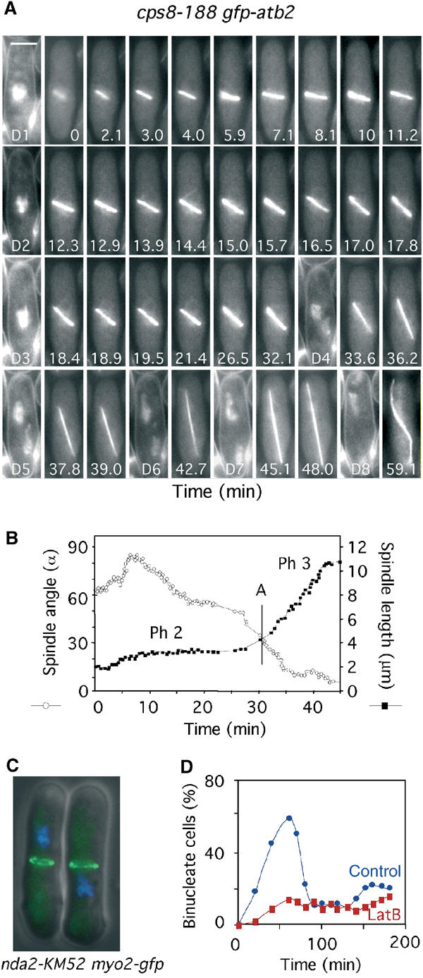
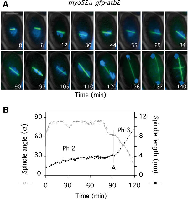
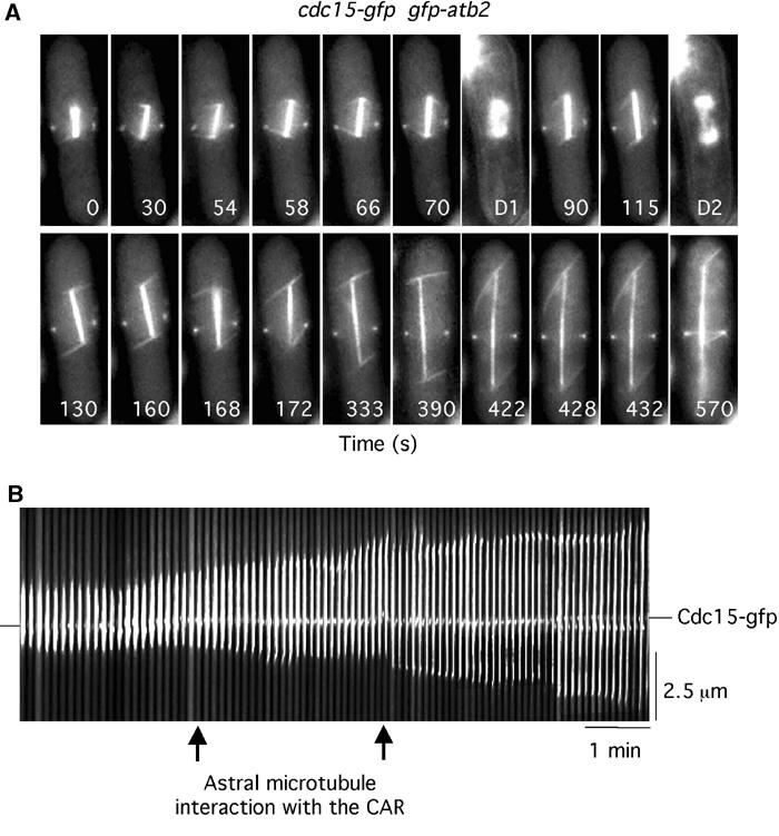
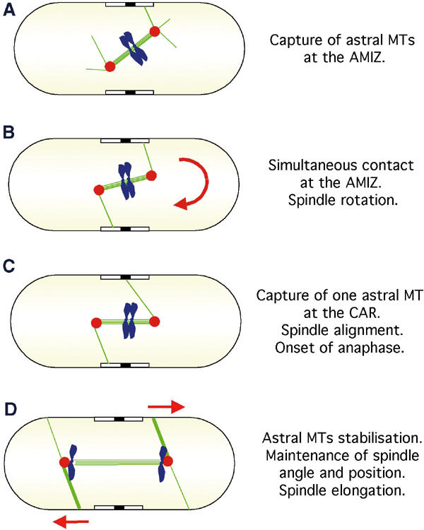
In animal cells, the mitotic spindle is aligned perpendicular to the axis of cell division. This ensures that sister chromatids are separated to opposite sides of the cytokinetic actomyosin ring (CAR). We show that, in fission yeast, spindle rotation is dependent on the interaction of astral microtubules with the cortical actin cytoskeleton. Interaction initially occurs with a region surrounding the nucleus, which we term the astral microtubule interaction zone (AMIZ). Simultaneous contact of astral microtubules from both poles with the AMIZ directs spindle rotation and this requires both actin and two type V myosins, Myo51 and Myo52. Astral microtubules from one pole only then contact the CAR, which is located at the centre of the AMIZ. We demonstrate that the anillin homologue Mid1, which dictates correct placement of the CAR, is necessary to stabilise the mitotic spindle perpendicular to the axis of cell division. Finally, we show that the position of the mitotic spindle is monitored by a checkpoint that regulates the timing of sister chromatid separation.


Figures

Figure 1
Figure 1
Microtubule dynamics and timing of chromosome separation are affected in the presence of latrunculin B. (A) Sychronous gfp-atb2 cells were placed in fresh medium containing DAPI either in the absence (blue circles) or presence (red squares) of 10 μM latrunculin B. The percentage of binucleate cells at various times is shown. (B) Microtubule dynamics in gfp-atb2 cells in the absence (upper panel) (Supplementary Movie 1) or presence (lower panel) (Supplementary Movie 2) of latrunculin B. Numbers indicate time in minutes from the start of phase 2. Bar=4 μm. (C) Analysis of spindle angle relative to the long cell axis and (D) spindle length in control cells (blue circles) and latrunculin B-treated cells (red squares) (from Supplementary Movies 1 and 2). The onset of chromosome segregation (anaphase A) is shown by a vertical blue bar in control cells and a vertical red bar in latrunculin B-treated cells.
Figure 2
Figure 2
Spindle orientation requires balanced interaction of astral microtubules with the medial cell cortex. (A) Microtubule dynamics in gfp-atb2 cells in medium lacking DAPI (Supplementary Movie 3). Numbers indicate time in seconds from the beginning of the movie. (B) Analysis of astral microtubule length (upper panel) and spindle angle (open circles, lower panel) was carried out as depicted in Supplementary Movie 3. Astral microtubule length from the upper SPB (red) or lower SPB (blue) to the cell cortex is shown. Three periods are highlighted: period I (t=60–130 s), period II (t=130–172 s) and period III (t=172–245 s). (C) Cartoon showing astral microtubule configurations during periods I–III. The point of interaction of the astral microtubules with the cell cortex defines an AMIZ surrounding the nucleus. (D) Frames 5, 153 and 235 from Supplementary Movie 3 were enlarged to illustrate the point of interaction of the astral microtubules from the upper and lower SPBs with the medial cell cortex (Bar=2 μm). (E) Microtubule dynamics in gfp-atb2 cells in the presence of 10μM latrunculin B (Supplementary Movie 4). (F) Astral microtubule length (upper panel) and spindle angle (lower panel) were determined from Supplementary Movie 4 as detailed above.
Figure 3
Figure 3
Astral microtubules regulate spindle orientation, spindle elongation and the timing of anaphase onset. (A) cdc11-123 gfp-atb2 cells were filmed in medium containing Hoechst (Supplementary Movie 5). Numbers indicate the time from when the spindle attained 2 μm. Bar=4 μm. D1 (t=0) and D2 (t=13.6) are images of chromatin staining. (B) Analysis of the changes in spindle angle (open circles) and spindle length (closed squares) (Supplementary Movie 5). The vertical bar denotes the time of chromosome separation (anaphase A).
Figure 4
Figure 4
The medial cortical actin cytoskeleton regulates spindle orientation, spindle elongation and the timing of anaphase onset. (A) cps8-188 gfp-atb2 cells were filmed in medium containing Hoechst (Supplementary Movie 6). Numbers indicate the time from when the spindle attained 2 μm. Bar=4 μm. D1–D8 are images of chromatin staining. (B) Analysis of the changes in spindle angle (open circles) and spindle length (closed squares) in cps8-188 gfp-atb2 cells (Supplementary Movie 6). The vertical bar denotes the time of chromosome separation (anaphase A). (C) nda2-KM52 myo2-gfp cells arrested at 20°C for 6 h. Cells were visualised for GFP (green) and chromatin (blue). (D) nda2-KM52 cells were arrested at 20°C for 6 h and released at the permissive temperature (29°C) in the presence (red squares) or absence (blue circles) of 10 μM latrunculin B. The percentage of binucleate cells at various times was determined.
Figure 5
Figure 5
The spindle orientation checkpoint is activated by disruption of the CAR. (A) myo52Δ gfp-atb2 cells were filmed in medium containing Hoechst (Supplementary Movie 7). Numbers indicate the time from when the spindle attained 2 μm. Bar=4 μm. (B) Analysis of spindle angle (open circles) and spindle length (closed squares) in myo52Δ gfp-atb2 cells (Supplementary Movie 7). The vertical bar denotes the time of chromosome separation (anaphase A).
Figure 6
Figure 6
Astral microtubules interact with the CAR. (A) cdc15-gfp gfp-atb2 cells were filmed in medium containing Hoechst. Numbers indicate the time from the beginning of the movie (Supplementary Movie 8). D1 and D2 are images of chromatin staining. (B) Kymographic analysis of spindle position relative to the CAR in cdc15-gfp gfp-atb2 cells. In all, 100 images are shown (each 4 pixels wide). Arrows indicate the point of interaction of the astral microtubule from the lower SPB with the cortex.
Figure 7
Figure 7
Mid1 stabilises spindle alignment to the longitudinal axis of the cell. (A) mid1Δ gfp-atb2 cells were filmed in medium containing Hoechst. Numbers indicate the time from when the spindle attained 2 μm. D1–D4 are images of chromatin staining. (B) Analysis of the changes in spindle angle (open circles) and spindle length (closed squares) from (A). The vertical bar denotes the time of chromosome separation (anaphase A). (C) Synchronous mid1-gfp cdc11-cfp cells placed in fresh medium containing Hoechst either in the absence (left panels) or presence (right panels) of 10 μm latrunculin B. Images were taken to visualise Cdc11-CFP (red), Mid1-GFP (green) or chromatin (blue) when cells were in G2 (single SPB), metaphase (separated SPBs but unseparated chromatin), anaphase (separated SPBs and separated chromatin) and telophase.
Figure 8
Figure 8
Model illustrating the function of astral microtubules in fission yeast. (A) In wild-type cells, the spindle forms at a random angle to the cell axis. (B) Astral microtubules (green lines) that polymerise from the extranuclear face of the SPBs (red circles) interact with a zone that surrounds the nucleus, which we term an AMIZ (open box). Spindle rotation is driven by simultaneous contact on both sides of the cell cortex. (C) An astral microtubule from one SPB contacts the CAR (closed box). This stabilises alignment of the spindle along the longitudinal axis of the cell. (D) Spindle elongation (anaphase B) and separation of sister chromatids (blue) occur co-incidentally. Astral microtubules maintain spindle angle and contribute to the rate of spindle elongation during anaphase B.

References

    1. Adames NR, Cooper JA (2000) Microtubule interactions with the cell cortex causing nuclear movements in Saccharomyces cerevisiae. J Cell Biol 149: 863–874 - PMC - PubMed
    1. Adames NR, Oberle JR, Cooper JA (2001) The surveillance mechanism of the spindle position checkpoint in yeast. J Cell Biol 153: 159–168 - PMC - PubMed
    1. Aist JR, Bayles CJ, Tao W, Berns MW (1991) Direct experimental evidence for the existence, structural basis and function of astral forces during anaphase B in vivo. J Cell Sci 100: 279–288 - PubMed
    1. Ayscough KR, Stryker J, Pokala N, Sanders M, Crews P, Drubin DG (1997) High rates of actin filament turnover in budding yeast and roles for actin in establishment and maintenance of cell polarity revealed using the actin inhibitor latrunculin-A. J Cell Biol 137: 399–416 - PMC - PubMed
    1. Bahler J, Steever AB, Wheatley S, Wang Y, Pringle JR, Gould KL, McCollum D (1998) Role of polo kinase and Mid1p in determining the site of cell division in fission yeast. J Cell Biol 143: 1603–1616 - PMC - PubMed

Publication types

MeSH terms