. 2004 Mar 24;23(6):1257-66.
doi: 10.1038/sj.emboj.7600148. Epub 2004 Mar 11.

Structure of the translocator domain of a bacterial autotransporter

Affiliations

Structure of the translocator domain of a bacterial autotransporter

Clasien J Oomen et al. EMBO J. .

Abstract

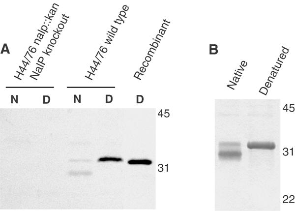
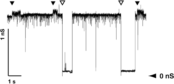
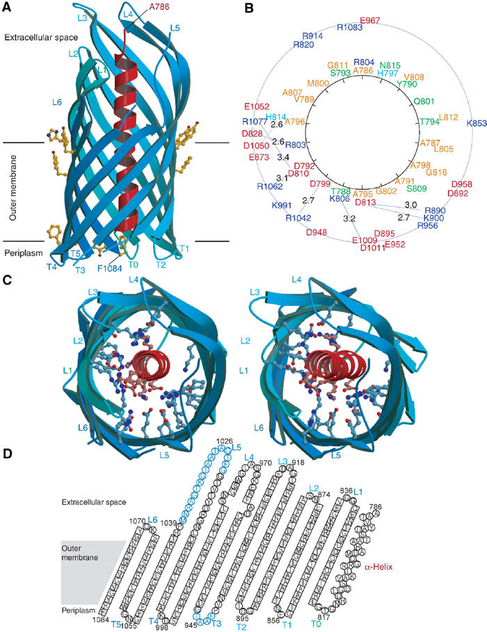
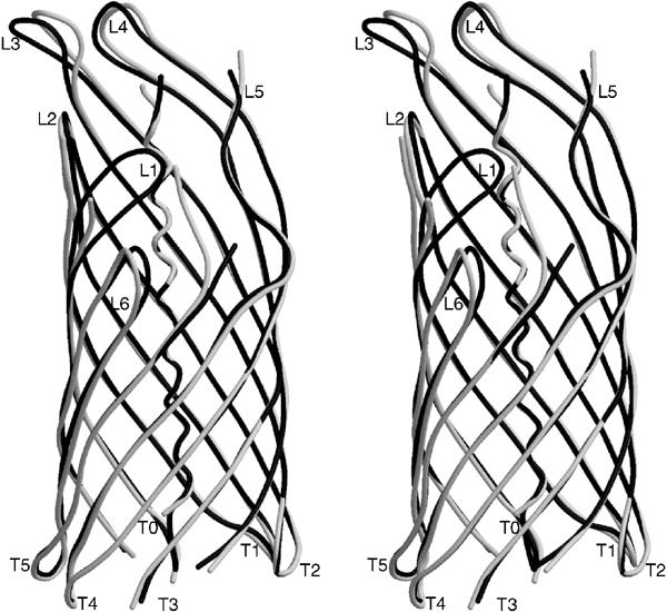
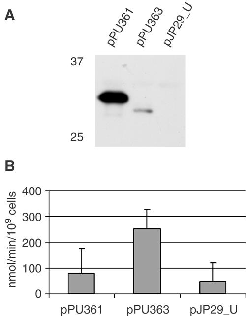
Autotransporters are virulence-related proteins of Gram-negative bacteria that are secreted via an outer-membrane-based C-terminal extension, the translocator domain. This domain supposedly is sufficient for the transport of the N-terminal passenger domain across the outer membrane. We present here the crystal structure of the in vitro-folded translocator domain of the autotransporter NalP from Neisseria meningitidis, which reveals a 12-stranded beta-barrel with a hydrophilic pore of 10 x 12.5 A that is filled by an N-terminal alpha-helix. The domain has pore activity in vivo and in vitro. Our data are consistent with the model of passenger-domain transport through the hydrophilic channel within the beta-barrel, and inconsistent with a model for transport through a central channel formed by an oligomer of translocator domains. However, the dimensions of the pore imply translocation of the secreted domain in an unfolded form. An alternative model, possibly covering the transport of folded domains, is that passenger-domain transport involves the Omp85 complex, the machinery required for membrane insertion of outer-membrane proteins, on which autotransporters are dependent.


Figures

Figure 1
Figure 1
Biochemical characterisation of the NalP translocator domain. (A) Western blot analysis with NalPβ antiserum of membrane preparations of a N. meningitidis nalP knockout mutant and of the wild-type strain H44/76, incubated at room temperature (N) or 100°C (D) before electrophoresis, and of recombinant NalPβ produced in inclusion bodies in E. coli. (B) Heat modifiability of refolded NalPβ was analysed on Coomassie-stained SDS–PAGE gels. Samples were incubated at room temperature (native) or 100°C (denatured) prior to electrophoresis. The positions of molecular size markers (in kDa) are indicated at the right in both panels. These figures demonstrate that recombinant NalPβ is similar to the processed translocator domain of NalP that remains in the meningococcal outer membrane after secretion of the passenger domain.
Figure 2
Figure 2
Recording of NalPβ pores formed in planar lipid bilayers at an applied potential of 100 mV. The recordings show conductance steps of 1.3 nS (open arrowhead) and 0.15 nS (filled arrowhead). The horizontal arrowhead shows the zero-conductance level.
Figure 3
Figure 3
Stereo picture of the final 2m∣Fo∣-D∣Fc∣ electron density map of space group P6122 at a σ-level of 1.0 around the α-helix. The protein model is depicted in ball-and-stick representation.
Figure 4
Figure 4
Crystal structure of NalPβ. (A) Side view of NalPβ in space group P6122 shows a 12-stranded β-barrel (blue ribbon representation) with a shear number of 14. The hydrophobic membrane-embedded region is flanked by aromatic residues (yellow ball-and-stick). The periplasmic side is characterised by short turns (T0–T5) and the extracellular side by longer loops (L1–L6) connecting the alternating β-strands. An α-helical ‘plug' (red ribbon representation) is connected to the barrel via T0 and positions the N-terminus of the translocator domain (Ala 786) at the extracellular side. (B) Schematic representation of the mixed character of the α-helix and its interactions with the β-barrel wall with the α-helical residues depicted on a helical wheel. Colour coding: positively charged residues in blue, negatively charged residues in red, hydrophilic residues in green and nonpolar residues in orange. The distances between charged groups of the helix and the barrel wall are indicated in Å. (C) Stereo top view of the NalPβ β-barrel in the same orientation as in (B). The barrel interior is highly hydrophilic due to the presence of many charged amino acids (ball-and-stick representation). (D) Topology model of NalPβ; residues pointing outwards from the β-barrel are indicated in grey. Amino acids in β-strands are indicated as squares, in α-helix as hexagons and in loops as circles. Amino acids not visible in the electron density of space group P6122 are indicated in blue.
Figure 5
Figure 5
Stereo view of the differences in backbone conformation for space groups P6122 (black coil representation) and C2221 (white coil representation). In space group C2221, no interpretable electron density was observed for residues 816–817, 834–836, 915–917, 943–945, 972–973 and 1024–1036. The channel size in space group P6122 is reduced to 6.5 × 12.5 Å (10.5 × 12.5 Å in space group C2221), because loop L1 is pushed inwards due to a crystal packing contact.
Figure 6
Figure 6
Uptake of the β-lactam antibiotic nitrocefin by E. coli CE1265 cells expressing NalPβ variants. CE1265 (pPU361) and CE1265 (pPU363) express NalPβ and NalPβΔhelix, respectively, both fused to the signal sequence of PhoE, and CE1265 (pJP29_U) expresses just the signal sequence of PhoE. (A) Western blot analysis with NalPβ antiserum of CE1265 cells expressing the NalPβ variants. Plasmids used are indicated above the lanes, and molecular weight markers on the left of the panel. Only the relevant part of the gel, showing that expression of NalPβΔhelix is much lower than that of NalPβ, is shown. (B) The rate of nitrocefin uptake mediated by the NalPβ variants in CE1265 is given in nmol/min/109 cells. Experiments were performed three times in triplicate, and the values given are the means and the standard deviation. Differences between CE1265 (pPU363) on the one hand and CE1265 (pPU361) and CE1265 (pJP29_U) on the other hand were statistically significant (P<0.005).

References

    1. Bosch D, Leunissen J, Verbakel J, de Jong M, van Erp H, Tommassen J (1986) Periplasmic accumulation of truncated forms of outer-membrane PhoE protein of Escherichia coli K-12. J Mol Biol 189: 449–455 - PubMed
    1. Bradley P, Cowen L, Menke M, King J, Berger B (2001) BETAWRAP: successful prediction of parallel β-helices from primary sequence reveals an association with many microbial pathogens. Proc Natl Acad Sci USA 98: 14819–14824 - PMC - PubMed
    1. Brandon LB, Goldberg MB (2001) Periplasmic transit and disulfidebond formation of the autotransported Shigella protein IcsA. J Bacteriol 183: 951–958 - PMC - PubMed
    1. Brunger AT, Adams PD, Clore GM, DeLano WL, Gros P, Grosse-Kunstleve RW, Jiang JS, Kuszewski J, Nilges M, Pannu NS, Read RJ, Rice LM, Simonson T, Warren GL (1998) Crystallography & NMR system: a new software suite for macromolecular structure determination. Acta Crystallogr D 54: 905–921 - PubMed
    1. CCP4 (1994) The CCP4 suite: programs for protein crystallography. Acta Crystallogr D 50: 760–763 - PubMed

Publication types

MeSH terms