

Methanoarchaeal sulfolactate dehydrogenase: prototype of a new family of NADH-dependent enzymes
- PMID: 15014443
- PMCID: PMC381418
- DOI: 10.1038/sj.emboj.7600147
Methanoarchaeal sulfolactate dehydrogenase: prototype of a new family of NADH-dependent enzymes
Abstract
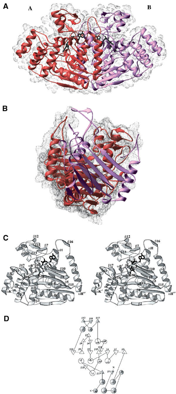
The crystal structure of the sulfolactate dehydrogenase from the hyperthermophilic and methanogenic archaeon Methanocaldococcus jannaschii was solved at 2.5 A resolution (PDB id. 1RFM). The asymmetric unit contains a tetramer of tight dimers. This structure, complexed with NADH, does not contain a cofactor-binding domain with 'Rossmann-fold' topology. Instead, the tertiary and quaternary structures indicate a novel fold. The NADH is bound in an extended conformation in each active site, in a manner that explains the pro-S specificity. Cofactor binding involves residues belonging to both subunits within the tight dimers, which are therefore the smallest enzymatically active units. The protein was found to be a homodimer in solution by size-exclusion chromatography, analytical ultracentrifugation and small-angle neutron scattering. Various compounds were tested as putative substrates. The results indicate the existence of a substrate discrimination mechanism, which involves electrostatic interactions. Based on sequence homology and phylogenetic analyses, several other enzymes were classified as belonging to this novel family of homologous (S)-2-hydroxyacid dehydrogenases.
Figures


Similar articles
-
The crystal structure of the apoenzyme of the iron-sulphur cluster-free hydrogenase.J Mol Biol. 2006 May 5;358(3):798-809. doi: 10.1016/j.jmb.2006.02.035. Epub 2006 Mar 2. J Mol Biol. 2006. PMID: 16540118
-
The X-ray structure of Escherichia coli enoyl reductase with bound NAD+ at 2.1 A resolution.J Mol Biol. 1998 Dec 18;284(5):1529-46. doi: 10.1006/jmbi.1998.2271. J Mol Biol. 1998. PMID: 9878369
-
Glycerol dehydrogenase. structure, specificity, and mechanism of a family III polyol dehydrogenase.Structure. 2001 Sep;9(9):789-802. doi: 10.1016/s0969-2126(01)00645-1. Structure. 2001. PMID: 11566129
-
Biosynthesis of riboflavin: structure and properties of 2,5-diamino-6-ribosylamino-4(3H)-pyrimidinone 5'-phosphate reductase of Methanocaldococcus jannaschii.J Mol Biol. 2006 Jun 23;359(5):1334-51. doi: 10.1016/j.jmb.2006.04.045. Epub 2006 May 6. J Mol Biol. 2006. PMID: 16730025
-
Structure of glyceraldehyde-3-phosphate dehydrogenase from the archaeal hyperthermophile Methanocaldococcus jannaschii.Acta Crystallogr Sect F Struct Biol Cryst Commun. 2009 Dec 1;65(Pt 12):1227-33. doi: 10.1107/S1744309109047046. Epub 2009 Nov 27. Acta Crystallogr Sect F Struct Biol Cryst Commun. 2009. PMID: 20054117 Free PMC article.
Cited by
-
A functionally uncharacterized type-2 malate/L-lactate dehydrogenase family protein from Thermus thermophilus HB8 catalyzes stereospecific reduction of 2-keto-3-deoxy-D-gluconate.Extremophiles. 2022 Nov 23;26(3):37. doi: 10.1007/s00792-022-01282-z. Extremophiles. 2022. PMID: 36416985
-
Gene Encoding a Novel Enzyme of LDH2/MDH2 Family is Lost in Plant and Animal Genomes During Transition to Land.J Mol Evol. 2019 Jan;87(1):52-59. doi: 10.1007/s00239-018-9884-2. Epub 2019 Jan 4. J Mol Evol. 2019. PMID: 30607448
-
Expression, Purification, and Characterization of (R)-Sulfolactate Dehydrogenase (ComC) from the Rumen Methanogen Methanobrevibacter millerae SM9.Archaea. 2017 Nov 6;2017:5793620. doi: 10.1155/2017/5793620. eCollection 2017. Archaea. 2017. PMID: 29234237 Free PMC article.
-
Comparative transcriptome analysis of Methylibium petroleiphilum PM1 exposed to the fuel oxygenates methyl tert-butyl ether and ethanol.Appl Environ Microbiol. 2007 Nov;73(22):7347-57. doi: 10.1128/AEM.01604-07. Epub 2007 Sep 21. Appl Environ Microbiol. 2007. PMID: 17890343 Free PMC article.
-
The Characterization of Ancient Methanococcales Malate Dehydrogenases Reveals That Strong Thermal Stability Prevents Unfolding Under Intense γ-Irradiation.Mol Biol Evol. 2024 Dec 6;41(12):msae231. doi: 10.1093/molbev/msae231. Mol Biol Evol. 2024. PMID: 39494471 Free PMC article.
References
-
- Abrahams JP, Leslie AGW (1996) Methods used in the structure determination of bovine mitochondrial F1 ATPase. Acta Crystallogr D 52: 30–42 - PubMed
-
- Baker PJ, Britton KL, Engel PC, Farrants GW, Lilley KS, Rice DW, Stillman TJ (1992a) Subunit assembly and active site location in the structure of glutamate dehydrogenase. Proteins 12: 75–86 - PubMed
-
- Baker PJ, Britton KL, Rice DW, Rob A, Stillman TJ (1992b) Structural consequences of sequence patterns in the fingerprint region of the nucleotide binding fold. J Mol Biol 228: 662–671 - PubMed
-
- Bhat TN (1988) Calculation of an OMIT map. J Appl Crystallogr 21: 279–281
-
- Brünger AT, Adams PD, Clore GM, DeLano WL, Gros P, Grosse-Kunstleve RW, Jiang J-S, Kuszewski J, Nilges M, Pannu NS, Read RJ, Rice LM, Simonson T, Warren GL (1998) Crystallography & NMR System: a new software suite for macromolecular structure determination. Acta Crystallogr D 54: 905–921 - PubMed
Publication types
MeSH terms
Substances
LinkOut - more resources
Full Text Sources
Miscellaneous
