. 2004 Mar;19(5):1305-16.
doi: 10.1111/j.1460-9568.2004.03239.x.

The actions of monoamines and distribution of noradrenergic and serotoninergic contacts on different subpopulations of commissural interneurons in the cat spinal cord

Affiliations

The actions of monoamines and distribution of noradrenergic and serotoninergic contacts on different subpopulations of commissural interneurons in the cat spinal cord

Ingela Hammar et al. Eur J Neurosci. 2004 Mar.

Abstract

Modulatory actions of monoamines were investigated on spinal commissural interneurons which coordinate left-right hindlimb muscle activity through direct projections to the contralateral motor nuclei. Commissural interneurons located in Rexed lamina VIII, with identified projections to the contralateral gastrocnemius-soleus motor nuclei, were investigated in deeply anaesthetized cats. Most interneurons had dominant input from either the reticular formation or from group II muscle afferents; a small proportion of neurons had input from both. Actions of ionophoretically applied serotonin and noradrenaline were examined on extracellularly recorded spikes evoked monosynaptically by group II muscle afferents or reticulospinal tract fibres. Activation by reticulospinal fibres was facilitated by both serotonin and noradrenaline. Activation by group II afferents was also facilitated by serotonin but was strongly depressed by noradrenaline. To investigate the possible morphological substrates of this differential modulation, seven representative commissural interneurons were labelled intracellularly with tetramethylrhodamine-dextran and neurobiotin. Contacts from noradrenergic and serotoninergic fibres were revealed by immunohistochemistry and analysed with confocal microscopy. There were no major differences in the numbers and distributions of contacts among the interneurons studied. The findings suggest that differences in modulatory actions of monoamines, and subsequent changes in the recruitment of subpopulations of commissural interneurons in various behavioural situations, depend on intrinsic interneuron properties rather than on the patterns of innervation by monoaminergic fibres. The different actions of noradrenaline on different populations of interneurons might permit reconfiguration of the actions of the commissural neurons according to behavioural context.


Figures

Fig. 1
Fig. 1
Locations of stimulation sites in the reticular formation and of the sampled commissural interneurons. (A) Transverse section of the medulla in the plane of the insertion of the electrodes at the level of the inferior olive. Open circles indicate stimulation sites in the MLF. Vertical scale shows Horsley-Clarke coordinates. (B) Summary diagrams showing the locations of intracellularly labelled commissural neurons in the L4 and L5 segments of the spinal cord. ○, ●, cells with monosynaptic input from group II afferents and from RF, respectively. The three ◇ represent the locations of three interneurons with short-latency input from both. Arrows indicate the interneurons in which the distribution of 5-HT and NA contacts was analysed in detail. IO, inferior olive.
Fig. 2
Fig. 2
Similar facilitatory effects of 5-HT on commissural interneurons activated from RF and by group II afferents. (A and C) Top pair of records are single sweep records from an interneuron monosynaptically activated by RF fibres and from the cord dorsum. The traces below are peri-stimulus time histograms (PSTH) of responses to 20 stimuli. They were complied from stimuli delivered before (control), during (15s, 1 and 2min) and after ionophoresis of 5-HT (Rec., 5 min after withdrawal of the ionophoretic pipette). (B and D) The same format as in A and C but for an interneuron activated by stimulation of group II afferents in the Q nerve. Dotted vertical lines in C and D indicate the minimal latency in the control records. The dotted horizontal lines in A and B indicate the discrimination levels; only spikes crossing these lines were used for construction of PSTHs. (E and F) Collision tests for the neurons in A and B, respectively. The records show that antidromic spikes from the contralateral GS motor nucleus collided with synaptically evoked responses at intervals shorter than twice the conduction time from this nucleus. Shock artefacts truncated. Time calibrations as indicated.
Fig. 3
Fig. 3
Facilitation of responses to RF stimulation and depression of responses to group II activation by NA. (A and B) Extracellular records of monosynaptic responses to stimulation in the RF and of group II afferents in the Sart nerve in two different commissural interneurons and records from cord dorsum. Shock artefacts have been truncated. (B and D) PSTHs of responses evoked by 20 consecutive stimuli recorded before (control), during and after ionophoresis of NA. Note that in B only the third stimulus evoked spikes during the control period, whereas the first and second stimulus also evoked spikes during ionophoresis. The facilitated responses which appeared after the first stimulus were used for the pooled data of Fig. 4. The dotted horizontal lines in A and B indicate the discrimination level; only spikes crossing these lines were used for constructing the histograms. (E and F) Spike potentials evoked from GS motor nucleus; latencies < 1 ms show that they were evoked antidromically.
Fig. 4
Fig. 4
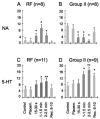
Effects of monoamines on two subpopulations of commissural interneurons. The plots show changes in the number of spikes evoked during a series of 20 consecutive stimuli following ionophoresis of noradrenaline (NA; upper panels) or serotonin (5-HT; lower panels) on lamina VIII commissural interneurons activated by either the reticulospinal tract fibres (left panels) or by group II afferents (right panels). The ordinates show the mean number of responses (± SEM) before, during (dark grey) or after ionophoresis at the times indicated. Control and placement (Placem.), responses evoked before and after placement of the drug-containing microelectrode, respectively. Other data are for the time periods indicated below the bars during ionophoresis and after up to 10 min of recovery (Rec.). *P = 0.05–0.01, *P < 0.01 vs. control levels.
Fig. 5
Fig. 5
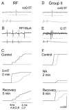
Similar effects of 5-HT and NA on responses evoked by coQ and from ipsilateral Q afferents or RF. Responses of two commissural interneurons are shown, one (A–C) activated by RF and the other (D–F) monosynaptically activated by ipsilateral group II afferents. A and D show responses evoked by stimulation of coQ at 5 × T during ionophoresis (two stimuli in A, two in D). B and E show responses of the same neurons following stimulation of the ipsilateral reticulospinal tract fibres and ipsilateral Q at 5 × T, respectively. (C) Superimposed cumulative sums of responses, from RF (black) and from coQ (grey) show the number of responses evoked before (control) during (2 min of ionophoresis) and after ionophoresis of 5-HT. Cumulative sums were constructed from the bin values of PSTHs like those in Figs 2 and 3. Note that stimulation of coQ did not evoke responses before the ionophoresis began, and that the number of responses was greatly reduced and the latency increased 5 min after the ionophoresis ended. (F), as (C), showing superimposed cumulative sums of responses from the ipsilateral Q (black) and the contralateral Q (grey). Dotted horizontal line indicates the discrimination level; only spikes crossing this line were used for constructing the histograms.
Fig. 6
Fig. 6
Examples of intracellular records from labelled interneurons. The three rows show intracellular records (upper traces) from three interneurons (nos 2, 6 and 3 from Table 1), together with records of afferent or descending volleys (lower traces). The left column shows effects of stimulation of group II afferents in the Q nerve; (A) monosynaptic and disynaptic EPSPs, (D) an EPSP that is either monosynaptic or disynaptic, and (G) a disynaptic IPSP (possibly preceded by a very small EPSP as in D). The dotted vertical lines indicate the onset of monosynaptic and most probably disynaptic PSPs. The middle column shows the effects of stimulation of reticulospinal tract fibres: evoking a monosynaptic EPSP in H, a monosynaptic EPSP followed by IPSP in E and no clear response in B. Dotted lines indicate the first components of the RF descending volleys and onset latencies of the earliest monosynaptic EPSPs. The right-hand column shows the effects of stimuli applied in GS motor nuclei: small initial segment and soma-dendritic spike in I and blocked spikes in C and F. These impaled neurons ceased generating full actions potentials briefly after penetration. The dotted vertical line indicates onset of the two earliest spikes.
Fig. 7
Fig. 7
Examples of contacts between monoaminergic fibres and commissural interneurons. (A) A projected image compiled from 85 × 0.5-μm optical sections through the soma of a cell (with RF input; cell 6 in Table 1, records in Fig. 6G-I), showing the abundance of 5-HT-immunoreactive axons bearing varicosities (red) in the vicinity of the cell soma and the relative infrequency of d.b.h.-immunoreactive axons (green) in the same area. (B) A projected image (compiled from 26 × 0.5-μm optical sections) of a dendrite from the cell shown in A. Boutons immunoreactive for 5-HT (shown in red) and d.b.h. (green) can be seen close to the dendrite. (C–E). Single optical sections from the series shown in B illustrate contacts made by the boutons labelled by arrowheads in B onto the dendrite. (F) A further projected image (compiled from 16 × 0.5-μm optical sections) showing a 5-HT-containing axon forming four boutons along a labelled dendrite (originating from a cell with input from both RF and group II afferents; cell 4 in Table 1). (G) and (H) show the terminals labelled 1 and 2 in (F) at higher magnification. Scale bars, 10 μm (A), 5 μm (B–F), 2.5 μm (G–H).
Fig. 8
Fig. 8
Distribution of 5-HT- and NA-immunoreactive terminals on three commissural interneurons. (A, D and E) Reconstructions of the dendritic trees of three commissural interneurons, with input from group II afferents, from RF and from both (nos 2, 6 and 3, respectively, in Table 1) and distributions of 5-HT (red circles) and d.b.h. (blue triangles) contacts throughout their dendritic trees. Insets show the positions of cell bodies within spinal grey matter. (B, C and G) Sholl plots for the two types of varicosity showing the distribution (number per 100 μm dendritic length) of contacts at 25-μm intervals throughout the dendritic tree. Data sets in red represent 5HT-immunoreactive boutons, in blue d.b.h.-immunopositive boutons. Records from these cells are shown in Fig. 6A–C, D–F and H and I, respectively. Scale bars, 20 μm.

Similar articles

Cited by

References

    1. Aggelopoulos NC, Bawa P, Edgley SA. Activation of midlumbar neurones by afferents from anterior hindlimb muscles in the cat. J Physiol (Lond) 1996a;497:795–802. - PMC - PubMed
    1. Aggelopoulos NC, Burton MJ, Clarke RW, Edgley SA. Characterization of a descending system that enables crossed group II inhibitory reflex pathways in the cat spinal cord. J Neurosci. 1996b;16:723–729. - PMC - PubMed
    1. Aggelopoulos NC, Edgley SA. Segmental localisation of the relays mediating crossed inhibition of hindlimb motoneurones from group II afferents in the anaesthetized cat spinal cord. Neurosci Lett. 1995;185:60–64. - PubMed
    1. Arya T, Bajwa S, Edgley SA. Crossed reflex actions from group II muscle afferents in the lumbar spinal cord of the anaesthetized cat. J Physiol (Lond) 1991;444:117–131. - PMC - PubMed
    1. Bannatyne BA, Edgley SA, Hammar I, Jankowska E, Maxwell D. Networks of inhibitory and excitatory commissural interneurons mediating crossed reticulospinal actions. Eur J Neurosci. 2003;18:2273–2284. - PMC - PubMed

Publication types