. 2004 May 21;279(21):22514-21.
doi: 10.1074/jbc.M400536200. Epub 2004 Mar 10.

A novel marker of tissue junctions, collagen XXII

Affiliations

A novel marker of tissue junctions, collagen XXII

Manuel Koch et al. J Biol Chem. .

Abstract

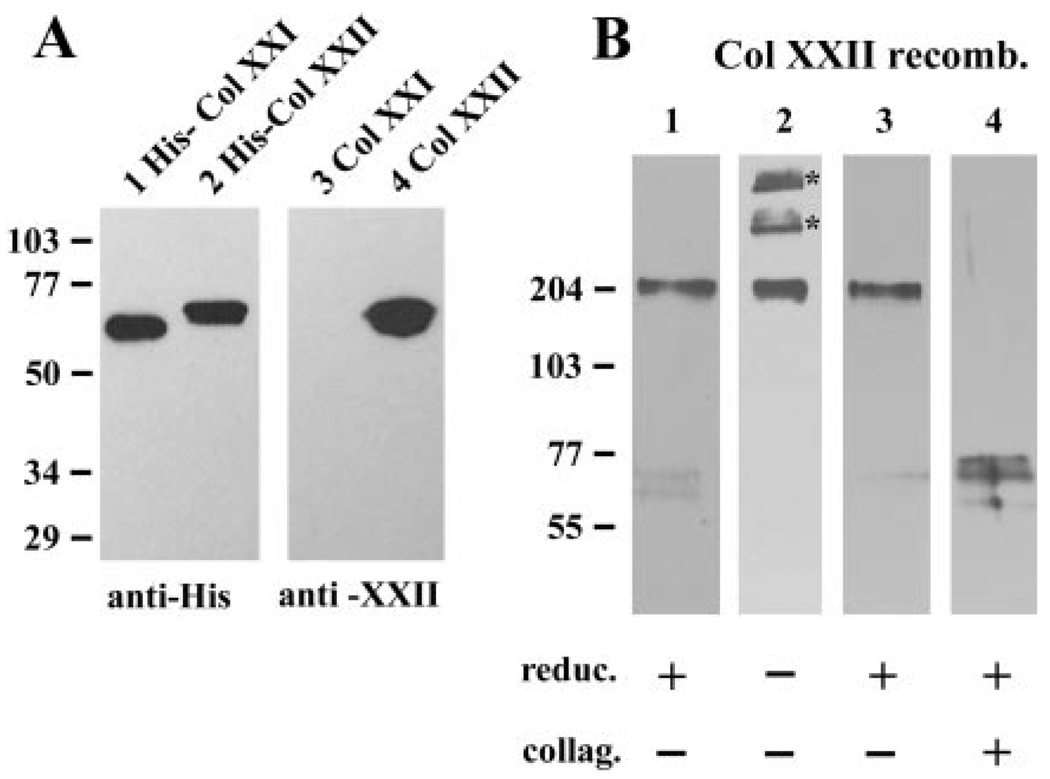
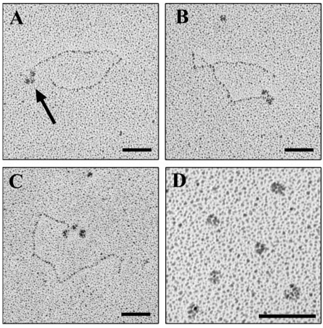
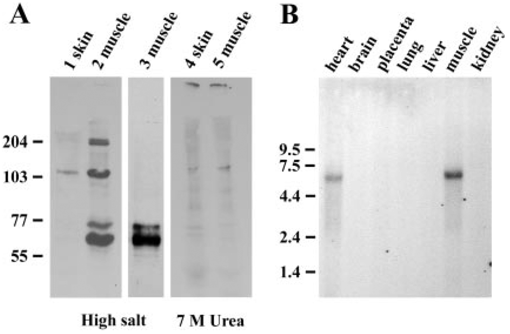
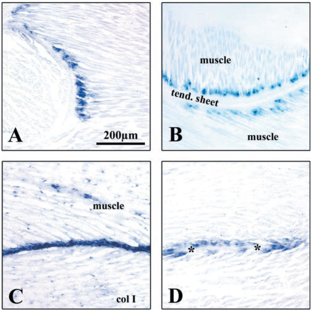
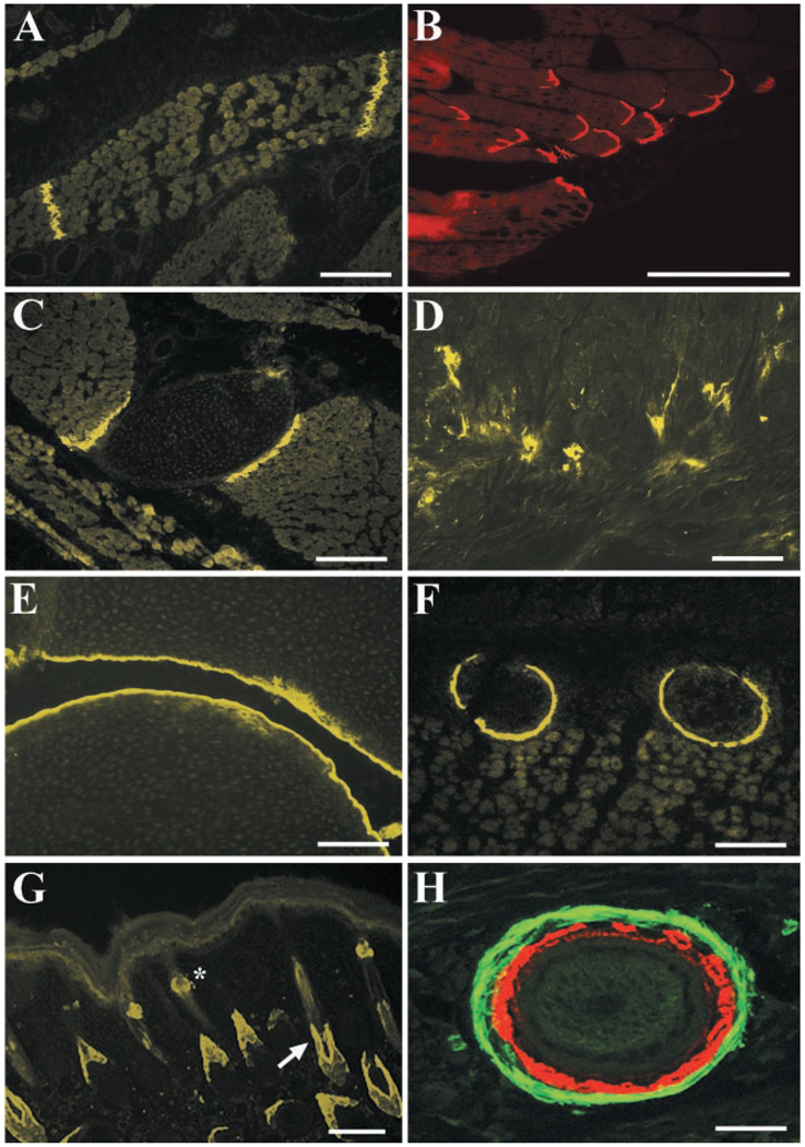
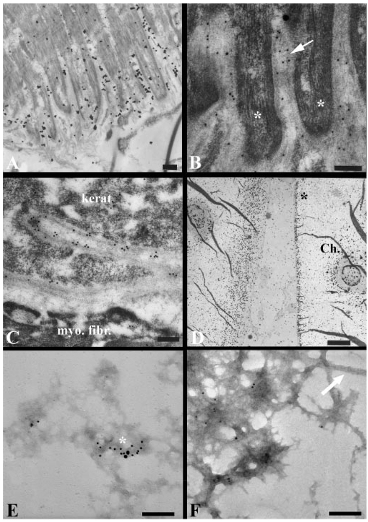
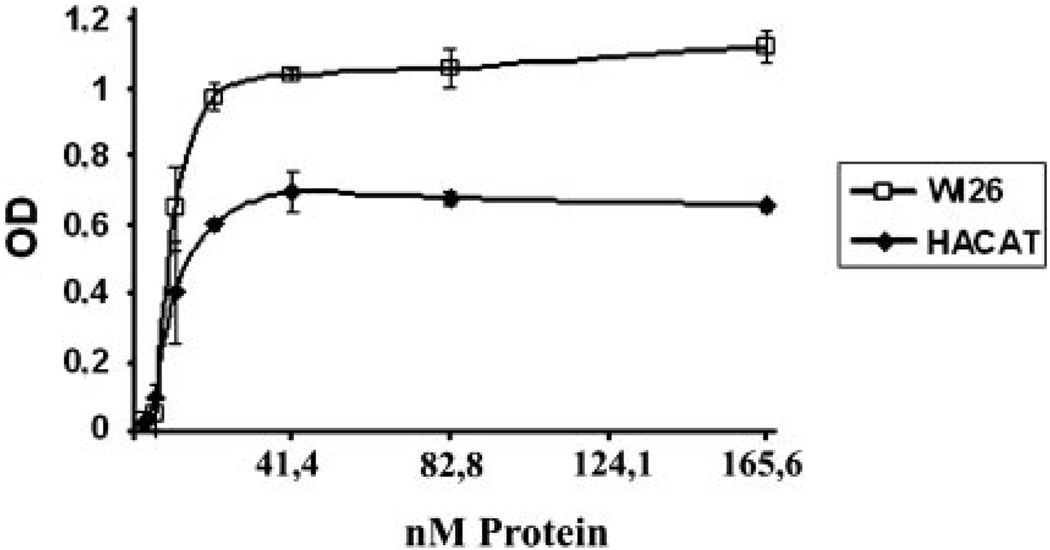
Here we describe a novel specific component of tissue junctions, collagen XXII. It was first identified by screening an EST data base and subsequently expressed as a recombinant protein and characterized as an authentic tissue component. The COL22A1 gene on human chromosome 8q24.2 encodes a collagen that structurally belongs to the FACIT protein family (fibril-associated collagens with interrupted triple helices). Collagen XXII exhibits a striking restricted localization at tissue junctions such as the myotendinous junction in skeletal and heart muscle, the articular cartilage-synovial fluid junction, or the border between the anagen hair follicle and the dermis in the skin. It is deposited in the basement membrane zone of the myotendinous junction and the hair follicle and associated with the extrafibrillar matrix in cartilage. In situ hybridization of myotendinous junctions revealed that muscle cells produce collagen XXII, and functional tests demonstrated that collagen XXII acts as a cell adhesion ligand for skin epithelial cells and fibroblasts. This novel gene product, collagen XXII, is the first specific extracellular matrix protein present only at tissue junctions.


Figures

FIG. 1
FIG. 1. The complete amino acid sequence of human Col XXII as predicted from the corresponding cDNA sequence
A 28-amino acid signal peptide sequence precedes the N terminus (underlined). The different domains are outlined; a VWA domain is followed by the TSPN domain. In the TSPN two potential N-glycosylation sites are circled. Between the globular domains and the collagenous domain a linker region with a polyproline stretch is present. The collagenous region is interrupted by several larger amino acid stretches and six smaller imperfections (circled) of the Gly-X-Y repeats.
FIG. 2
FIG. 2. Schematic presentation of the domain structure of collagen XXI and XXII
At the N terminus, the VWA domain (circle, gray) and a TSPN domain (square, gray) are the building blocks of the NC1 domain. The Gly-X-Y triplets Col1-Col5 (open squares) are interrupted several times (black squares) by small NC2-NC5 linker domains. Smaller imperfections of the collagen triplets are indicated by thin lines. The putative N-linked glycosylation sites are marked by asterisks. The cysteines (C), which are presumably involved in the inter-chain stabilization, are marked above the schematic drawings. The double arrow indicates the region that was used to generate the polyclonal antibody R34. Collagens XXI and XXII are quite similar in their domain structure. However, the region spanning the collagenous domains Col1–Col3 and the non-collagenous domains NC1–NC4 of collagen XXII is missing in collagen XXI (connecting lines between the two drawings).
FIG. 3
FIG. 3. Genetic relationship of the VWA domains of matrilins and collagens
The VWA domains of matrilins form one subgroup and can be further separated into two branches. FACIT collagens constitute the second group. Collagens XII and XIV are closely related and form one subgroup. Similarly, collagens XXI and XXII are closely related and form another subgroup. This tree was calculated with two different methods; both calculations yielded the same prediction.
FIG. 4
FIG. 4. Immunoblot analysis of recombinant Col XXII
A, aliquots of culture media from 293-EBNA cells expressing the NC1 globular domain of collagens XXI and XXII were electrophoretically separated, and the histidine-tagged proteins were detected using an anti-His antibody (1, 2) and the affinity-purified antibody pAbR34 (3, 4). pAbR34 recognizes the NC1 protein of Col XXII but not of Col XXI. B, full-length Col XXII was expressed in 293-EBNA cells, and the medium was immunoblotted (pAbR34). A single band of about 200 kDa was seen under reducing conditions (lane 1). Under non-reducing conditions, also dimeric and trimeric forms were present (lane 2, asterisk). Treatment of recombinant Col XXII with collagenase abolished the full-length form and yielded the collagenase-resistant NC1 domain (lane 4). Lane 3, control incubation without collagenase. Molecular mass standards in kDa are shown on the left.
FIG. 5
FIG. 5. Rotary shadowing images of purified recombinant Col XXII
A–C, three examples of a full-length molecule are shown. The thin rod-like structure represents the collagenous region, and the kinks represent the non-collagenous interruptions. The N terminus contains three globular domains (arrow). D, recombinant NC1 domain forms single globules. Bars, 50 nm.
FIG. 6
FIG. 6. Expression of Col XXII in tissues
A, immunoblot with pAb R34 of proteins extracted from mouse skin and muscle using high salt (lanes 1–3) and urea (lanes 4 and 5). The skin extracts contained relatively small amounts of Col XXII (lane 1). In contrast, in high salt extract of muscle, four distinct bands were detectable (lane 3). Treatment of the extract shown in lane 2 with collagenase abolished the bands with higher molecular weight, indicating that they had a collagenous structure. Only the NC1 domain resisted the protease digestion (lane 3). As seen here, the NC1 domain occasionally migrated as a double band, probably due to differential glycosylation of the molecules (lane 2 and 3). Additional extraction of the tissues with 7 m urea did not result in recovery of more Col XXII from the skin (lane 4) or muscle (lane 5). B, Northern blot of Col XXII in different tissues. Only a single mRNA of 6.4 kb was detected on a human tissue blot. A strong signal was found in the heart and skeletal muscle but not in other organs such as brain, placenta, lung, liver, or kidney. In A molecular weight standards on the left are shown in kDa; in B the RNA size standards are in kb.
FIG. 7
FIG. 7. In situ hybridization
A, Col XXII mRNA was detected in the muscle close to the rib. The staining is restricted to a narrow band of cells, and no labeling was detected within the bone or in other locations within the muscle. B, muscle cells at the attachment site to aponeurosis, which separates muscle compartments, express Col XXII mRNA. C, for comparison, collagen I mRNA is mainly synthesized by fibroblasts of the aponeurosis between the muscles. D, hybridization of a parallel section of C for Col XXII mRNA shows that Col XXII is not synthesized by the tendon fibroblasts (asterisks) but can be detected only in the muscle cells.
FIG. 8
FIG. 8. Col XXII is deposited at tissue junctions
A, in the muscle of newborn mice, Col XXII staining is seen in tendinous sheets separating muscle compartments. B, at the insertion sites of muscle into tendon, Col XXII immunoreactivity is limited to the MTJ. C, at the outer body wall Col XXII is localized at their attachment sites of intercostal muscles to the ribs. D, in the heart, Col XXII is present at the chordae tendineae insertion sites into the muscle of the right ventricle. E, on both sides of the joint a narrow band close to the articular surface is positive for Col XXII. F, the cartilaginous part of ribs, close to the sternum, is positive for Col XXII staining (cross-section). G, at the lower portion of hair follicles (arrow), a limited, sheath-like Col XXII staining is observed. The staining of sebaceous gland is also evident (asterisk). H, cross-section through the lower part of the dermis reveals ring-like Col XXII staining (red) around a hair follicle. The myofibroblasts surrounding the hair follicle were stained with antibodies to α-actinin (yellow). Bars: A–G, 100 µm; H, 20 µm.
FIG. 9
FIG. 9. Ultrastructural localization of Col XXII in situ
Ultrathin sections were incubated with pAb R34, and the bound antibodies were visualized with a colloidal gold-labeled second antibody. A and B, the basement membrane zone of the myotendinous junction contains Col XXII. In A, the finger-like insertions of the myocytes into the tendon (asterisk) are surrounded by a basement membrane, which is decorated with gold particles (arrow). In B, a larger magnification shows the gold labeling of the lamina densa of the basement membrane. C, around the hair follicles, Col XXII is localized along the partially interdigitated basement membrane along the follicle wall, between the myofibroblasts and the keratinocytes (kerat., keratinocytes; myo. fibr., myofibroblast). Most gold particles are associated with the lamina densa. D, in the joint, Col XXII is deposited close to the articular surface (Ch., chondrocyte). E and F, native fibrils were extracted from articular cartilage. Immunogold particles representing Col XXII were not associated directly with cross-banded collagen-containing fibrils (F, white arrow) but were localized to the amorphous fibrillin-containing microfibrillar material (E: white asterisk, large particles: Col XXII, small particles, fibrillin). Bars: A–D, 200 nm; E and F, 250 nm.
FIG. 10
FIG. 10. Cell binding of Col XXII
The fibroblast cell line Wi-26 and the keratinocyte cell line HACAT were tested for their binding to recombinant Col XXII. The wells were coated with different amounts of full-length Col XXII, and the cells were allowed to attach for 30 min. After washing, the adherent cells were quantitated as described under “Experimental Procedures.” At a coating concentration of 40 nm Col XXII, cell binding reached saturation levels. OD, optical density.

Similar articles

Cited by

References

    1. Myllyharju J, Kivirikko KI. Ann. Med. 2001;33:7–21. - PubMed
    1. Sertie AL, Sossi V, Camargo AA, Zatz M, Brahe C, Passos-Bueno MR. Hum. Mol. Genet. 2000;9:2051–2058. - PubMed
    1. Hashimoto T, Wakabayashi T, Watanabe A, Kowa H, Hosoda R, Nakamura A, Kanazawa I, Arai T, Takio K, Mann DM, Iwatsubo T. EMBO J. 2002;21:1524–1534. - PMC - PubMed
    1. Dogic D, Eckes B, Aumailley M. Curr. Top. Pathol. 1999;93:75–85. - PubMed
    1. Koch M, Bohrmann B, Matthison M, Hagios C, Trueb B, Chiquet M. J. Cell Biol. 1995;130:1005–1014. - PMC - PubMed

Publication types

MeSH terms

Associated data