. 2004 Apr;78(7):3502-13.
doi: 10.1128/jvi.78.7.3502-3513.2004.

High-throughput screening of the yeast kinome: identification of human serine/threonine protein kinases that phosphorylate the hepatitis C virus NS5A protein

Affiliations

High-throughput screening of the yeast kinome: identification of human serine/threonine protein kinases that phosphorylate the hepatitis C virus NS5A protein

Carlos Coito et al. J Virol. 2004 Apr.

Abstract

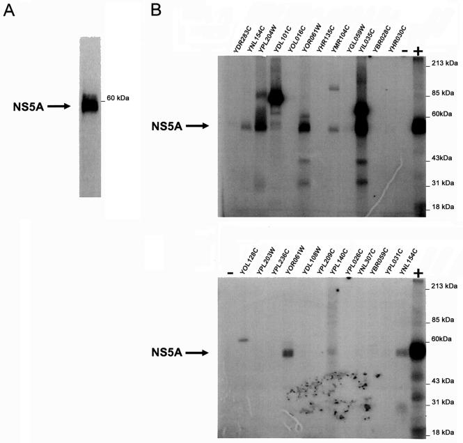
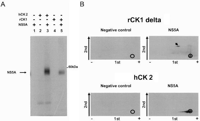
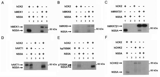
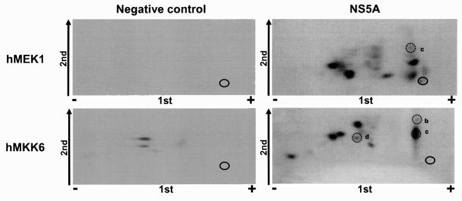
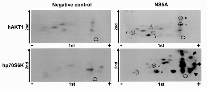
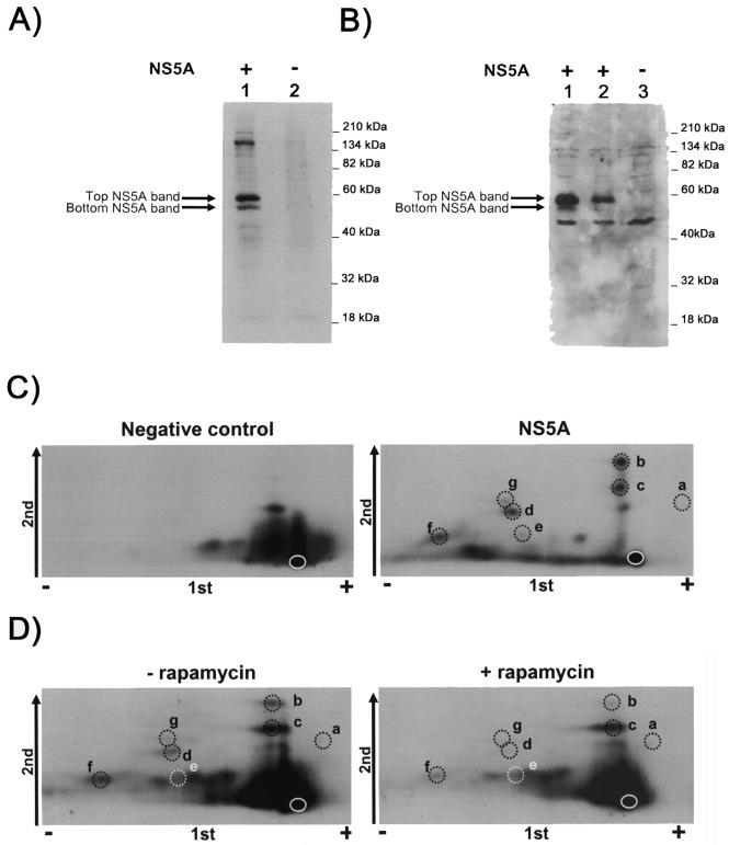
The hepatitis C virus NS5A protein plays a critical role in virus replication, conferring interferon resistance to the virus through perturbation of multiple intracellular signaling pathways. Since NS5A is a phosphoprotein, it is of considerable interest to understand the role of phosphorylation in NS5A function. In this report, we investigated the phosphorylation of NS5A by taking advantage of 119 glutathione S-transferase-tagged protein kinases purified from Saccharomyces cerevisiae to perform a global screening of yeast kinases capable of phosphorylating NS5A in vitro. A database BLAST search was subsequently performed by using the sequences of the yeast kinases that phosphorylated NS5A in order to identify human kinases with the highest sequence homologies. Subsequent in vitro kinase assays and phosphopeptide mapping studies confirmed that several of the homologous human protein kinases were capable of phosphorylating NS5A. In vivo phosphopeptide mapping revealed phosphopeptides common to those generated in vitro by AKT, p70S6K, MEK1, and MKK6, suggesting that these kinases may phosphorylate NS5A in mammalian cells. Significantly, rapamycin, an inhibitor commonly used to investigate the mTOR/p70S6K pathway, reduced the in vivo phosphorylation of specific NS5A phosphopeptides, strongly suggesting that p70S6 kinase and potentially related members of this group phosphorylate NS5A inside the cell. Curiously, certain of these kinases also play a major role in mRNA translation and antiapoptotic pathways, some of which are already known to be regulated by NS5A. The findings presented here demonstrate the use of high-throughput screening of the yeast kinome to facilitate the major task of identifying human NS5A protein kinases for further characterization of phosphorylation events in vivo. Our results suggest that this novel approach may be generally applicable to the screening of other protein biochemical activities by mechanistic class.


Figures

FIG. 1.
FIG. 1.
Purity of the NS5A protein and in vitro phosphorylation of NS5A by yeast kinases. (A) The NS5A substrate used during this study was tested for its purity. Native recombinant NS5A (1 μg), purified from Sf9 cells, was resolved by SDS-PAGE and visualized by silver staining. The arrow indicates the NS5A protein. (This panel is reprinted with the permission of the publisher of reference and is shown here to demonstrate the purity of the substrate, which was an essential prerequisite for our analyses.) (B) In a regular yeast kinase assay, baculovirus recombinant NS5A purified from Sf9 cells (100 ng) was phosphorylated in kinase buffer containing GST-yeast kinase bound to glutathione beads as described in Materials and Methods. The reaction was monitored by autoradiography of 32P-labeled NS5A resolved by SDS-PAGE. Two autoradiography films from two regular yeast kinase assays are shown. The arrows show the NS5A protein. Human recombinant casein kinase 2 was used as a positive control to demonstrate phosphorylation of NS5A under the conditions described here. Yeast extracts from cells transfected with an empty glutathione vector were used as a negative control to demonstrate that phosphorylation of NS5A was not associated with proteins nonspecifically bound to either GST or the glutathione-agarose beads. The kinases present in the phosphorylation reaction were as follows: YDR283C (lane 1), YNL154C (lane 2), YPL204W (lane 3), YDL101C (lane 4), YOL016C (lane 5), YOR061W (lane 6), YHR135C (lane 7), YMR104C (lane 8), YGL059W (lane 9), YIL035C (lane 10), YBR028C (lane 11), YHR030C (lane 12), negative control (lane 13), positive control (lane 14), negative control (lane 15), YOL128C (lane 16), YPL203W (lane 17), YPL236C (lane 18), YOR061W (lane 19), YDL108W (lane 20), YPL209C (lane 21), YPL140C (lane 22), YPL026C (lane 23), YNL307C (lane 24), YBR059C (lane 25), YPL031C (lane 26), YNL154C (lane 27), and positive control (lane 28).
FIG. 2.
FIG. 2.
In vitro phosphorylation of NS5A with CK1 delta and CK2. (A) Autoradiography of 32P-labeled NS5A resolved by SDS-PAGE. The CK2 used in this experiment is a human kinase, and the CK1 delta is a rat kinase. The phosphorylation reactions carried out were as follows: NS5A minus kinase (lane 1), hCK2 minus NS5A (lane 2), hCK2 plus NS5A (lane 3), rCK1 minus NS5A (lane 4), and rCK1 plus NS5A (lane 5). (B) Phosphopeptide mapping of NS5A phosphorylated with either rCK1 or hCK2. In vitro-phosphorylated NS5A was resolved by SDS-PAGE, and the proteins were electroblotted onto nitrocellulose membranes. The radiolabeled NS5A bands were detected by autoradiography and subsequently sliced out and subjected to enzymatic digestion with trypsin and chymotrypsin. The peptides were applied on cellulose TLC plates at an origin shown by the black circle. The peptides were then separated in the first dimension by one-dimensional electrophoresis (1st). The positively charged peptides migrate toward the anode (negative plug [-]), and the negatively charged peptides migrate toward the cathode (the positive plug [+]). Using the same TLC plate, the peptides were resolved in the second dimension (shown by the arrow labeled 2nd) by ascending chromatography. The TLC plate was then exposed to film to generate the phosphopeptide map. The black dots show where the phosphopeptides are in the map. Phosphorylation reactions carried out in the absence of NS5A (shown in this panel) or in the presence of NS5A but not the kinase (not shown) were used as negative controls to test for peptides coming from potential contaminating proteins of molecular weight similar to that of NS5A or poorly resolved kinases. The NS5A map of the phosphorylation reaction carried out in the presence of NS5A but not the kinase gives an autoradiography without any signal (result not shown). The black arrow inside the CK1 panel shows the major NS5A peptide phosphorylated by CK1 delta.
FIG. 3.
FIG. 3.
In vitro phosphorylation of NS5A with members of the STE and AGC groups of human kinases. This figure shows the region of the autoradiography film around the molecular weight of NS5A incubated with or without different kinases. The kinase reactions were carried out with the related human kinases for the yeast kinases found to phosphorylate NS5A in this study. MEK1, MKK6, MKK7β1, AKT1, and p70S6K used in this study are human recombinant proteins. Baculovirus recombinant NS5A purified from Sf9 cells (100 ng) was phosphorylated in kinase buffer containing the appropriate human kinases as described under Materials and Methods. The reaction was monitored by autoradiography of 32P-labeled NS5A resolved by SDS-PAGE. In each experiment, hCK2 was used as a positive control, demonstrating that NS5A was phosphorylated under the conditions used here. Arrows show NS5A or the kinase autophosphorylation. (A) Phosphorylation with hMEK1. Phosphorylation reactions carried out with NS5A in the presence of either hCK2 (lane 1) or hMEK1 (lane 4) are shown. Negative controls including incubation of NS5A (lane 2) or kinase alone (lanes 3 and 5) are shown for comparison. The arrows in this panel show two phosphoproteins, NS5A phosphorylated by hCK2 or hMEK1 and the autophosphorylation band of MEK1. (B) Phosphorylation with hMKK6. Phosphorylation reactions carried out with NS5A in the presence of either hCK2 (lane 1) or hMKK6 (lane 4) are shown. Negative controls including incubation of NS5A (lane 3) or kinase alone (lanes 2 and 5) are shown for comparison. The arrows in this panel show two phosphoproteins, NS5A phosphorylated by hCK2 or hMKK6 and the autophosphorylation band of MKK6. (C) Phosphorylation with hMKK7β1. Phosphorylation reactions carried out with NS5A in the presence of either hCK2 (lane 1) or hMKK7β1 (lane 4) are shown. Negative controls including incubation of NS5A (lane 2) or kinase alone (lanes 3 and 5) are shown for comparison. The arrows in this panel show two phosphoproteins, the NS5A phosphorylated by hCK2 or hMKK7β1 and the autophosphorylation band of hMKK7β1. (D) Phosphorylation with hAKT1. Phosphorylation reactions carried out with NS5A in the presence of either hCK2 (lane 5) or hAKT1 (lane 2) are shown. Negative controls including incubation of NS5A (lane 4) or kinase alone (lanes 1 and 3) are shown for comparison. The arrows in this panel show two phosphoproteins, NS5A phosphorylated by hCK2 or hAKT1 and the autophosphorylation band of hAKT1. (E) Phosphorylation with hp70S6K. Phosphorylation reactions carried out with NS5A in the presence of either hCK2 (lane 5) or hp70S6K (lane 2) are shown. Negative controls including incubation of NS5A (lane 3) or kinase alone (lanes 1 and 4) are shown for comparison. The arrows in this panel show three phosphoproteins, two bands for NS5A and one autophosphorylation band for hp70S6K. The phosphorylation of NS5A by hp70S6K produced two phosphopeptides. (F) Phosphorylation with hCHK2. Phosphorylation reactions carried out with NS5A in the presence of either hCK2 (lane 1) or hCHK2 (lane 4) are shown. Negative controls including incubation of NS5A (lane 5) or kinase alone (lanes 2 and 3) are shown for comparison.
FIG. 4.
FIG. 4.
Phosphopeptide mapping of NS5A phosphorylated in vitro with either hMEK1 or hMKK6. NS5A was phosphorylated with either the hMEK1 or hMKK6 kinase from the MAPK kinase family and subjected to phosphopeptide mapping as described in the legend to Fig. 3. The negative control panel is the analysis of the phosphopeptide signal from the phosphorylation reactions using hMEK1 or hMKK6 carried out in the absence of NS5A. The dotted circles labeled with letters are in vitro-labeled NS5A phosphopeptides that match with the NS5A phosphopeptides generated in vivo (see Fig. 6B).
FIG. 5.
FIG. 5.
Phosphopeptide mapping of NS5A phosphorylated in vitro with either hAKT1 or hp70S6K. NS5A was phosphorylated with either the hAKT1 or hp70S6K kinase and subjected to phosphopeptide mapping as described in the legend to Fig. 3. The negative control panel is the analysis of the phosphopeptide signal from the phosphorylation reactions using hAKT1 or hp70S6K carried out in the absence of NS5A. The three black arrows inside the phosphopeptide map of NS5A phosphorylated by hp70S6K show negatively charged NS5A phosphopeptides, which run toward the positive plug during the first dimension of the phosphopeptide map. The dotted circles labeled with letters are in vitro-labeled NS5A phosphopeptides that match the NS5A phosphopeptides generated in vivo (see Fig. 6B).
FIG. 6.
FIG. 6.
Phosphopeptide mapping of NS5A phosphorylated in vivo. COS-1 cells were transiently transfected with 2 or 10 μg of plasmid encoding isogenic NS5A-1b used in this study for the in vitro NS5A maps. As a negative control, COS-1 cells were transfected with the empty plasmid vector. At 15 h posttransfection, cells were labeled with 32P for 4 h at 37°C and harvested, and an antibody against NS5A was added for 3 h. The proteins associated with the NS5A antibody were isolated on protein G-agarose beads for 2 h at 4°C. The samples were immediately eluted with protein sample buffer, analyzed by SDS-PAGE (10% gel), and transferred to a nitrocellulose membrane. (A) An autoradiograph of the nitrocellulose membrane that contains the proteins isolated with the NS5A antibody and the protein G beads. (B) Immunoblot analysis of NS5A expressed in COS-1 cells. Nonradioactive cell lysates (200 ng) from cells transfected with 10 μg (lane 1), 2 μg (lane 2), or 0 μg (lane 3) of pcDNA3 NS5A-1b were resolved by SDS-PAGE (10% gel) and transferred to a nitrocellulose membrane prior to blotting with an NS5A-specific monoclonal antibody.The two differentially phosphorylated species of NS5A are indicated by arrows. In addition, several nonspecifically reacting bands were detected, as evidenced by their presence in cell lysates in the presence or absence of transfection with pcDNA3 NS5A-1b. (C) In vivo phosphopeptide map of NS5A. Due to the clear resolution of the two NS5A bands, each band was analyzed individually. The autoradiograph of the map of the bottom NS5A band is shown here. As a negative control, the same-molecular-weight region of the immunoprecipitation from cells transfected with an empty plasmid was subjected to phosphopeptide mapping. Regular phosphopeptide mapping was done as described in Materials and Methods and in the legend to Fig. 3. The dotted circles show the specific in vivo NS5A phosphopeptides (labeled a to g). Each letter corresponds to a different phosphopeptide. This in vivo NS5A phosphopeptide autoradiograph was superimposed on each in vitro NS5A phosphopeptide map obtained in this study. The spots matching between the in vivo and in vitro maps have the same letters. (D) In vivo phosphopeptide map of NS5A expressed in COS-1 cells inhibited or not inhibited by rapamycin. This panel shows the phospho-NS5A-peptide map from cells inhibited or not inhibited with rapamycin. The autoradiograph of the map of the bottom NS5A band is shown here. The band from the same region of the immunoprecipitation from cells transfected with an empty plasmid and inhibited or not inhibited with rapamycin was subjected to phosphopeptide mapping and showed the same phosphopeptide pattern as the one seen in the negative control of panel C. The dotted circles show the localization of the phosphopeptides (labeled a to g) observed in the NS5A map in panel C. The ratios of the radioactivity counts between NS5A minus rapamycin and NS5A plus rapamycin for each phosphopeptide in the NS5A map in panel C have been calculated. These ratios are the following: a, 0.9; b, 5.0; c, 0.9; d, 11.5; e, 0.6; f, 4.4; g, 1.1. These ratios represent all experiments performed.

References

    1. Chan, T. O., S. E. Rittenhouse, and P. N. Tsichlis. 1999. AKT/PKB and other D3 phosphoinositide-regulated kinases: kinase activation by phosphoinositide-dependent phosphorylation. Annu. Rev. Biochem. 68:965-1014. - PubMed
    1. Deacon, K., and J. L. Blank. 1997. Characterization of the mitogen-activated protein kinase kinase 4 (MKK4)/c-Jun NH2-terminal kinase 1 and MKK3/p38 pathways regulated by MEK kinases 2 and 3. MEK kinase 3 activates MKK3 but does not cause activation of p38 kinase in vivo. J. Biol. Chem. 272:14489-14496. - PubMed
    1. Deacon, K., and J. L. Blank. 1999. MEK kinase 3 directly activates MKK6 and MKK7, specific activators of the p38 and c-Jun NH2-terminal kinases. J. Biol. Chem. 274:16604-16610. - PubMed
    1. Enomoto, N., I. Sakuma, Y. Asahina, M. Kurosaki, T. Murakami, C. Yamamoto, N. Izumi, F. Marumo, and C. Sato. 1995. Comparison of full-length sequences of interferon-sensitive and resistant hepatitis C virus 1b. Sensitivity to interferon is conferred by amino acid substitutions in the NS5A region. J. Clin. Investig. 96:224-230. - PMC - PubMed
    1. Enomoto, N., I. Sakuma, Y. Asahina, M. Kurosaki, T. Murakami, C. Yamamoto, Y. Ogura, N. Izumi, F. Marumo, and C. Sato. 1996. Mutations in the nonstructural protein 5A gene and response to interferon in patients with chronic hepatitis C virus 1b infection. N. Engl. J. Med. 334:77-81. - PubMed

Publication types

MeSH terms