. 2004 Apr;78(7):3601-20.
doi: 10.1128/jvi.78.7.3601-3620.2004.

Concurrent expression of latent and a limited number of lytic genes with immune modulation and antiapoptotic function by Kaposi's sarcoma-associated herpesvirus early during infection of primary endothelial and fibroblast cells and subsequent decline of lytic gene expression

Affiliations

Concurrent expression of latent and a limited number of lytic genes with immune modulation and antiapoptotic function by Kaposi's sarcoma-associated herpesvirus early during infection of primary endothelial and fibroblast cells and subsequent decline of lytic gene expression

Harinivas H Krishnan et al. J Virol. 2004 Apr.

Abstract

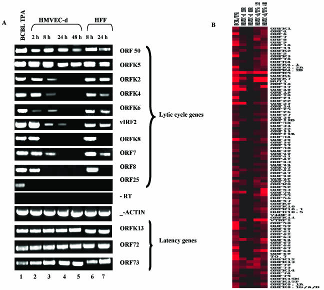
Kaposi's sarcoma-associated herpesvirus (KSHV) infection of in vitro target cells is characterized by the expression of the latency-associated open reading frame (ORF) 73 gene (LANA-1) and the absence of progeny virus production. This default latent infection can be switched into lytic cycle by phorbol ester and by the lytic cycle ORF 50 (RTA) protein. In this study, the kinetics of latent and lytic gene expression immediately following KSHV infection of primary human dermal microvascular endothelial (HMVEC-d) and foreskin fibroblast (HFF) cells were examined by real-time reverse transcriptase PCR and whole-genome array. Within 2 h postinfection (p.i.), high levels of ORF 50 transcripts were detected in both cell types, which declined sharply by 24 h p.i. In contrast, comparatively low levels of ORF 73 expression were detected within 2 h p.i., increased subsequently, were maintained at a steady state, and declined slowly by 120 h p.i. The RTA and LANA-1 proteins were detected in the majority of infected cells by immunoperoxidase assays. In genome array, only 29 of 94 (31%) KSHV genes were expressed, which included 11 immediate-early/early, 8 early, and 5 late lytic genes and 4 latency-associated genes. While the expression of latent ORF 72, 73, and K13 genes continued, nearly all of the lytic genes declined or were undetectable by 8 and 24 h p.i. in HMVEC-d and HFF cells, respectively. Only a limited number of RTA-activated KSHV genes were expressed briefly, and the majority of KSHV genes involved in viral DNA synthesis and structural proteins were not expressed. However, early during infection, the lytic K2, K4, K5, K6, and vIRF2 genes with immune modulation functions and the K7 gene with antiapoptotic function were expressed. Expression of K5 was detected for up to 5 days of observation, and vIRF2 was expressed up to 24 h p.i. The full complement of lytic cycle genes were expressed when 12-O-tetradecanoylphorbol-13-acetate was added to the HMVEC-d cells after 48 h p.i. These data suggest that in contrast to alpha- and betaherpesviruses and some members of gammaherpesviruses, gamma-2 KSHV in vitro infection is characterized by the concurrent expression of latent and a limited number of lytic genes immediately following infection and a subsequent decline and/or absence of lytic gene expression with the persistence of latent genes. Expression of its limited lytic cycle genes could be a "strategy" that evolved in KSHV allowing it to evade the immune system and to provide the necessary factors and time to establish and/or maintain latency during the initial phases of infection. These are unique observations among in vitro herpesvirus infections and may have important implications in KSHV biology and pathogenesis.


Figures

FIG. 1.
FIG. 1.
Kinetics of KSHV entry into HMVEC-d and HFF cells. HMVEC-d and HFF cells were infected at an MOI of 100 KSHV DNA copies per cell. At different time points, unbound KSHV was removed by washing and the bound, noninternalized virus was removed by treating the cells with trypsin-EDTA for 5 min. Infected and mock-infected cells were washed, and internalized viral DNA was isolated and quantified by real-time DNA-PCR with primers and Taqman probe specific for ORF 73 (Table 1). Viral DNA copy numbers were calculated from the standard graph generated by real-time PCR of known concentrations of a cloned ORF 73 gene. Each reaction was done in duplicate, and each point represents the average ± standard deviation of three experiments.
FIG. 2.
FIG. 2.
Kinetics of KSHV ORF 73 and 50 gene expression during primary infection of HMVEC-d (A) and HFF (B) cells. Cells were infected at an MOI of 100 KSHV DNA copies per cell. At different time points p.i. RNA was isolated and treated with DNase I, and 250 ng of DNase-treated RNA was subjected to real-time RT-PCR with ORF 73 and 50 gene-specific primers and Taqman probes. Known concentrations of DNase-treated in vitro-transcribed ORF 50 and ORF 73 transcripts were used in a real-time RT-PCR to construct a standard graph from which the relative copy numbers of viral transcripts were calculated and normalized, with GAPDH used as the internal control. Each reaction was done in duplicate, and each point represents the average ± standard deviation of three experiments.
FIG. 3.
FIG. 3.
Immunoperoxidase assay detecting KSHV ORF 73 (LANA-1) and ORF 50 (RTA) proteins in HMVEC-d and HFF cells. Cells in eight-well chamber slides were infected with KSHV for 2 h, washed, and incubated at 37°C. At different time points, cells were washed, fixed with cold acetone, incubated at 4°C overnight with a predetermined dilution of rabbit antibodies against LANA-1 or RTA or with preimmune antibodies, processed for immunostaining as described in Materials and Methods, and examined under a light microscope. (A) ORF 50 expression in HMVEC-d cells 2 h p.i. (B) ORF 50 expression in HMVEC-d cells 24 h p.i. (C) Uninfected HMVEC-d cells with anti-ORF 50 antibodies. (D) ORF 73 expression in HMVEC-d cells 48 h p.i. (E) Uninfected HMVEC-d cells with anti-ORF 73 antibodies. (F and G) ORF 50 expression in HFF cells at 8 and 24 h p.i., respectively. (H) Uninfected HFF cells with anti-ORF 50 antibodies. (I) ORF 73 expression in HFF cells 48 h p.i. (J) Uninfected HFF cells with anti-ORF 73 antibodies. Magnification: ×40 (A, C, E, F, H, and J); ×100 (B, D, and G).
FIG. 4.
FIG. 4.
Gene array analyses of KSHV latent and lytic genes in HMVEC-d and HFF cells. (A) Cluster diagram representing the expression of KSHV latent and lytic genes. HMVEC-d cells were infected with KSHV for 2 and 8 h, and HFF cells were infected for 8 and 24 h. KSHV-positive BCBL-1 cells induced with TPA for 72 h were used as a control for the gene array analyses. The RNA transcripts from the infected and uninfected samples were converted into cDNA in the presence of fluorescent dyes Cy-3 and Cy-5 dUTPs, respectively. All samples were used in separate reactions of real-time DNA-PCR (without RT) to confirm the absence of contaminating DNA. The fluorescently labeled cDNAs were hybridized with KSHV arrays spotted on ceramic chips and scanned using an Affymetrix scanner. The average intensity of three different spots of each individual gene was calculated, and background intensities were subtracted and normalized across different arrays using cellular housekeeping gene transcripts. The normalized infected Cy-3/uninfected Cy-5 expression intensity ratios were used to construct the cluster diagrams, and the intensity ratios closer to 1.0 and below were taken as an indication of nonexpressed genes. KSHV genes detected at higher levels during infection are shown in progressively darker shades of red. (B) KSHV genes expressed in HMVEC-d and HFF cells during early times after primary infection are represented in a genomic view. The KSHV genes are designated per the methods of Jenner et al. (22) and color coded to represent the latency and lytic (primary, secondary, and tertiary) cycle-associated genes.
FIG. 5.
FIG. 5.
(A) RT-PCR confirmation of KSHV transcription in HMVEC-d and HFF cells. Total RNA prepared from the infected or uninfected cells was treated with DNase I and amplified by RT-PCR. The products were separated by gel electrophoresis, visualized, and quantified. Lane 1, TPA-induced BCBL-1 cells; lanes 2 to 5, HMVEC-d cells after 2, 8, 24, and 48 h p.i., respectively; lanes 6 and 7, HFF cells after 8 and 24 h p.i., respectively. −RT, all samples were used in separate reactions of DNA-PCR (without RT) to confirm the absence of contaminating DNA, and an example is shown. (B) Cluster diagram representing KSHV latent and lytic gene transcription in the HMVEC-d cells after TPA stimulation. HMVEC-d cells were infected with KSHV and, after 48 h, cells were stimulated with 20 ng of TPA/ml for 12 or 48 h and analyzed by KSHV gene array as per the procedures described for Fig. 4A.
FIG. 6.
FIG. 6.
Clustering of KSHV genes expressed in HMVEC-d and HFF cells according to the predicted putative functions and demonstrated functions. The cluster diagram representing the expression of KSHV genes and the experimental procedures were as described for Fig. 4A. KSHV genes detected at higher levels are shown in progressively darker shades of red. The designations of KSHV genes are as per Jenner et al. (22).
FIG. 7.
FIG. 7.
Immunoperoxidase assay detecting KSHV K5, LANA1, and RTA proteins in HMVEC-d cells. Cells in eight-well chamber slides were infected with KSHV for 2 h, washed, and incubated at 37°C. At different time points, cells were washed, fixed with cold acetone, incubated at 4°C overnight with predetermined dilutions of antibodies against K5, ORF 73, or ORF 50 or with preimmune antibodies, processed for immunostaining as described in Materials and Methods, and examined under a light microscope. (A) LANA-1 expression in HMVEC-d cells after 5 days p.i. (B) RTA expression in HMVEC-d cells after 5 days p.i. (C to E) K5 expression in HMVEC-d cells after 24 h (C) and 5 days (D and E) p.i. (F) Uninfected HMVEC-d cells with anti-K5 antibodies. Magnification: ×20 (A); ×40 (B, C, D, and F); ×100 (E). The arrows indicate the cytoplasmic and perinuclear staining seen with anti-K5 antibodies.
FIG. 8.
FIG. 8.
Model for KSHV gene expression early during in vitro infection. Early events of KSHV infection and gene expression in the target cells are depicted in overlapping dynamic phases. KSHV binds to the cell surface via its interactions with heparan sulfate (HS) (3), integrins (2), and possibly another yet-to-be-identified molecule(s) (phase 1). In phase 2, virus enters into the target cells (1-3, 32), probably overlapping with the induction of host cell signal pathways (32). In phase 3, viral capsid and tegument moves in the cytoplasm, probably facilitated by the induced signal pathways, and probably overlaps with the signal pathways induced during phase 4 host cell gene transcription and expression (33). In phase 5, viral DNA enters into the nucleus, followed by concurrent latent and lytic cycle gene expression as shown in this study, which is probably influenced by the KSHV-induced signal pathways and expressed host cell genes. Phase 6 involves the overlapping viral gene-induced host cell gene expression, which also may exert an influence on subsequent viral gene expression. During the early time of primary infection of endothelial and fibroblast cells, KSHV expresses the lytic ORF 50 gene and the latent ORF 73 gene concurrently, with initially higher levels of ORF 50 expression followed by a rapid decline. Nearly all latency-associated and limited lytic genes are transcribed concurrently. While the expression of latent ORF 72, 73, and K13 genes continues, nearly all of the lytic genes decline or are undetectable by 24 h p.i. Only a limited number of RTA-activated KSHV genes are expressed briefly, and the majority of KSHV genes involved in viral DNA synthesis and structural proteins are not expressed. Early during infection, the lytic K2, K4, K5, K6, and vIRF2 genes with immune modulation functions and the K7 gene with antiapoptotic function are expressed, which may be providing sufficient survival time for the infected cells from the host immune surveillance apparatus and apoptosis and the sufficient advantage necessary for the establishment of latent infection during the initial time of infection.

Similar articles

Cited by

References

    1. Akula, S. M., P. P. Naranatt, N. S. Walia, F. Z. Wang, B. Fegley, and B. Chandran. 2003. Kaposi's sarcoma-associated herpesvirus (human herpesvirus 8) infection of human fibroblast cells occurs through endocytosis. J. Virol. 77:7978-7990. - PMC - PubMed
    1. Akula, S. M., N. P. Pramod, F.-Z. Wang, and B. Chandran. 2002. Integrin α3β1 (CD49c/28) is a cellular receptor for Kaposi's sarcoma associated herpesvirus (KSHV/HHV-8) entry into the target cells. Cell 108:407-419. - PubMed
    1. Akula, S. M., F.-Z. Wang, J. Vieira, and B. Chandran. 2001. Human herpesvirus 8 interaction with target cells involves heparan sulfate. Virology 282:245-255. - PubMed
    1. Barnes, B., B. Lubyova, and P. M. Pitha. 2002. On the role of IRF in host defense. J. Interferon Cytokine Res. 22:59-71. - PubMed
    1. Bechtel, J. T., Y. Liang, J. Hvidding, and D Ganem. 2003. Host range of Kaposi's sarcoma-associated herpesvirus in cultured cells. J. Virol. 77:6474-6481. - PMC - PubMed

Publication types

MeSH terms