. 2004 Apr;78(7):3621-32.
doi: 10.1128/jvi.78.7.3621-3632.2004.

Transgenic mice expressing the nucleoprotein of Borna disease virus in either neurons or astrocytes: decreased susceptibility to homotypic infection and disease

Affiliations

Transgenic mice expressing the nucleoprotein of Borna disease virus in either neurons or astrocytes: decreased susceptibility to homotypic infection and disease

Mathias Rauer et al. J Virol. 2004 Apr.

Abstract

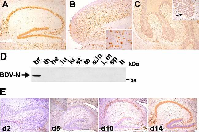
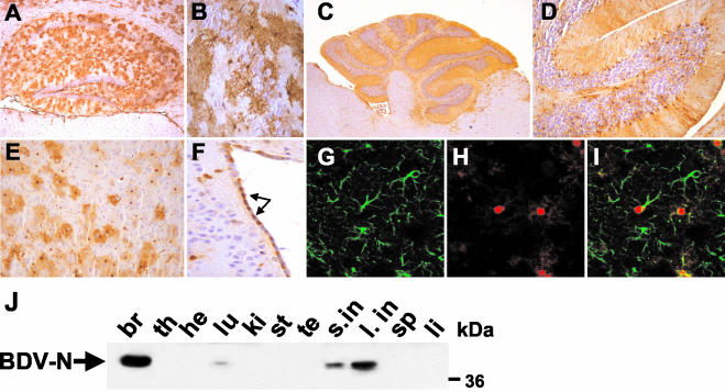
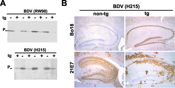
The nucleoprotein (N) of Borna disease virus (BDV) is the major target of the disease-inducing antiviral CD8 T-cell response in the central nervous system of mice. We established two transgenic mouse lines which express BDV-N in either neurons (Neuro-N) or astrocytes (Astro-N). Despite strong transgene expression, neurological disease or gross behavioral abnormalities were not observed in these animals. When Neuro-N mice were infected as adults, replication of BDV was severely impaired and was restricted to brain areas with a low density of transgene-expressing cells. Notably, the virus failed to replicate in the transgene-expressing granular and pyramidal neurons of the hippocampus (which are usually the preferred host cells of BDV). When Neuro-N mice were infected within the first 5 days of life, replication of BDV was not suppressed in most neurons, presumably because the onset of transgene expression in the brain occurred after these cells became infected with BDV. Astro-N mice remained susceptible to BDV infection, but they were resistant to BDV-induced neurological disorder. Unlike their nontransgenic littermates, Neuro-N mice with persistent BDV infection did not develop neurological disease after immunization with a vaccinia virus vector expressing BDV-N. In contrast to the situation in wild-type mice, this treatment also failed to induce N-specific CD8 T cells in the spleens of both transgenic mouse lines. Thus, while resistance to BDV infection in N-expressing neurons appeared to result from untimely expression of a viral nucleocapsid component, the resistance to BDV-induced neuropathology probably resulted from immunological tolerance.


Figures

FIG. 1.
FIG. 1.
Cell- and tissue-specific expression pattern of BDV-N in Neuro-N-44 transgenic mice. (A to C) Sagittal sections (8 μm in thickness) of paraffin-embedded brain hemispheres from 8- to 12-week-old Neuro-N-44 mice were stained with anti-N MAb Bo18. Sections were counterstained with hematoxylin. (A) Hippocampus; (B) frontal cortex. The insert shows a fourfold-higher magnification of the frontal cortex. Note the transgene-expressing and nonexpressing neurons in the immediate vicinity. (C) Cerebellum. The insert shows a 2.5-fold-higher magnification of one cerebellar lobe. The arrow indicates an N-expressing Purkinje cell. (D) Tissue-specific expression pattern of transgenic N. Organs were taken from a 7-week-old Neuro-N-44 male, homogenized, and subjected to SDS-PAGE. Immunoblotting (using MAb Bo18) for detection of N was performed. Samples are from brain (br), thymus (th), heart (he), lung (lu), kidney (ki), stomach (st), testis (te), small intestine (s.in), large intestine (l. in), spleen (sp), and liver (li). The arrow indicates the BDV-N-specific band at about 40 kDa. (E) Kinetics of modified Thy-1.2 promoter-controlled transgenic N expression in the CNS. Transgene expression in the hippocampus of Neuro-N-44 animals of the indicated ages was analyzed on sagittal brain sections by staining with MAb Bo18. The insert shows the CA3 region at a higher magnification.
FIG. 2.
FIG. 2.
Cell- and tissue-specific expression pattern of BDV-N in Astro-N-25 transgenic mice. (A to I) Sagittal sections (8 μm in thickness) of paraffin-embedded brain hemispheres from 8- to 12-week-old Astro-N-25 mice were stained with anti-N MAb Bo18 (A to F) or a mixture of Bo18 and rabbit anti-GFAP (G to I). Bound antibodies were visualized using a biotinylated secondary antibody (A to F) or a mixture of Cy2-labeled anti-rabbit antibody and Cy3-labeled anti-mouse antibody (G to I). Sections in panels A to F were counterstained with hematoxylin. (A) Hippocampus; (B) CA3 region of hippocampus; (C) cerebellum; (D) 2.5-fold-higher magnification image of cerebellum; (E) frontal cortex; (F) ventricle wall (arrows point to N-expressing ependymal cells). (G to I) Confocal laser-scanning microscopy analysis of double-labeled cortical section. (G) anti-GFAP staining; (H) anti-N staining; (I) overlaid images from panels G and H. (J) Tissue-specific expression pattern of transgenic N. Organs were taken from a 6-week-old Astro-N-25 male and homogenized, 80-μg samples were subjected to SDS-PAGE, and immunoblotting using MAb Bo18 for detection of N was performed. Samples are from brain (br), thymus (th), heart (he), lung (lu), kidney (ki), stomach (st), testis (te), small intestine (s.in), large intestine (l. in), spleen (sp), and liver (li). The arrow indicates the BDV-N-specific band at about 40 kDa.
FIG. 3.
FIG. 3.
Neurons of transgenic mice expressing BDV-N are resistant to BDV infection. (A) Transgenic (t) (+) and nontransgenic (−) Neuro-N-44 animals 3 to 5 weeks of age were infected by the intracerebral route with mouse-adapted variants of BDV strain RW98 or strain H215. At 6 weeks (H215) or 9 weeks (RW98) postinfection, mice were sacrificed and samples of brain homogenates were analyzed by SDS-PAGE and immunoblotting using a rabbit antiserum against BDV-P. (B) Immunohistochemical analysis of transgene expression and virus replication 5 weeks postinfection in the hippocampus of selected transgenic (tg+) and nontransgenic (tg−) Neuro-N-44 mice infected with H215 at the age of 4 weeks. Sagittal brain sections were stained with either MAb Bo18 (which selectively detects the transgene product) or MAb 21E7 (which detects viral P antigen in infected cells). The lower left panel shows the typical distribution of BDV-infected cells in the hippocampus of nontransgenic mice. Note that the majority of hippocampal neurons in brains of transgenic mice did not contain viral P antigen (lower right panel), indicating a lack of infection.
FIG. 4.
FIG. 4.
Age-dependent BDV resistance of dentate gyrus neurons in Neuro-N-44 mice. (A) Transgenic Neuro-N-44 animals were infected with mouse-adapted BDV strain H215 at the indicated ages (d0, day 0; d8, day 8; d21, day 21). Animals were sacrificed 5 weeks postinfection. Brain hemispheres were paraffin embedded, and 8-μm-thick sections were stained with anti-P MAb 21E7 (left panels) to visualize virus-infected cells or with MAb Bo18 (right panels) to visualize transgenically expressed N. Note the high number of infected neurons in the dentate gyrus of Neuro-N animals infected as newborns. The insert in the upper left panel shows a higher-magnification image of the CA3 region; arrows point to rare infected pyramidal CA3 neurons. The insert in the upper right panel shows a higher-magnification image of BDV-infected dentate gyrus neurons expressing transgenic N. Note the punctate staining pattern in most granular neurons, indicating a redistribution of transgenic N in infected cells. (B) Nontransgenic littermates were infected as newborns (left panel) or at the age of 25 days (right panel) and sacrificed 5 weeks postinfection. Brains were processed as described for panel A, and 8-μm-thick paraffin sections were stained with anti-P MAb 21E7 to visualize infected cells.
FIG. 5.
FIG. 5.
Mouse astrocytes can be infected with BDV. Newborn wild-type B10.BR mice were infected with BDV strain H215. At 5 weeks of age, the animals were sacrificed and sagittal sections (8 μm in thickness) of paraffin-embedded brain hemispheres were stained with a mixture of anti-P MAb 30H8 and rabbit anti-GFAP. Bound antibodies were visualized using a mixture of Cy2-labeled anti-rabbit antibody and Cy3-labeled anti-mouse antibody. An uninfected animal served as the control.
FIG. 6.
FIG. 6.
Transgenic N expression in astrocytes does not impair the efficacy of BDV infection. Astro-N-25 transgenic mice and nontransgenic littermates were infected with BDV at the age of 3 weeks, and brain hemispheres were taken at 9 weeks postinfection for immunoblot analysis (A) and immunohistological analysis (B to D) of brain homogenates. (A) Cerebella (cb) and the remaining parts of the brain hemispheres (ce) of three transgenic (tg) (+) and three nontransgenic (−) animals were separately homogenized, and samples were analyzed by immunoblotting using a rabbit antiserum against BDV-P (-P). Sagittal sections (8 μm in thickness) of paraffin-embedded brain hemispheres (used as described for panel A) from selected animals were double stained with a rabbit polyclonal serum against P (αP) to detect virus infection and with MAb Bo18 detecting transgenically expressed N. Bound antibodies were visualized using a mixture of Cy2-labeled anti-rabbit antibody and Cy3-labeled anti-mouse antibody. Confocal laser-scanning microscopy showed virus-infected, P-antigen-positive cells (B) and BDV-N transgene-expressing cells (C); panel D shows overlaid images from panels B and C. Double-positive cells appear in yellow, and uninfected transgene-expressing cells appear in red. The border between the molecular layer (top) and the granular layer (bottom) is indicated by a dotted line.
FIG. 7.
FIG. 7.
N-specific CD8+ T-cell response is impaired in Neuro-N-44 mice. (A and B) Spleens from two VV-N-immunized nontransgenic (non-tg) (A) and five Neuro-N-44 (B) mice were recovered 7 days post-VV infection, and single cell suspensions were restimulated for 9 days with TELEISSI peptide. Cytolytic activity of restimulated cultures was determined in a 51chromium release assay using L929 target cells coated with TELEISSI (closed symbols) or the irrelevant control peptide FEANGNLI (open symbols). Results are representative of two independent experiments. (C and D) Severity of disease (C) and degree of meningoencephalitis (D) in persistently BDV-infected Neuro-N-mice and nontransgenic littermates after infection with VV-N are shown as mean values for the following groups of mice: two Neuro-N-44 mice and five nontransgenic littermates infected with BDV strain H215 at the age of 5 days (expt. 1) and five Neuro-N-44 mice and three nontransgenic littermates infected as newborns (expt. 2). VV-N infection was performed at day 65 (expt. 1) or 64 (expt. 2). Animals were observed for clinical symptoms until day 13 post-VV infection (C). Severity of disease was scored in a range from 0 to 3 (0, no symptoms; 1, low degree of ataxia and increased anxiety; 2, clear ataxia, torticollis, uncontrolled movements of extremities when the animal was held up by the tail, rough fur or hunched posture, and characteristic position of hind limbs when animal was lifted by the tail; 3, pronounced weight loss, severe ataxia and torticollis, paraparesis, apathy, and morbidity). (D) Meningoencephalitis was scored on an arbitrary scale from 0 to 3 (0, no infiltrates; 1, up to two perivascular infiltrates per brain section, with one or two layers of cells and some mononuclear cells in meninges; 2, three to five perivascular infiltrates per brain section (mostly with multilayer appearance), incidental spread into parenchyma, and intermediate meningitis; 3, more than five perivascular infiltrates per brain section (with multiple layers of cells), strong infiltration of parenchyma at multiple sites, and strong meningitis.
FIG. 8.
FIG. 8.
Impaired BDV-N-specific CD8 T-cell response in Astro-N-25 mice. (A and B) Spleen cells from immunized nontransgenic (A) and Astro-N-25 (B) mice were restimulated with TELEISSI peptide-loaded antigen-presenting cells. Cytolytic activity levels of restimulated cultures were determined in a 51chromium release assay using L929 target cells coated with TELEISSI (closed symbols) or the irrelevant control peptide FEANGNLI (open symbols). (C) Five (Astro-N-25 × MRL)F1 mice (solid line) and three nontransgenic littermates (dashed line) were infected with BDV strain H215 at the age of 16 days. They were observed for clinical symptoms until 20 weeks postinfection.

Similar articles

Cited by

References

    1. Adams, R. H., and D. T. Brown. 1985. BHK cells expressing Sindbis virus-induced homologous interference allow the translation of nonstructural genes of superinfecting virus. J. Virol. 54:351-357. - PMC - PubMed
    1. Bilzer, T., and L. Stitz. 1994. Immune-mediated brain atrophy. CD8+ T cells contribute to tissue destruction during borna disease. J. Immunol. 153:818-823. - PubMed
    1. Borrow, P., J. L. Cornell, M. D. Ruppe, and L. Mucke. 1995. Immunization-induced inflammatory infiltration of the central nervous system in transgenic mice expressing a microbial antigen in astrocytes. J. Neuroimmunol. 61:133-149. - PubMed
    1. Bothe, K., A. Aguzzi, H. Lassmann, A. Rethwilm, and I. Horak. 1991. Progressive encephalopathy and myopathy in transgenic mice expressing human foamy virus genes. Science 253:555-557. - PubMed
    1. Brenner, M., W. C. Kisseberth, Y. Su, F. Besnard, and A. Messing. 1994. GFAP promoter directs astrocyte-specific expression in transgenic mice. J. Neurosci. 14:1030-1037. - PMC - PubMed

Publication types

MeSH terms

LinkOut - more resources