. 2004 Apr;78(7):3704-9.
doi: 10.1128/jvi.78.7.3704-3709.2004.

Movement protein of a closterovirus is a type III integral transmembrane protein localized to the endoplasmic reticulum

Affiliations

Movement protein of a closterovirus is a type III integral transmembrane protein localized to the endoplasmic reticulum

Valera V Peremyslov et al. J Virol. 2004 Apr.

Abstract

Cell-to-cell movement of beet yellows closterovirus requires four structural proteins and a 6-kDa protein (p6) that is a conventional, nonstructural movement protein. Here we demonstrate that either virus infection or p6 overexpression results in association of p6 with the rough endoplasmic reticulum. The p6 protein possesses a single-span, transmembrane, N-terminal domain and a hydrophilic, C-terminal domain that is localized on the cytoplasmic face of the endoplasmic reticulum. In the infected cells, p6 forms a disulfide bridge via a cysteine residue located near the protein's N terminus. Mutagenic analyses indicated that each of the p6 domains, as well as protein dimerization, is essential for p6 function in virus movement.


Figures
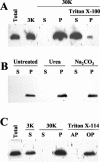
FIG. 1.
FIG. 1.
Immunoblot analyses, using anti-p6 serum, of the extracts from BYV-infected plants. Lanes: Total, protein extract prior to fractionation; 3K, supernatant following extract centrifugation at 3,000 × g; S and P, supernatant and pellet, respectively, following centrifugation at 30,000 × g; AP and OP, aqueous and organic phases, respectively, following extraction with Triton X-114. Samples treated with Triton X-100, urea, or Na2CO3 buffer are marked accordingly.
FIG. 2.
FIG. 2.
Immunoblot analyses of the protein extracts following separation in the sucrose gradients, with the fraction numbers shown at the top. The types of antisera used for analysis are indicated at left, and MgCl2 concentrations are shown at right. BiP, ER-resident marker protein.
FIG. 3.
FIG. 3.
(A to C) Confocal laser scanning microscopy analysis of the 16c transgenic plants that express ER-targeted GFP (A), the p6/GFP fusion (B), or the GFP fusion to the C-terminal, hydrophilic domain of p6 (C). The green corresponds to the GFP fluorescence, and the occasional red spots represent the autofluorescent chloroplasts. (D) Amino acid sequence (top) and membrane topology (bottom) of BYV p6. A1 to A12 and arrows indicate the alanine-scanning mutations introduced into indicated positions of p6. Red hexagons indicate premature stop codon mutations replacing the residues shown above.
FIG. 4.
FIG. 4.
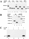
(A) Analysis of dimerization of wild-type p6 and three alanine mutants targeting each of the cysteine residues present in p6 (see also Fig. 3D and Table 1). Lanes: p6, agrobacterium-mediated expression of p6; BYV, virus-infected plants. The presence or absence of DTT in the protein dissociation buffer is indicated above the lanes with a plus or minus sign, respectively. D, p6 dimer; M, p6 monomer. Positions of the protein markers are shown at left. In both panels A and B, p6 was detected using immunoblotting and p6 antiserum raised against the C-terminal hydrophilic domain of p6. (B) Dimerization of the p6/GFP fusion product. The designations are the same as in panel A, except for M* and D*, which correspond to a monomer and a dimer formed by the p6/GFP fusion product, respectively. (C) Treatment of the resuspended P30 fraction of the p6-containing protein extracts with proteinase K (PrK) in the presence or absence of Triton X-100 as indicated at the top.

Similar articles

Cited by

References

    1. Ahlquist, P., A. O. Noueiry, W. M. Lee, D. B. Kushner, and B. T. Dye. 2003. Host factors in positive-strand RNA virus genome replication. J. Virol. 77:8181-8186. - PMC - PubMed
    1. Alzhanova, D. V., Y. Hagiwara, V. V. Peremyslov, and V. V. Dolja. 2000. Genetic analysis of the cell-to-cell movement of beet yellows closterovirus. Virology 268:192-200. - PubMed
    1. Alzhanova, D. V., A. Napuli, R. Creamer, and V. V. Dolja. 2001. Cell-to-cell movement and assembly of a plant closterovirus: roles for the capsid proteins and Hsp70 homolog. EMBO J. 20:6997-7007. - PMC - PubMed
    1. Beachy, R. N., and M. Heinlein. 2000. Role of p30 in replication and spread of TMV. Traffic 1:540-544. - PubMed
    1. Bordier, C. 1981. Phase separation of integral membrane proteins in Triton X-114 solution. J. Biol. Chem. 256:1604-1607. - PubMed

Publication types

MeSH terms