. 2004 Apr;78(7):3710-21.
doi: 10.1128/jvi.78.7.3710-3721.2004.

The subcellular distribution of multigene family 110 proteins of African swine fever virus is determined by differences in C-terminal KDEL endoplasmic reticulum retention motifs

Affiliations

The subcellular distribution of multigene family 110 proteins of African swine fever virus is determined by differences in C-terminal KDEL endoplasmic reticulum retention motifs

Christopher Netherton et al. J Virol. 2004 Apr.

Abstract

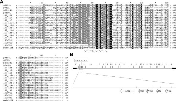
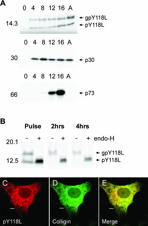
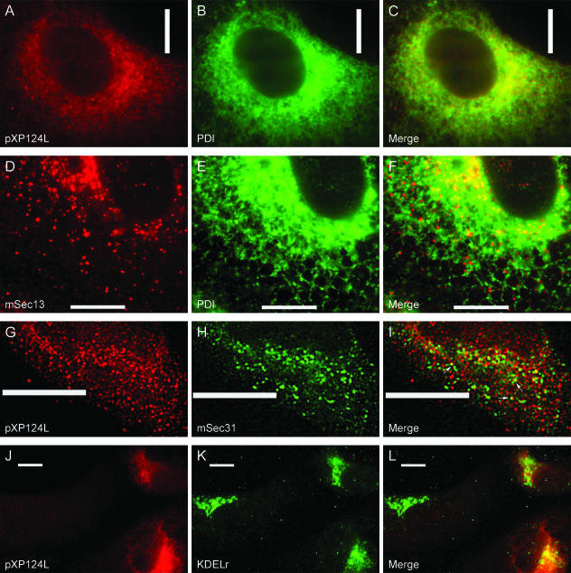
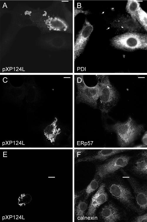
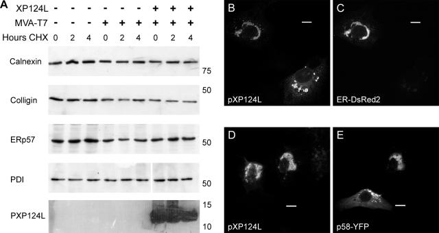
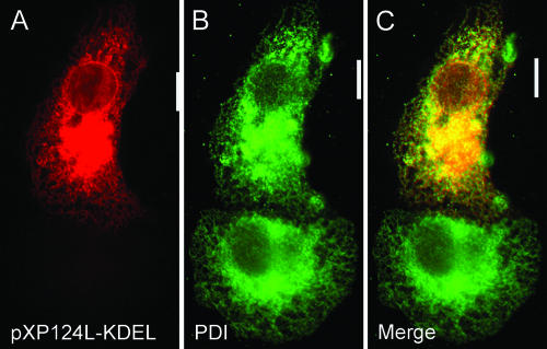
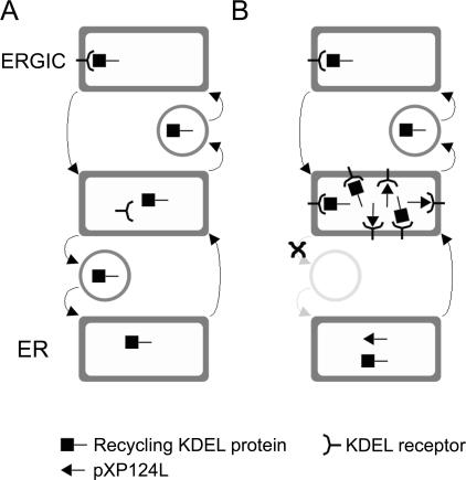
African swine fever virus (ASFV) is a large double-stranded DNA virus that replicates in discrete areas in the cytosol of infected cells called viral factories. Recent studies have shown that assembling virions acquire their internal envelopes through enwrapment by membranes derived from the endoplasmic reticulum (ER). However, the mechanisms that underlie the formation of viral factories and progenitor viral membranes are as yet unclear. Analysis of the published genome of the virus revealed a conserved multigene family that encodes proteins with hydrophobic signal sequences, indicating possible translocation into the ER lumen. Strikingly, two of these genes, XP124L and Y118L, encoded proteins with KDEL-like ER retention motifs. Analysis of XP124L and Y118L gene product by biochemical and immunofluorescence techniques showed that the proteins were localized to pre-Golgi compartments and that the KEDL motif at the C terminus of pXP124L was functional. XP124L expression, in the absence of other ASFV genes, had a dramatic effect on the contents of the ER that was dependent precisely on the C-terminal sequence KEDL. The normal subcellular distribution of a number of proteins resident to this important, cellular organelle was drastically altered in cells expressing wild-type XP124L gene product. PXP124L formed unusual perinuclear structures that contained resident ER proteins, as well as proteins of the ER-Golgi intermediate compartment. The data presented here hint at a role for MGF110 gene product in preparing the ER for its role in viral morphogenesis; this and other potential functions are discussed.


Figures

FIG. 1.
FIG. 1.
MGF110 proteins are conserved between different ASFV isolates. (A) Soluble MGF110 protein sequences (2, 13, 15, 39) from four ASFV isolates were aligned by using the PILEUP program of GCG10.1. Protein sequences from the Ba71v isolate have a “p” prefix, sequences from the LIS57 isolate have an “l57” prefix, sequences from the Lisbon 60 Vero cell-adapted isolate have an “l60v” prefix, and a sequence from the Malawi Lil20/1 isolate is indicated by a “malw” prefix. Shading of the alignment was carried out with the Genedoc program and indicates the degree of similarity between residues, with black, dark gray shading, and light gray shading indicating 100, 80, and 60%, respectively. The position of the MGF110 cysteine motif is also highlighted. (B) EcoRI restriction map of the Ba71v isolate of ASFV, with the positions of MGF110 genes shown in an enlarged section below.
FIG. 2.
FIG. 2.
pY118L is an early ASFV protein localized to the ER. (A) Control Vero cells (lanes 0), cells infected with Ba71v for increasing times (in hours) as indicated, or infected for 16 h in the presence of cytosine β-d-arabinofuranoside (lanes A) were pulsed for 30 min with [35S]methionine and [35S]cysteine, lysed, and immunoprecipitated with antibodies specific for pY118L (R30), p30 (C18), or p73 (17LD3). (B) Vero cells were infected for 12 h and then pulse-labeled as described above before being chased in complete medium for the indicated lengths of time. PY118L was immunoprecipitated with R30 and subjected to endo-H digestion (lanes +). Proteins were resolved by SDS-PAGE and subjected to autofluorography. Sizes of molecular mass markers are indicated in kilodaltons on the left. (C to E) Vero cells infected for 8 h with Ba71v were fixed with 4% paraformaldehyde and probed with antibody R30 (pY118L) (C) and anti-colligin (D). Primary antibody was visualized with appropriate goat antibodies conjugated to Alexa-488 (D) or Alexa-594 (C). Panel E is a digitally merged image of panels C and D. Digital sections (0.2 μm) were captured at ×100 magnification and digitally deconvolved by using Openlab 2.1.3. Bars, 10 μm.
FIG. 3.
FIG. 3.
pXP124L localizes to pre-Golgi compartments. Vero cells infected for 8 h with Ba71v were fixed with either glutaraldehyde solution (A to C) or 4% paraformaldehyde (D to L). Cells were then probed with either antibodies R29 (pXP124L in A, G, and J) or 1D3 (PDI in B and E) and antibodies recognizing either mSec13 (D), mSec31 (H), or the KDEL receptor (K). Primary antibody was visualized with appropriate goat antibodies conjugated to Alexa-488 (B, E, H, and K) or Alexa-594 (A, D, G, and J). Panels C, F, I, and L are digitally merged images of panels A and B, D and E, G and H, and J and K, respectively. Digital sections (0.2 μm) were visualized at ×60 (A to F and J to L) or ×100 (G to I) magnification and digitally deconvolved by using Openlab 2.1.3. Bars, 10 μm.
FIG. 4.
FIG. 4.
Role of the C-terminal sequences in the cellular localization of pXP124L and pY118L. (A) Uninfected (N) and Vero cells infected for 12 h with ASFV were pulse-labeled for 30 min with [35S]methionine and [35S]cysteine (P) and then chased in complete medium for 2 (C) or 4 (4C) h. Cells were lysed and then immunoprecipitated with antibody R29 and digested with endo-H (lanes +). (B) Wild-type XP124L and XP124LΔKEDL were expressed in Vero cells by using MVA-T7. Cells were pulse-labeled for 30 min (P) and then chased for 2 h (C). Cell lysates (P and C) and the medium from the chase (M) were immunoprecipitated with antibody R29 and digested with endo-H (“+” lanes). Proteins were resolved by SDS-PAGE and subjected to autofluorography. Sizes of molecular mass markers are indicated in kilodaltons on the left. (C to E and H to M) Vero cells expressing XP124LΔKEDL (C to E), Y118L (H to J), or Y118LΔKDEL (K to M) were fixed with 4% paraformaldehyde and then probed with antibody R29 (C) or R30 (H and K) and antibody 1D3 (PDI in panels D, I, and L). Primary antibody was visualized with appropriate goat antibodies conjugated to Alexa-488 (D, I, and L) or Alexa-594 (C, H, and K). Images were captured at ×60 magnification and processed as described in the legend to Fig. 2. Bars, 10 μm. (F) Cells infected with MVA-T7 were pulse-labeled for 4 h in the presence (lane “+”) or absence (lane “−”) of 200 μg of cycloheximide/ml (lane CHX). Cell lysates were resolved by SDS-PAGE and subjected to autofluorography. (G) Control cells or cells expressing Y118L or Y118LΔKDEL were incubated in the presence of cycloheximide (200 μg/ml) for the indicated lengths of time (in hours), and the levels of pY118L and pY118LΔKDEL were determined by Western blotting with antibody R30.
FIG. 5.
FIG. 5.
Effect of pXP124L on the early secretory pathway. Vero cells expressing XP124L were fixed with methanol (C to F) or 4% paraformaldehyde (A and B). Cells were then probed with antibody R29 (pXP124L in panel A) or mouse anti-pX124L (pXP124L in panels C and E) and antibody 1D3 (PDI in panel B), anti-ERp57 (D), or anti-calnexin (F). Primary antibody was visualized with appropriate goat antibodies conjugated to Alexa-488 (B, D, and F) or Alexa-594 (A, C, and E), and images were captured at ×60 magnification and processed as described in the legend to Fig. 2. Bars, 10 μm.
FIG. 6.
FIG. 6.
Effect of pXP124L on distribution of resident ER proteins. (A) Control Vero cells, cells infected with MVA-T7, or cells infected with MVA-T7 and transfected with XP124L were incubated in the presence of cycloheximide (200 μg/ml) for the indicated lengths of time (in hours). The levels of ER proteins and pXP124L were determined by Western blotting. Sizes of molecular weight markers are indicated on the right of each panel in kDa. (B to E) Vero cells transiently expressing ER-DsRed2 (B and C) or p58-YF (D and E) were infected with MVA-T7 and then transfected with a construct encoding wild-type XP124L. Cells were fixed with 4% paraformaldehyde and then probed with antibody R29 (pXP124L in panels B and D). Primary antibody was visualized with goat anti-rabbit antibody conjugated to Alexa-488 (B) or Alexa-594 (D). YFP and DsRed2 were observed through their natural fluorescence. Digital sections (0.2 μm) were captured at ×60 magnification and digitally deconvolved by using Openlab 2.1.3. Bars, 10 μm.
FIG. 7.
FIG. 7.
Expression of XP124L-KDEL. Cells expressing XP124L-KDEL were fixed with 4% paraformaldehyde and then stained with R29 (A) and antibody 1D3 (PDI) (B). Primary antibody was visualized with appropriate goat antibodies conjugated to Alexa-488 (B) or Alexa-594 (A). Digital sections (0.2 μm) were captured at ×60 magnification and digitally deconvolved by using Openlab 2.1.3. Bars, 10 μm.
FIG. 8.
FIG. 8.
Expression of MGF110 proteins and redistribution of ER proteins in LIS57-infected cells. (A) Uninfected or LIS57-infected bone marrow cells were lysed and subjected to SDS-PAGE. Protein was transferred to nitrocellulose membranes, and proteins were detected by Western blotting. The sizes of molecular mass markers are indicated on the left of each panel in kilodaltons. (B to G) Porcine bone marrow cells (PBMC) (B to D) and PAECs (E to G) infected overnight with LIS57 were fixed with 4% paraformaldehyde and then stained with TW34 (B and E), 1D3 (C and F), and DAPI (B, D, E, and G). Primary antibody was visualized by using appropriate goat antibodies conjugated to Alexa-488 (C, D, F, and G) or Alexa-594 (B and E). Digital sections (0.2 μm) were captured at ×100 magnification and digitally deconvolved by using Openlab 3.1. Merged images of TW34 and DAPI staining (B and E) or 1D3 and DAPI staining (D and G) were created digitally. Bars, 10 μm.
FIG. 9.
FIG. 9.
Possible mechanism for pXP124L induced redistribution of resident ER proteins. (A) In resting cells, escaped KDEL proteins are bound in the ERGIC by the KDEL receptor. They are then moved to areas specialized for concentrating retrograde cargo, where the receptor dissociates, and then returned to the ER in COPI-coated vesicles. (B) In cells overexpressing XP124L, KDEL proteins leave the ER normally, bind the KDEL receptor, but then become caught while returning to the ER, potentially by slow release of the KDEL bearing pXP124L from the KDEL receptor. The high levels of the viral protein may therefore inhibit the recycling of cellular proteins, and this would explain the colocalization of pXP124L with resident ER proteins in perinuclear regions of the cell.

Similar articles

Cited by

References

    1. Afonso, C. L., J. G. Neilan, G. F. Kutish, and D. L. Rock. 1996. An African swine fever virus bcl-2 homolog, 5-HL, suppresses apoptotic cell death. J. Virol. 70:4858-4863. - PMC - PubMed
    1. Almendral, J. M., F. Almazán, R. Blasco, and E. Viñuela. 1990. Multigene families in African swine fever virus: family 110. J. Virol. 64:2064-2072. - PMC - PubMed
    1. Andrés, G., R. García-Escudero, C. Simón-Mateo, and E. Viñuela. 1998. African swine fever virus is enveloped by a two-membraned collapsed cisterna derived from the endoplasmic reticulum. J. Virol. 72:8988-9001. - PMC - PubMed
    1. Beinert, H., R. H. Holm, and E. Münck. 1997. Iron-sulfur clusters: nature's modular, multipurpose structures. Science 277:653-659. - PubMed
    1. Benham, A. M., A. Cabibbo, A. Fassio, N. Bulleid, R. Sitia, and I. Braakman. 2000. The CXXCXXC motif determines the folding, structure and stability of human Ero1-Lα. EMBO J. 19:4493-4502. - PMC - PubMed

Publication types

MeSH terms

LinkOut - more resources