. 2004 Apr;78(7):3733-41.
doi: 10.1128/jvi.78.7.3733-3741.2004.

Second sialic acid binding site in Newcastle disease virus hemagglutinin-neuraminidase: implications for fusion

Affiliations

Second sialic acid binding site in Newcastle disease virus hemagglutinin-neuraminidase: implications for fusion

Viatcheslav Zaitsev et al. J Virol. 2004 Apr.

Abstract

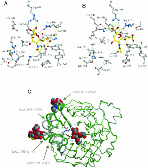
Paramyxoviruses are the leading cause of respiratory disease in children. Several paramyxoviruses possess a surface glycoprotein, the hemagglutinin-neuraminidase (HN), that is involved in attachment to sialic acid receptors, promotion of fusion, and removal of sialic acid from infected cells and progeny virions. Previously we showed that Newcastle disease virus (NDV) HN contained a pliable sialic acid recognition site that could take two states, a binding state and a catalytic state. Here we present evidence for a second sialic acid binding site at the dimer interface of HN and present a model for its involvement in cell fusion. Three different crystal forms of NDV HN now reveal identical tetrameric arrangements of HN monomers, perhaps indicative of the tetramer association found on the viral surface.


Figures

FIG. 1.
FIG. 1.
Thiosialoside ligand and its appearance in the electron density maps. (A) The thiosialoside Neu5Ac-2-S-α(2,6)Gal1OMe. (B) Stereo Fo-Fc electron density map for one of the active sites of the orthorhombic crystal form. (C) Stereo Fo-Fc electron density map at one of the symmetrical sialic acid binding sites at the dimer interface. Both electron density maps were contoured at 2.5 σ, and the refined ligand is superimposed.
FIG. 2.
FIG. 2.
HN dimer and relationship between the ligand binding sites viewed down the twofold axis. (A) Schematic drawing of the HN dimer showing the location of the active sites with Neu5Ac2en bound and the sialic acid binding site with Neu5Ac or the complete thiosialoside bound. (B) GRASP image, with the monomers in different colors.
FIG. 3.
FIG. 3.
Stereo view of the sialic acid binding site. The thiosialoside is shown with yellow bonds. Residues belonging to monomer A are shown with cyan bonds, and residues belonging to monomer B are shown with grey bonds. Water molecules are shown as red spheres.
FIG. 4.
FIG. 4.
Two views of the HN tetramer. (Top) View looking down towards the viral surface. (Bottom) Side view of the tetramer, with the viral surface indicated in grey. The active sites contain Neu5Ac2en (green). The sialic acid binding sites contain Neu5Ac (yellow) or complete thiosialoside. The blue spheres show the locations of residue 124 at the N terminus of each monomer; the red spheres show the locations of the C terminus of each monomer. The arrows indicate the direction of the twofold axes of symmetry relating the monomers within a dimer and relating two dimers.
FIG. 5.
FIG. 5.
Changes in the two states of the HN monomer. (A) The pH 4.6 active site with the β-anomer of sialic acid bound. (B) The pH 6.3 active site with Neu5Ac2en bound. (C) Superposition of the pH 4.6 (grey) and pH 6.3 (green) monomers. Residues Asp198, Arg174, Glu547, and Tyr526 are shown, as are Neu5Ac2en at the active site and Neu5Ac or the complete thiosialoside at the dimer interface. The loops whose positions changed significantly are drawn with thicker lines and labeled.
FIG. 6.
FIG. 6.
Model for fusion. (Left) F and HN hold each other in the switched-off state, with the region from 124 to 151 binding to the membrane-proximal HR2a region of F (18). HN binds to sialic acid receptors. (Middle) The sialic acid is released from the sialic acid-containing receptor, Neu5Ac2en is bound at the active site, the dimer association changes, and a new sialic acid binding site is created (F was removed for clarity). (Right) The changes in HN promote F into its fusogenic state, releasing its fusion peptide into the cell membrane while the virus is held proximal to the membrane by the sialic acid binding site. The structure(s) adopted by the HN stalk region is unknown.

Similar articles

Cited by

References

    1. Amaya, M. F., A. Buschiazzo, T. Nguyen, and P. M. Alzari. 2003. The high resolution structures of free and inhibitor-bound Trypanosoma rangeli sialidase and its comparison with T. cruzi trans-sialidase. J. Mol. Biol. 325:773-784. - PubMed
    1. Baker, K. A., R. E. Dutch, R. A. Lamb, and T. S. Jardetzky. 1999. Structural basis for paramyxovirus-mediated membrane fusion. Mol. Cell 3:309-319. - PubMed
    1. Bousse, T., T. Takimoto, W. L. Gorman, T. Takahashi, and A. Portner. 1994. Regions on the hemagglutinin-neuraminidase proteins of human parainfluenza virus type-1 and Sendai virus important for membrane-fusion. Virology 204:506-514. - PubMed
    1. Bousse, T., T. Takimoto, and A. Portner. 1995. A single amino-acid change enhances the fusion promotion activity of human parainfluenza virus type-1 hemagglutinin-neuraminidase glycoprotein. Virology 209:654-657. - PubMed
    1. Brunger, A. T., P. D. Adams, G. M. Clore, W. L. DeLano, P. Gros, R. W. Grosse-Kunstleve, J. S. Jiang, J. Kuszewski, M. Nilges, N. S. Pannu, R. J. Read, L. M. Rice, T. Simonson, and G. L. Warren. 1998. Crystallography & NMR system: a new software suite for macromolecular structure determination. Acta Crystallogr. Sect. D Biol. Crystallogr. 54:905-921. - PubMed

Publication types

LinkOut - more resources