. 2004 Apr;78(7):3753-62.
doi: 10.1128/jvi.78.7.3753-3762.2004.

Low pH is required for avian sarcoma and leukosis virus Env-induced hemifusion and fusion pore formation but not for pore growth

Affiliations

Low pH is required for avian sarcoma and leukosis virus Env-induced hemifusion and fusion pore formation but not for pore growth

G B Melikyan et al. J Virol. 2004 Apr.

Abstract

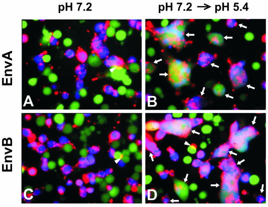
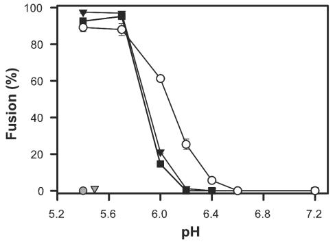
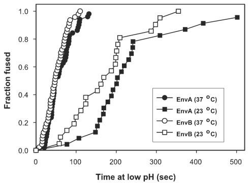
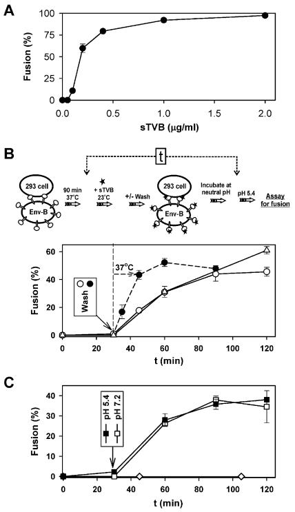
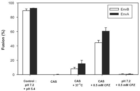
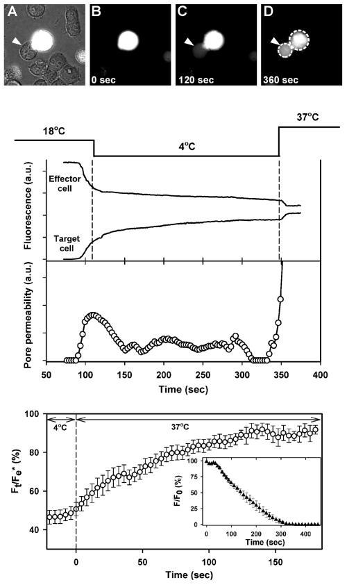
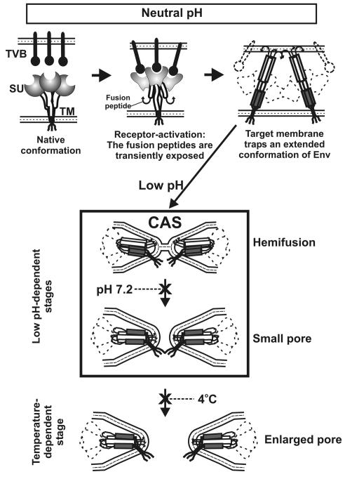
Binding of avian sarcoma and leukosis virus (ASLV) to its cognate receptor on the cell surface causes conformational changes in its envelope protein (Env). It is currently debated whether low pH is required for ASLV infection. To elucidate the role of low pH, we studied the association between ASLV subgroup B (ASLV-B) and liposomes and fusion between effector cells expressing Env from ASLV-A and ASLV-B and target cells expressing cognate receptors. Neither EnvA nor EnvB promoted cell-cell fusion at neutral pH, but lowering the pH resulted in quick and extensive fusion. As expected for a low-pH-triggered reaction, fusion was a steep function of pH. Steps that required low pH were identified. Binding a soluble form of the receptor caused ASLV-B to hydrophobically associate with liposome membranes at neutral pH, indicating that low pH is not required for insertion of Env's fusion peptides into membranes. But both cell-cell hemifusion and fusion pore formation were pH dependent. It is proposed that fusion peptide insertion stabilizes the conformation of ASLV Env into a form that can be acted upon by low pH. At this point, but not before, low pH can induce fusion and is in fact required for fusion to occur. However, low pH is no longer necessary after formation of the initial fusion pore: pore enlargement does not require low pH.


Figures

FIG. 1.
FIG. 1.
ASLV Env-mediated fusion monitored by a three-color fluorescence assay. 3T3/EnvA (A and B) or 3T3/EnvB cells (C and D) were loaded with calcein (green) and cocultured with 293(TVA950) or 293(TVB) target cells, respectively, colabeled with CMAC (blue) and DiI (red). Cells were coincubated for 2 h at 37°C (pH 7.2) followed by exposure to neutral pH (A and C) or to a pH 5.4 solution (B and D) for 15 min. Fusion occurred only when pH was lowered (B and D). Fused cells were positive for all three dyes and are marked by arrows. Regions of cells that partially overlap (marked by arrowheads) are readily distinguished from those of fused cells.
FIG. 2.
FIG. 2.
The pH dependence of cell-cell fusion. 3T3/EnvA cells were preincubated with either 293(TVA-800) (filled squares) or 293(TVA-950) cells (filled triangles), and 3T3/EnvB cells (open circles) were preincubated with 293(TVB) cells. In all cases, preincubation was for 1 h at 37°C, after which cells were exposed to solutions of different acidity levels for 10 min. Fusion was quantified by the transfer of aqueous dye 30 min after reneutralizing the external solution at 37°C. EnvA did not promote fusion when paired with target cells expressing TVB (shaded triangle); EnvB did not support fusion when paired with TVA-950-expressing cells (shaded circle).
FIG. 3.
FIG. 3.
Kinetics of low pH-induced fusion. 3T3/EnvA (filled symbols) and 3T3/EnvB (open symbols) cells were coincubated with 293(TVA-950) and 293(TVB) cells, respectively, for 1 h at neutral pH and 37°C before a pH 5.4 solution was introduced at either 23°C (squares) or at 37°C (circles). The waiting times from application of the low-pH solution to the onset of fluorescent dye redistribution were measured, ranked, and plotted as cumulative distributions.
FIG. 4.
FIG. 4.
Receptor-activated ASLV-B associates with liposomes through a hydrophobic interaction. Association between ASLV and liposomes was monitored as levels of comigration in sucrose gradients. (A) ASLV-A and ASLV-B particles were incubated with or without sTVB and mixed with liposomes at 37°C. After ultracentrifugation on a sucrose step gradient, the top (lanes T) and bottom (lanes B) fractions were subjected to SDS-polyacrylamide gel electrophoresis and the ASLV capsid protein and sTVB were detected in immunoblot analysis using an anti-ASLV capsid antibody and SUBrIgG, respectively. (B) The stability of the receptor-activated ASLV-B particle-liposome interaction was assessed (following liposome association) by incubation in the presence of 1 M NaCl, 10 mM Na2CO3, 4 M urea, or 0.5% Triton X-100.
FIG. 5.
FIG. 5.
(A) Fusion between cells expressing EnvB and receptor-deficient 293T cells in the presence of various concentrations of sTVB. Effector and target cells were coincubated for 1 h at 37°C with the indicated concentrations of sTVB. Fusion was triggered by exposure to pH 5.4 for 10 min, and dye spread was monitored after an additional incubation of 15 min at neutral pH (37°C). (B) Time dependence for acquisition of the ability to fuse at low pH for soluble receptor-activated effector cells that are in contact with receptor-deficient target cells. 3T3/EnvB and 293T cells were preincubated at 37°C for 1.5 h, exposed to 1.5 μg of sTVB/ml for 30 min at 23°C, washed, and incubated for various times at 23°C (open circles) or 37°C (filled circles). Alternatively, sTVB was never removed during the 23°C incubation (open triangles). Fusion was subsequently induced by lowering the pH to 5.4 for 2 min at 37°C and was quantified after an additional incubation at 37°C at neutral pH for 15 min. The indicated time (t) values refer to the times of acidification after addition of sTVB (time = 0), as shown in the panel illustrating the experimental protocol. (C) Effector-target cell pairs were incubated with sTVB for 30 min, and then sTVB was removed by washing. Cells were acidified to pH 5.4 for 2 min and reneutralized, and (at the indicated times) the pH was again lowered for 2 min (closed squares). Alternatively, cells were maintained at neutral pH and then the pH was lowered at the indicated time for 2 min followed by reneutralization (open squares). Fusion was not observed when 3T3/EnvB cells were preincubated with sTVB, washed, and only then incubated with target 293T cells for 1 h (open diamonds).
FIG. 6.
FIG. 6.
Cold-arrested intermediate of ASLV Env-induced fusion. 3T3/EnvA and 3T3/EnvB cells were preincubated with 293(TVA950) cells and 293(TVB) cells, respectively, for 1 h at neutral pH and 37°C. Bound effector and target cells were treated with pH 5.4 for 15 min at 4°C and then reneutralized at the same temperature. Neither lipid (DiI [data not shown]) nor aqueous (calcein [bars]) dye had mixed at this point (CAS). Raising the temperature to 37°C after the CAS was reached led to some fusion (CAS + 37°C), but the level of fusion fell far short of the full extent (Control: pH 7.2 + pH 5.4). The precise extent of fusion upon raising temperature from that of the CAS depended on the density of Env that was expressed on the effector cells. The addition of 0.5 mM CPZ at the CAS for 1 min at 23°C (CAS + 0.5 mM CPZ) led to significant aqueous dye mixing. In control experiments, dye did not spread when CPZ was added to bound cells that had not been exposed to low pH (pH 7.2 + 0.5 mM CPZ).
FIG. 7.
FIG. 7.
Pore growth proceeds at neutral pH. Effector (containing calcein and GFP) and target cells were bound together at 37°C and then placed in a 4°C chamber (image A, phase contrast overlaid with fluorescence; image B, fluorescence). Cell pairs were locally exposed to pH 5.4 at 18°C by ejection of solution from a micropipette; ejection was stopped after 1 min. Immediately after aqueous dye was observed to spread (image C), the temperature was lowered to 4°C. Middle panel: the rate of dye spread (fluorescence [upper graph]) and the calculated pore permeabilities (open circles [lower graph]) decreased. Raising temperature to 37°C at neutral pH led to rapid dye transfer due to growth of the pore. Dye concentration quickly reached a steady state (image D and upper graph). The temperature protocol is shown above the graphs. In this particular experiment, the fluorescence of the effector cell remained somewhat greater than that of the target cell. After saponin was added, the majority of the fluorescence of the effector cell quickly (within seconds) decayed but a steady-state level remained above background. Therefore, a small fraction of dye within the effector cell was immobile; the pore was not the cause of the fluorescence inequality. In general, the morphology of the cells changed after pore formation. In the illustrated experiment, the cell boundaries did not appreciably change after fusion, aiding visual clarity, although there was a small immobile fraction of dye. Bottom panel: the ratio of the fluorescence of the target cell (Ft) to the final florescence of the effector cell after all dye movement ceased (Fe*) as a function of time (averaged for nine fusion experiments). Individual fluorescence traces were aligned at the time of the temperature jump to 37°C (defined as time = 0). Inset: the time course for release of dye from isolated effector cells after adding saponin at 4°C (averaged forseven cells). F/F0 represents the ratio of the cell fluorescence (F) at times after adding saponin normalized to the initial fluorescence level (F0). Error bars represent standard errors of the means.
FIG. 8.
FIG. 8.
A model for ASLV Env-induced fusion. (Top panel) Upon binding receptors (situated within the target membrane), the SU subunits of Env (shaded circles) undergo conformational changes (denoted by altered shaded shapes) and the internal fusion peptides (curved arrows of the TM subunit) are released. After insertion of the fusion peptides into the target membrane, the N-terminal heptad repeats (open boxes) and the C-terminal helices (gray boxes) are exposed, creating an extended conformation of Env. Receptor has dissociated from Env. (Center panel) After insertion of the fusion peptides into the target membrane, low pH-induced conformational changes lead to hemifusion. Hemifusion can occur at 4°C. Subsequent conformation changes that lead to pore formation also require acidic pH, but a higher temperature is needed than that for hemifusion. Because peptides corresponding to the C-terminal repeats of the TM subunit of ASLV Env inhibit ASLV infectivity and cell-cell fusion (15), it is almost certain that ASLV Env forms a six-helix bundle. A particular sequence by which Env may fold into a six-helix bundle is illustrated, but the actual sequence is not yet known. (Bottom panel) Pore enlargement can proceed at neutral pH. The processes of hemifusion, pore formation, and pore enlargement proceed as Env undergoes conformational changes.

Similar articles

Cited by

References

    1. Adkins, H. B., S. C. Blacklow, and J. A. Young. 2001. Two functionally distinct forms of a retroviral receptor explain the nonreciprocal receptor interference among subgroups B, D, and E avian leukosis viruses. J. Virol. 75:3520-3526. - PMC - PubMed
    1. Adkins, H. B., J. Brojatsch, J. Naughton, M. M. Rolls, J. M. Pesola, and J. A. T. Young. 1997. Identification of a cellular receptor for subgroup E avian leukosis virus. Proc. Natl. Acad. Sci. USA 94:11617-11622. - PMC - PubMed
    1. Adkins, H. B., J. Brojatsch, and J. A. Young. 2000. Identification and characterization of a shared TNFR-related receptor for subgroup B, D, and E avian leukosis viruses reveal cysteine residues required specifically for subgroup E viral entry. J. Virol. 74:3572-3578. - PMC - PubMed
    1. Bates, P., J. A. Young, and H. E. Varmus. 1993. A receptor for subgroup A Rous sarcoma virus is related to the low density lipoprotein receptor. Cell 74:1043-1051. - PubMed
    1. Blumenthal, R., A. Bali-Puri, A. Walter, D. Covell, and O. Eidelman. 1987. pH-dependent fusion of vesicular stomatitis virus with Vero cells. Measurement by dequenching of octadecyl rhodamine fluorescence. J. Biol. Chem. 262:13614-13619. - PubMed

Publication types

MeSH terms