. 2004 Apr;78(7):3763-76.
doi: 10.1128/jvi.78.7.3763-3776.2004.

A peptide from autoantigen La blocks poliovirus and hepatitis C virus cap-independent translation and reveals a single tyrosine critical for La RNA binding and translation stimulation

Affiliations

A peptide from autoantigen La blocks poliovirus and hepatitis C virus cap-independent translation and reveals a single tyrosine critical for La RNA binding and translation stimulation

Raquel E Izumi et al. J Virol. 2004 Apr.

Abstract

La, a 52-kDa autoantigen in patients with systemic lupus erythematosus, was one of the first cellular proteins identified to interact with viral internal ribosome entry site (IRES) elements and stimulate poliovirus (PV) and hepatitis C virus (HCV) IRES-mediated translation. Previous results from our laboratory have shown that a small, yeast RNA (IRNA) could selectively inhibit PV and HCV IRES-mediated translation by sequestering the La protein. Here we have identified an 18-amino-acid-long sequence from the N-terminal "La motif" which is required for efficient interaction of La with IRNA and viral 5' untranslated region (5'-UTR) elements. A synthetic peptide (called LAP, for La peptide) corresponding to this sequence (amino acids 11 to 28) of La was found to efficiently inhibit viral IRES-mediated translation in vitro. The LAP efficiently enters Huh-7 cells and preferentially inhibits HCV IRES-mediated translation programmed by a bicistronic RNA in vivo. The LAP does not bind RNA directly but appears to block La binding to IRNA and PV 5'-UTR. Competition UV cross-link and translation rescue experiments suggested that LAP inhibits IRES-mediated translation by interacting with proteins rather than RNA. Mutagenesis of LAP demonstrates that single amino acid changes in a highly conserved sequence within LAP are sufficient to eliminate the translation-inhibitory activity of LAP. When one of these mutations (Y23Q) is introduced into full-length La, the mutant protein is severely defective in interacting with the PV IRES element and consequently unable to stimulate IRES-mediated translation. However, the La protein with a mutation of the next tyrosine moiety (Y24Q) could still interact with PV 5'-UTR and stimulate viral IRES-mediated translation significantly. These results underscore the importance of the La N-terminal amino acids in RNA binding and viral RNA translation. The possible role of the LAP sequence in La-RNA binding and stimulation of viral IRES-mediated translation is discussed.


Figures

FIG. 1.
FIG. 1.
(A) Schematic representation of wt La and its deletion mutants (map not to scale). The wt La protein consists of 408 amino acids. The mutants ΔN10, ΔN22, and ΔN28 lack the N-terminal 10, 22, and 28 amino acids, respectively. ΔC214 and ΔC158 are carboxy-terminal deletions of 214 and 158 residues, respectively. (B) Coomassie stain of purified recombinant wt La and its deletion mutants. The wt and mutant proteins were expressed in E. coli and purified by using DEAE Sephacel and fast-performance liquid chromatography-heparin-agarose column chromatography as detailed in Materials and Methods.
FIG. 2.
FIG. 2.
UV cross-link analysis of wt and mutant La binding to IRNA, PV 5′-UTR, and HCV 5′-UTR. 32P-labeled IRNA (A), PV 5′-UTR (B), and HCV 5′-UTR (C) were incubated with 100 ng of purified La or various La mutants. The RNA-protein complexes were analyzed by SDS-PAGE after digestion with a mixture of RNases. In lane 8 of panels A and B and in lane 7 of panel C, column-purified proteins from bacteria expressing the plasmid without the La insert were examined.
FIG. 3.
FIG. 3.
(A) N-terminal amino acid sequence of wt La. The capital letters correspond to the sequence of LAP (residues 11 to 18) and NSP (residues 71 to 88). (B) LAP inhibits IRES-mediated translation in vitro. The effect of LAP and NSP on in vitro translation of p2CAT RNA in HeLa lysates is shown. In vitro translation reaction mixtures contained 1 μg of uncapped in vitro-transcribed p2CAT RNA in the absence of peptide (lane 1) and with either 40 or 60 μM LAP (lanes 2 and 3) or NSP (lanes 4 and 5) or buffer alone (lane 6). (C) Effect of LAP on cap-independent versus cap-dependent translation in vitro. In vitro translation reactions in HeLa cell-free lysates programmed with a bicistronic capped CAT-SL-PV 5′-UTR-luciferase (Luc) (where SL indicates a thermodynamically stable stem-loop [51]) RNA was carried out in the presence of buffer (lane 1), 60 μM NSP (lane 2), and 40 (lane 3) and 60 μM (lane 4) LAP. The arrowheads indicate Luc and CAT proteins.
FIG. 4.
FIG. 4.
LAP inhibits La binding to RNA. (A) Two hundred nanograms of purified La protein and uniformly 32P-labeled IRNA were used in a gel mobility shift assay in the absence (lane 2) and presence of 60 and 40 μM LAP (lanes 3 and 4, respectively) or 40 and 60 μM NSP (lanes 5 and 6, respectively), or buffer alone (lane 7). Lane 1 shows [32P]IRNA without added La. (B) UV cross-link analysis of PV 5′-UTR-La complex. Two hundred nanograms of purified La was incubated with 32P-labeled PV 5′-UTR in the presence of buffer alone (lane 2), 60 μM LAP (lane 3), or 60 μM NSP (lane 4). Lane 1 is a negative control without La.
FIG. 5.
FIG. 5.
Reversal of LAP-mediated inhibition of p2CAT RNA translation by HeLa cell lysates. (A) Uncapped p2CAT RNA was translated in 50 μg of HeLa cell-free lysate in the absence (lanes 1 to 4) or presence (lanes 5 to 8) of 60 μM LAP. Additional HeLa cell extracts in the amount of 15 μg (lanes 2 and 6), 30 μg (lanes 3 and 7), and 60 μg (lanes 4 and 8) were included in the reaction mixtures. (B) Uncapped p2CAT RNA was translated in the absence (lanes 1 to 4) or presence (lanes 5 to 8) of 60 μM LAP. An additional twofold (lanes 2 and 6), fourfold (lanes 3 and 7), and sixfold (lanes 4 and 8) molar excess of p2CAT RNA was added in the translation reaction mixtures.
FIG. 6.
FIG. 6.
LAP efficiently enters the cytoplasm of Huh-7 cells. Huh-7 cells were incubated overnight with 5 μM FITC-labeled LAP (green) (A), 5 μM unconjugated FITC (C), or 5 μM FITC-NSP (D). The cell membrane is stained (orange) with DiI. Cells were visualized by confocal microscopy as described in Materials and Methods. (B) Huh-7 cells were incubated overnight with LAP-FITC (green) as described above. The nuclei were stained with Hoechst dye (blue). In this sample, cell membrane was not stained with DiI. (E) Kinetics of LAP cell entry. HeLa cells were incubated with 5 μM LAP-FITC (diamonds) or NSP-FITC (squares). At various time points the cells were washed and harvested and then analyzed by flow cytometry. The graph shows an average of three sample wells per time point.
FIG. 6.
FIG. 6.
LAP efficiently enters the cytoplasm of Huh-7 cells. Huh-7 cells were incubated overnight with 5 μM FITC-labeled LAP (green) (A), 5 μM unconjugated FITC (C), or 5 μM FITC-NSP (D). The cell membrane is stained (orange) with DiI. Cells were visualized by confocal microscopy as described in Materials and Methods. (B) Huh-7 cells were incubated overnight with LAP-FITC (green) as described above. The nuclei were stained with Hoechst dye (blue). In this sample, cell membrane was not stained with DiI. (E) Kinetics of LAP cell entry. HeLa cells were incubated with 5 μM LAP-FITC (diamonds) or NSP-FITC (squares). At various time points the cells were washed and harvested and then analyzed by flow cytometry. The graph shows an average of three sample wells per time point.
FIG. 7.
FIG. 7.
LAP inhibits HCV IRES-mediated translation in vivo. Huh-7 cells were preincubated with various concentrations of LAP (A) or the HIV-1 Tat peptide (B). After 2.5 h, the cells were washed free of peptides and transfected with the capped bicistronic RNA template. Duplicate samples of cells treated with FITC-LAP or FITC-Tat peptides were examined to confirm peptide cell entry. At 6 h posttransfection, the cell lysates were harvested and measured for Renilla and firefly luciferase activities. Representative data from three separate transfections are shown.
FIG. 8.
FIG. 8.
Effects of various amino acid substitutions on the translation-inhibitory activity of LAP. Various amino acid substitution mutants of LAP were synthesized chemically and purified to near homogeneity. The mutated amino acids are underlined and are shown in Table 2. The effects of 20, 40, and 60 μM concentrations of wt and mutant peptides were tested using uncapped p2CAT RNA template in HeLa cell-free lysates as described in Materials and Methods. The numbers at the bottom of each panel indicate relative translation as determined by quantification of the CAT polypeptide.
FIG. 9.
FIG. 9.
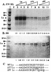
Mutations in full-length La corresponding to LAP mutants 771 and 772 interfere with RNA binding. (A) 32P-labeled PV 5′-UTR RNA was UV cross-linked to 0.5, 1, or 1.5 μg of wt La (lanes 3 to 5), ΔYY (Y23Q Y24Q; lanes 6 to 8), or ΔFF (F25Q F28Q; lanes 9 to 11). Lane 1 shows the migration of molecular weight markers. Lane 2 contains the labeled probe but no protein. (B) The lower panel is a silver-stained gel corresponding to the amount of protein used in the top panel. (C) Amino acid sequences of the wt and mutant La. The numbers at the bottom of each panel indicate relative band intensity as measured by densitometric scanning using the NIH Image J program.
FIG. 10.
FIG. 10.
A single amino acid change in full-length La interferes with RNA binding and translation stimulation. (A) PV 5′-UTR was UV cross-linked to 0.5 μg of wt La (lane 2), 0.5 and 1 μg of Y23 (Y23Q; lanes 3 and 4), or 0.5 and 1 μg of Y24 (Y24Q; lanes 5 and 6). Lane 1 contains no protein. (B) Silver-stained gel corresponding to the amount of protein used in the UV cross-link analysis. (C) Effects of wt and mutants 741 (Y23Q) and 633 (Y24Q) on p2CAT translation in reticulocyte lysates. In vitro translation from the p2CAT RNA was performed in the absence (lane 1) or presence of 0.5 (lanes 2, 5, and 8), 1.0 (lanes 3, 6, and 9), or 1.5 (lanes 4, 7, and 10) μg of wt La (lanes 2 to 4), Y23Q (lanes 5 to 7), and Y24Q (lanes 8 to 10). The numbers at the bottom of each lane indicate the fold stimulation of translation compared to the control (lane 1) without added La. (D) Sequences of the wt and mutant La. The Y23Q and Y24Q mutations were confirmed by sequencing the entire La cDNA.

Similar articles

Cited by

References

    1. Ali, N., and A. Siddiqui. 1997. Molecular mechanisms of translation initiation in eukaryotes. Proc. Natl. Acad. Sci. USA 94:2249-2254. - PubMed
    1. Ali, N., G. J. M. Prujin, D. J. Kenan, J. D. Keene, and A. Siddiqui. 2000. Human La antigen is required for the hepatitis C virus internal ribosome entry site-mediated translation. J. Biol. Chem. 275:27531-27540. - PubMed
    1. Ali, N., and A. Siddiqui. 1995. Interaction of polypyrimidine tract binding protein with the 5′ non-coding region of the hepatitis C virus RNA genome and its functional requirement in internal initiation of translation. J. Virol. 69:6367-6375. - PMC - PubMed
    1. Banerjee, R., M. Igo, R. Izumi, U. Datta, and A. Dasgupta. 2000. In vitro replication of RNA viruses, p. 158-164. In A. Cann (ed.), RNA viruses: a practical approach. Oxford University Press, New York, N.Y.
    1. Belsham, G. J., and N. Sonenberg. 1996. RNA-protein interactions in regulation of picornavirus RNA translation. Microbiol. Rev. 60:499-511. - PMC - PubMed

Publication types

MeSH terms