. 2004 Mar 23;101(12):4047-52.
doi: 10.1073/pnas.0400615101. Epub 2004 Mar 11.

Ataxin 1, a SCA1 neurodegenerative disorder protein, is functionally linked to the silencing mediator of retinoid and thyroid hormone receptors

Affiliations

Ataxin 1, a SCA1 neurodegenerative disorder protein, is functionally linked to the silencing mediator of retinoid and thyroid hormone receptors

Chih-Cheng Tsai et al. Proc Natl Acad Sci U S A. .

Abstract

Ataxin 1 (Atx1) is a foci-forming polyglutamine protein of unknown function, whose mutant form causes type 1 spinocerebellar ataxia in humans and exerts neurotoxicity in transgenic mouse and fly expressing mutant Atx1. In this study, we demonstrate that Atx1 interacts with the transcriptional corepressor SMRT (silencing mediator of retinoid and thyroid hormone receptors) and with histone deacetylase 3. Atx1 binds chromosomes and mediates transcriptional repression when tethered to DNA. Interaction with SMRT-related factors is a conserved feature of Atx1, because Atx1 also binds SMRTER, a Drosophila cognate of SMRT. Significantly, mutant Atx1 forms aggregates in Drosophila, and such mutant Atx1-mediated aggregates sequester SMRTER. Consistently, the neurodegenerative eye phenotype caused by mutant Atx1 is enhanced by a Smrter mutation and, conversely, is suppressed by a chromosomal duplication that contains the wild type Smrter gene. Together, our results suggest that Atx1 is a transcriptional factor whose mutant form exerts its deleterious effects in part by perturbing corepressor-dependent transcriptional pathways.


Figures

Fig. 1.
Fig. 1.
Atx1 forms nuclear foci and associates with SMRT independent of its glutamine repeat length. (A) Diagram of cytomegalovirus promoter-driven CFP-tagged Atx1 variant constructs used in the transfection experiments. Atx1(30Q), Atx1(82Q), and Atx1(0Q) represent wild-type Atx1, mutant (expanded) Atx1, and glutamine repeat-deleted Atx1, respectively; the polyglutamine tract is indicated by a dark gray box. (B) Restriction enzyme digestion patterns for CFP-Atx1 constructs. (CE) Nuclear focal patterns for different CFP-Atx1s and their effects on SMRT expression. HEK-293 cells were transfected with CFP-Atx1(30Q) (C), CFP-Atx1(82Q) (D), or CFP-Atx1(0Q) (E), respectively. CFP signal was captured in black and white and then rendered in green by using photoshop (Adobe Systems, Mountain View, CA). The endogenous SMRT protein was detected by an indirect immunostaining method using an anti-SMRT antibody and Texas red-conjugated secondary antibody (C′–E′).
Fig. 2.
Fig. 2.
Atx1 interacts with SMRT and SMRTER in yeast. (A) Summary of yeast two-hybrid assays for Atx1 and SMRT. The diagram shows the regions of SMRT used in the assays and also the known functional domains of SMRT, including SWI3/Ada 2/N-CoR/TFIIIB′ domains, repression domains (RD I, RD II, RD III, RD IV), and nuclear receptor-interacting domains (NRID). Yeast 190 cells were transformed with GAD-Atx1 (4–703) along with each of the GBT-SMRT variants. The Atx1-interacting domain was mapped to amino acids 1755–2518. Positive β-galactosidase activities were based on color reaction from the lifting assays. (B) Yeast two-hybrid assays for Atx1 and SMRT or SMRTER. Yeast 190 cells were transformed with GAD-Atx1 (4–703)(30Q), along with an empty GBT9 vector, with GBT-SMRT(1755–2518), or with GBT-SMRTER(2094–3040). Positive interactions revealed by 5-bromo-4-chloro-3-indolyl-β-dgalactoside reaction are shown as blue dots. (C) Expression patterns of CFP-Atx1 and YFP-SMRTER in the transfected cells. HEK-293 cells were transfected with plasmids expressing both YFP-SMRTER(2094–3040) and CFP-Atx1(30Q). (D) Yeast two-hybrid assays for Atx1 variants and SMRT or SMRTER. Yeast 190 cells were transformed with GBT-SMRT(1755–2518) or GBT-SMRTER(2094–3040), along with GAD-Atx1(0Q), GAD-Atx1(30Q), or GAD-Atx1(82Q). The β-galactocidase activities from the liquid assays are shown on the right.
Fig. 3.
Fig. 3.
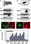
Atx1 interacts selectively with HDAC3 in vitro and in vivo. (A) Diagram of FLAG-tagged Atx1 and HDAC1 constructs used in the coimmunoprecipitation experiments. Lanes 1 and 6, FLAG alone; lane 2, FLAG-Atx1(0Q); lane 3, FLAG-Atx1(30Q); lane 4, FLAG-Atx1(82Q); lane 5, FLAG-HDAC1. The glutamine repeat tract is indicated by a black box. (B) Western blot analysis for FLAG-Atx1s and FLAG-HDAC1 expression. Whole-cell extracts (WCE) prepared from the transfected cells with plasmids corresponding to A were subjected to Western blot analysis by using the anti-FLAG M2 antibody. (C) Coimmunoprecipitation experiments to identify Atx1-associating proteins. WCE and immunoprecipitated complex (IP) prepared from the FLAG-Atx1 transfected cells were subjected to Western blot analysis by using anti-SMRT, anti-Sin3A, anti-CtBP, anti-HDAC1, anti-HDAC3, or anti-HDAC8 antibodies, respectively. (D and E) HDAC3 and HDAC1 staining patterns in cells transfected with CFP-Atx1. HEK-293 cells (D Inset and E) or MCF-7 cells (D) were transfected with CFP-Atx1(82Q) and were immunostained by using anti-HDAC3 antibody (D′) or anti-HDAC1 antibody (E′). (F) The Gal4 reporter assays for Gal4-Atx1 fusions. HEK-293 cells were cotransfected with a Gal4 responsive luciferase reporter (MH100x4), along with three different concentrations of plasmids corresponding to empty Gal4, Gal4-Atx1(0Q), Gal4-Atx1(30Q), Gal4-Atx1(82Q), Gal4-SMRT, or Gal4-E52. Whereas Gal4-SMRT is used here as a positive control, both Gal4-DBD and Gal4-E52 are used as negative control. Luciferase reporter activity was normalized with a β-galactosidase expressing a CMX-lacZ control construct.
Fig. 4.
Fig. 4.
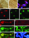
Atx1(82Q) binds chromosomes and sequesters SMRTER into its aggregates. (A and B) Mutant Atx1 causes salivary gland deformity. Shown are Nomarski images corresponding to salivary gland cells isolated from the Hsp70-Gal4;UAS-Atx1(82Q) larvae (A) and from the control UAS-Atx1(82Q) (B). (C) Atx1(82Q) forms aggregates in salivary gland. Nonsquashed salivary gland isolated from Hsp70-Gal4;UAS-Atx1(82Q) larvae were immunostained with anti-Atx1 (Texas red) antibody. The salivary gland is also counterstained with 4′,6-diamidino-2-phenylindole. (D and E) Atx1(82Q)-mediated aggregates sequester SMRTER. Squashed salivary gland cells isolated from Hsp70-Gal4;UAS-Atx1(82Q) larvae (D) or from the control UAS-Atx1(82Q) larvae (E) were immunostained with anti-Atx1 (Texas red) (D) and with anti-SMRTER (FITC) antibodies (D′ and E). An arrow indicates the region where Atx1 aggregates form. (F and G) Atx1 and SMRTER localize to overlapping chromosomal loci. The polytene chromosomes isolated from Hsp70-Gal4;UAS-Atx1(82Q) salivary glands (F) or from the control UAS-Atx1(82Q) salivary glands (G) were subjected to indirect immunostaining by using anti-Atx1 (Texas red) (F and G) and anti-SMRTER (FITC) (F′ and G′′). F′′ is a merged image. G′′ shows the chromosomes counterstained with 4′,6-diamidino-2-phenylindole.
Fig. 5.
Fig. 5.
Smrter mutation modulates the Atx1(82Q)-mediated eye phenotype. (A) Scheme of genetic crosses using indicated females and males to produce male progeny with corresponding genetic background. The numbered rows (1–6) correspond to the images shown in B. (B1B6) Smrter and Atx1 interact genetically. Shown are scanning electron microscopy images of retinas corresponding to adult wild-type (w1118/Y)(B1), SmrterBG1648/Y (B2), w1118/Y;GMR-Gal4,UAS-Atx1(82Q)/+ (B3), SmrterBG1648/Y;GMR-Gal4,UAS-Atx1(82Q)/+ (B4), w1118/Dp(1;Y)BSC5; GMR-Gal4,UAS-Atx1(82Q)/+ (B5), and w1118/Y;GMR-Gal4,UAS-Atx1(82Q)/UAS-dHdj1 (B6).

Similar articles

Cited by

References

    1. Orr, H. T., Chung, M. Y., Banfi, S., Kwiatkowski, T. J., Jr., Servadio, A., Beaudet, A. L., McCall, A. E., Duvick, L. A., Ranum, L. P. & Zoghbi, H. Y. (1993) Nat. Genet. 4, 221–226. - PubMed
    1. Banfi, S., Servadio, A., Chung, M. Y., Kwiatkowski, T. J., Jr., McCall, A. E., Duvick, L. A., Shen, Y., Roth, E. J., Orr, H. T. & Zoghbi, H. Y. (1994) Nat. Genet. 7, 513–520. - PubMed
    1. Zoghbi, H. Y. & Orr, H. T. (2000) Annu. Rev. Neurosci. 23, 217–247. - PubMed
    1. Burright, E. N., Clark, H. B., Servadio, A., Matilla, T., Feddersen, R. M., Yunis, W. S., Duvick, L. A., Zoghbi, H. Y. & Orr, H. T. (1995) Cell 82, 937–948. - PubMed
    1. Fernandez-Funez, P., Nino-Rosales, M. L., de Gouyon, B., She, W. C., Luchak, J. M., Martinez, P., Turiegano, E., Benito, J., Capovilla, M., Skinner, P. J., et al. (2000) Nature 408, 101–106. - PubMed

Publication types

MeSH terms