

doi: 10.1126/science.1091031.
Columnar architecture sculpted by GABA circuits in developing cat visual cortex
Affiliations
- PMID: 15017001
- PMCID: PMC2562723
- DOI: 10.1126/science.1091031
Item in Clipboard
Columnar architecture sculpted by GABA circuits in developing cat visual cortex
Science.
.
Abstract
The mammalian visual cortex is organized into columns. Here, we examine cortical influences upon developing visual afferents in the cat by altering intrinsic gamma-aminobutyric acid (GABA)-mediated inhibition with benzodiazepines. Local enhancement by agonist (diazepam) infusion did not perturb visual responsiveness, but did widen column spacing. An inverse agonist (DMCM) produced the opposite effect. Thus, intracortical inhibitory circuits shape the geometry of incoming thalamic arbors, suggesting that cortical columnar architecture depends on neuronal activity.
Figures
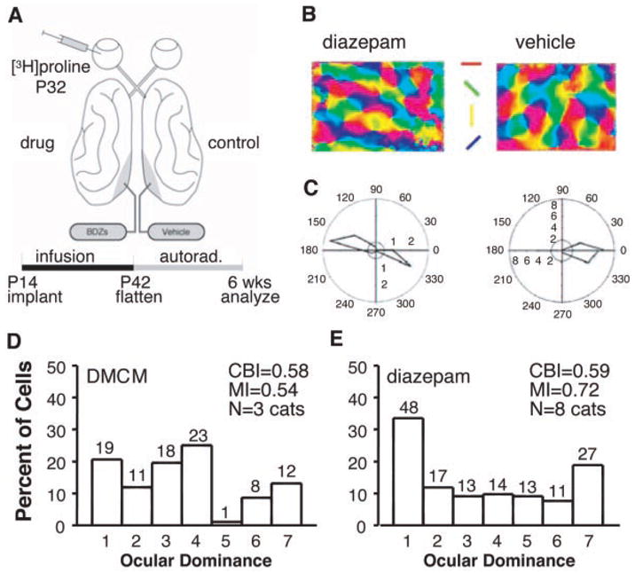
(A) Protocol for local modulation of intrinsic inhibition in vivo. Local benzodiazepine agonist (diazepam), inverse agonist (DMCM), or vehicle (propylene glycol) was infused from osmotic minipumps (0.2 or 2.5 μl/hour) into kitten striate cortex for 4 weeks starting at 14 to 17 days after birth (10). Monocular [3H]proline (2 mCi) was injected 10 days before electrophysiological recording. Labeled columns were measured in tangential sections through layer 4 of unfolded and flattened visual cortex (10). (B and C) Physiological properties of diazepam-treated cortical neurons. Robust orientation tuning in optical imaging angle maps (B) and single-unit responses (C). Spontaneous activity and response strength are indicated as respective concentric-circle radii. The absence of directionality in diazepam-treated cortex is apparent in this example (fig. S2). (D and E) Ocular dominance distribution after DMCM infusion was binocular, as in controls (D) (1, 2), but diazepam-treated hemispheres were significantly less binocular (E) (P < 0.001, χ2 test, DMCM versus diazepam). CBI, contralateral bias index; MI, monocularity index. The number of cells per ocular dominance group is as indicated.
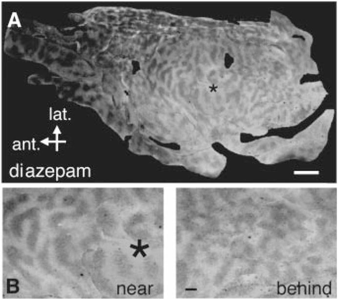
(A) Chronic diazepam infusion widens ocular dominance columns locally. Flatmount of overall layout of labeled (dark) and unlabeled (white) ocular dominance bands in layer 4 of diazepam-infused (*) (3.5 mM, 2.5 μl/hour) kitten visual cortex. (B) Wide, crisp columns just anterior to the cannula site compared to an area behind and away from the infusion. Scale bars: 5 mm (A); 1 mm (B).
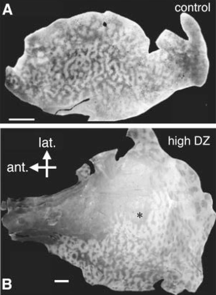
(A) Control hemisphere exhibits relatively homogeneous column spacing across a homologous extent of area 17. (B) Graded effect of diazepam treatment. A high dose (35 mM, 0.2 μl/hour) yields an area of column desegregation (anterolateral to cannula site) (*) reminiscent of direct GABAA receptor activation with muscimol (3). Scale bar: 5 mm.
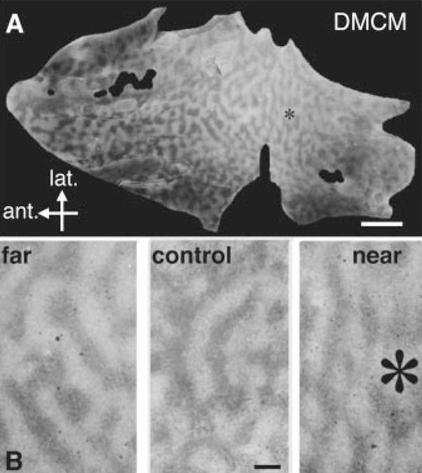
(A) Chronic inverse agonist infusion locally reduces column size. Overall layout of labeled ocular dominance bands (dark) surrounding a DMCM infusion site (*) (50 μM, 2.5 μl/hour). (B) Columns near a DMCM source were blurred and narrow (right) compared to distal (left) or homologous regions in control hemispheres (middle). Scale bars, 5 mm (A); 1 mm (B).
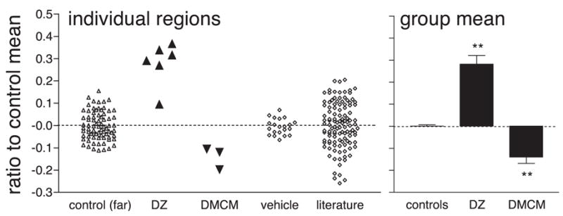
Bidirectional control of developing column size by cortical inhibition. Wavelet measurements (10, 20) of spacing in individual regions were normalized to control mean far from the cannula, or across vehicle and untreated control hemispheres from literature (3, 22, 23). Statistical comparison is by group. **P < 0.001, t test versus control.
Comment in
-
Neuroscience. Blocking plasticity in the visual cortex.Science. 2004 Mar 12;303(5664):1619-21. doi: 10.1126/science.1096224. Science. 2004. PMID: 15016985 No abstract available.
References
Publication types
MeSH terms
Substances
Grants and funding
LinkOut - more resources
Full Text Sources
Other Literature Sources
Miscellaneous
