

SARS: radiological features
- PMID: 15018128
- PMCID: PMC7169195
- DOI: 10.1046/j.1440-1843.2003.00519.x
SARS: radiological features
Abstract
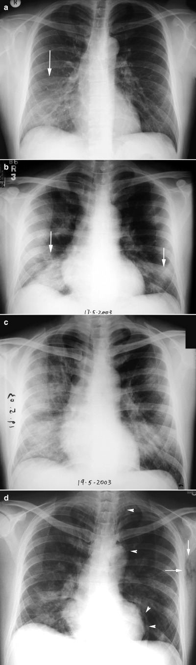
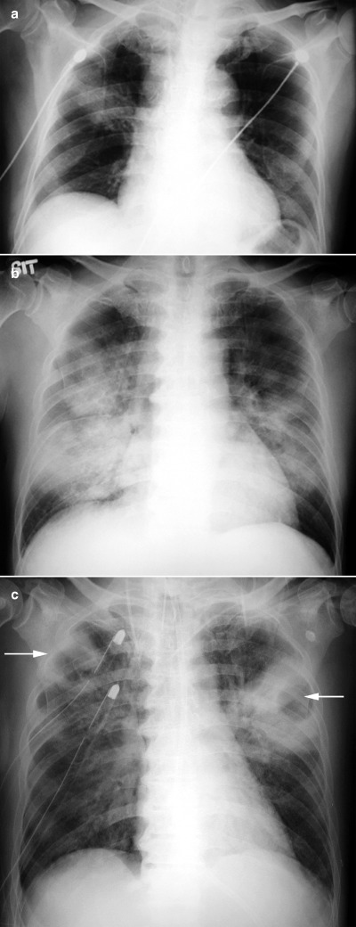
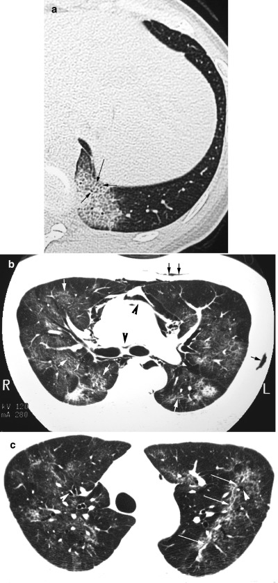
Air-space disease is typical in severe acute respiratory syndrome (SARS) and may be indistinguishable from pneumonia of other causes. In the majority of patients, ground glass opacities on chest radiographs progress rapidly to focal, multifocal or diffuse consolidation. Unilateral involvement is common in the early acute phase, becoming bilateral at maximal lung involvement. Generally, radiographic opacities peak between 8 and 10 days after onset of illness, with radiographic scores reflecting temporal changes in clinical and laboratory parameters such as oxygen saturation (SaO2) and liver transaminases. Pleural effusions, cavitating consolidation and mediastinal lymphadenopathy are not typical radiographic features. Pneumomediastinum and pneumothoraces are complications that are associated with extensive disease, with or without assisted ventilation. The utility of high resolution computed tomography (HRCT) and CT scans lies in the confirmation of airspace opacities in cases with normal initial chest radiographs that have strong contact history and signs and symptoms highly suspicious of SARS during the outbreak, allowing early treatment and prompt isolation. The characteristic HRCT feature in the acute phase is ground-glass opacities with smooth interlobular septal thickening, sometimes with consolidation in a subpleural location, which progress rapidly to involve other areas of the lungs. Temporal lung changes documented on HRCT suggest that some residual opacities found may not be reversible.
Figures
Similar articles
-
Comparison of initial high resolution computed tomography features in viral pneumonia between metapneumovirus infection and severe acute respiratory syndrome.Eur J Radiol. 2012 May;81(5):1083-7. doi: 10.1016/j.ejrad.2011.02.050. Epub 2011 Mar 25. Eur J Radiol. 2012. PMID: 21439753 Free PMC article.
-
Severe acute respiratory syndrome: temporal lung changes at thin-section CT in 30 patients.Radiology. 2004 Mar;230(3):836-44. doi: 10.1148/radiol.2303030853. Radiology. 2004. PMID: 14990845
-
Early X-ray and CT appearances of severe acute respiratory syndrome: an analysis of 28 cases.Chin Med J (Engl). 2003 Jun;116(6):823-6. Chin Med J (Engl). 2003. PMID: 12877787
-
Imaging of unusual diffuse lung diseases.Curr Opin Pulm Med. 2004 Sep;10(5):383-9. doi: 10.1097/01.mcp.0000134389.50775.b2. Curr Opin Pulm Med. 2004. PMID: 15316437 Review.
-
Similarities and Differences of Early Pulmonary CT Features of Pneumonia Caused by SARS-CoV-2, SARS-CoV and MERS-CoV: Comparison Based on a Systemic Review.Chin Med Sci J. 2020 Sep 30;35(3):254-261. doi: 10.24920/003727. Chin Med Sci J. 2020. PMID: 32972503 Free PMC article.
Cited by
-
A comparison of COVID-19, SARS and MERS.PeerJ. 2020 Aug 19;8:e9725. doi: 10.7717/peerj.9725. eCollection 2020. PeerJ. 2020. PMID: 32879801 Free PMC article.
-
Follow-up chest radiographic findings in patients with MERS-CoV after recovery.Indian J Radiol Imaging. 2017 Jul-Sep;27(3):342-349. doi: 10.4103/ijri.IJRI_469_16. Indian J Radiol Imaging. 2017. PMID: 29089687 Free PMC article.
-
Prevalence and risk factors for lung involvement on low-dose chest CT (LDCT) in a paucisymptomatic population of 247 patients affected by COVID-19.Insights Imaging. 2020 Nov 17;11(1):117. doi: 10.1186/s13244-020-00939-7. Insights Imaging. 2020. PMID: 33201409 Free PMC article.
-
Chest imaging of H7N9 subtype of human avian influenza.Radiol Infect Dis. 2015 Mar;1(2):51-56. doi: 10.1016/j.jrid.2015.02.001. Epub 2015 Feb 27. Radiol Infect Dis. 2015. PMID: 32289064 Free PMC article.
-
Coronavirus Disease 2019 in the Perioperative Period of Lung Resection: A Brief Report From a Single Thoracic Surgery Department in Wuhan, People's Republic of China.J Thorac Oncol. 2020 Jun;15(6):1065-1072. doi: 10.1016/j.jtho.2020.04.003. Epub 2020 Apr 11. J Thorac Oncol. 2020. PMID: 32289516 Free PMC article.
References
-
- Tsang KW, Ho PL, Ooi GC et al. A cluster of cases of severe acute respiratory syndrome in Hong Kong. N. Engl. J. Med. 2003; 348: 1977–85. - PubMed
-
- Lee N, Hui D, Wu A et al. A major outbreak of severe acute respiratory syndrome in Hong Kong. N. Engl. J. Med. 2003; 348: 1986–94. - PubMed
-
- World Health Organization. Case definitions for surveillance of severe acute respiratory syndrome (SARS). Revised May 1, 2003. [Cited 1 May, 2003.] Available from URL: http://www.who.int/csr/sars/casedefinition/en/
-
- Ksiazek TG, Erdman D, Goldsmith CS et al. A novel coronavirus associated with severe acute respiratory syndrome. N. Engl. J. Med. 2003; 348: 1953–66. - PubMed
Publication types
MeSH terms
LinkOut - more resources
Full Text Sources
Miscellaneous
