

Analysis of multimerization of the SARS coronavirus nucleocapsid protein
- PMID: 15020242
- PMCID: PMC7111152
- DOI: 10.1016/j.bbrc.2004.02.074
Analysis of multimerization of the SARS coronavirus nucleocapsid protein
Abstract
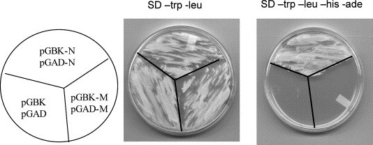
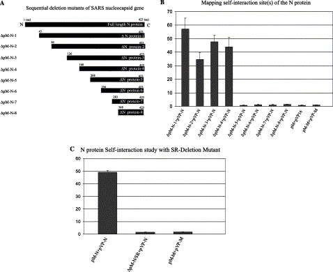
Severe Acute Respiratory Syndrome (SARS), an emerging disease characterized by atypical pneumonia, has recently been attributed to a novel coronavirus. The genome of SARS Coronavirus (SARS-CoV) has recently been sequenced, and a number of genes identified, including that of the nucleocapsid protein (N). It is noted, however, that the N protein of SARS-CoV (SARS-CoV N) shares little homology with nucleocapsid proteins of other members of the coronavirus family [Science 300 (2003) 1399; Science 300 (2003) 1394]. N proteins of other coronavirus have been reported to be involved in forming the viral core and also in the packaging and transcription of the viral RNA. As data generated from some viral systems other than coronaviruses suggested that viral N-N self-interactions may be necessary for subsequent formation of the nucleocapsid and assembly of the viral particles, we decided to investigate SARS-CoV N-N interaction. By using mammalian two-hybrid system and sucrose gradient fractionations, a homotypic interaction of N, but not M, was detected by the two-hybrid analysis. The mammalian two-hybrid assay revealed an approximately 50-fold increase in SEAP activity (measurement of protein-protein interaction) in N-N interaction compared to that observed in either M-M or mock transfection. Furthermore, mutational analyses characterized that a serine/arginine-rich motif (SSRSSSRSRGNSR) between amino acids 184 and 196 is crucial for N protein oligomerization, since deletion of this region completely abolished the N protein self-multimerization. Finally, the full-length nucleocapsid protein expressed and purified from baculovirus system was found to form different levels of higher order structures as detected by Western blot analysis of the fractionated proteins. Collectively, these results may aid us in elucidating the mechanism pertaining to formation of viral nucleocapsid core, and designing molecular approaches to intervene SARS-CoV replication.
Figures


References
-
- Marra M.A., Jones S.J.M., Astell C.R., Holt R.A., Brooks-Wilson A., Butterfield Y.S.N., Kattra J., Asano J.K., Barber S.A., Chan S.Y., Cloutier A., Coughlin S.M., Freeman D., Girn N., Griffith O.L., Leach S.R., Mayo M., McDonald H., Montgomery S.B., Pandoh P.K., Petrescu A.S., Robertson A.G., Schein J.E., Siddiqui A., Smailus D.E., Stott J.M., Yang G.S., Plummer F., Andonov A., Artsob H., Bastien N., Bernard K., Booth T.F., Bowness D., Czub M., Drebot M., Fernando L., Flick R., Garbutt M., Gray M., Grolla A., Jones S., Feldmann H., Meyers A., Kabani A., Li Y., Normand S., Stroher U., Tipples G.A., Tyler S., Vogrig R., Ward D., Watson B., Brunham R.C., Krajden M., Petric M., Skowronski D.M., Upton C., Roper R.L. The genome sequence of the SARS-associated coronavirus. Science. 2003;300(5624):1399–1404. - PubMed
-
- Rota P.A., Oberste M.S., Monroe S.S., Nix W.A., Campagnoli R., Icenogle J.P., Penaranda S., Bankamp B., Maher K., Chen M., Tong S., Tamin A., Lowe L., Frace M., DeRisi J.L., Chen Q., Wang D., Erdman D.D., Peret T.C.T., Burns C., Ksiazek T.G., Rollin P.E., Sanchez A., Liffick S., Holloway B., Limor J., McCaustland K., Olsen-Rasmussen M., Fouchier R., Gunther S., Osterhaus A.D.M.E., Drosten C., Pallansch M.A., Anderson L.J., Bellini W.J. Characterization of a novel coronavirus associated with Severe Acute Respiratory Syndrome. Science. 2003;300(5624):1394–1399. - PubMed
-
- Holmes K.V. Chapter 36: Coronaviruses. In: Knipe D.M., Howley P.M., editors. vol. 1. Williams & Wilkins; Baltimore, MD: 2001. pp. 1187–1203. (Fields Virology).
-
- Drosten C., Gunther S., Preiser W., van der Werf S., Brodt H.-R., Becker S., Rabenau H., Panning M., Kolesnikova L., Fouchier R.A.M., Berger A., Burguiere A.-M., Cinatl J., Eickmann M., Escriou N., Grywna K., Kramme S., Manuguerra S., Muller S., Rickerts V., Sturmer M., Vieth S., Klenk H.-D., Osterhaus A.D.M.E., Schmitz H., Doerr H.-W. Identification of a novel coronavirus in patients with Severe Acute Respiratory Syndrome. N. Engl. J. Med. 2003;348(20):1967–1976. - PubMed
MeSH terms
Substances
LinkOut - more resources
Full Text Sources
Other Literature Sources
Molecular Biology Databases
Research Materials
Miscellaneous
