. 2004 Apr;63(4):354-9.
doi: 10.1136/ard.2003.008458.

Joint erosion in rheumatoid arthritis: interactions between tumour necrosis factor alpha, interleukin 1, and receptor activator of nuclear factor kappaB ligand (RANKL) regulate osteoclasts

Affiliations

Joint erosion in rheumatoid arthritis: interactions between tumour necrosis factor alpha, interleukin 1, and receptor activator of nuclear factor kappaB ligand (RANKL) regulate osteoclasts

D O' Gradaigh et al. Ann Rheum Dis. 2004 Apr.

Abstract

Background: Osteoclasts, specialised bone resorbing cells regulated by RANKL and M-CSF, are implicated in rheumatoid joint erosion. Lymphocyte-monocyte interactions activate bone resorption, this being attributed to tumour necrosis factor alpha (TNFalpha) and interleukin 1 beta (IL1beta) enhanced osteoblast expression of RANKL. In animal studies, TNF potently increases osteoclast formation in the presence of RANKL. RANKL-independent osteoclastogenesis also occurs, though IL1 is required for resorptive function in most studies. These inflammatory cytokines have a pivotal role in rheumatoid arthritis,

Objective: To study the interactions of TNFalpha and IL1beta with RANKL, particularly the time course of the interactions and the role of lymphocytes.

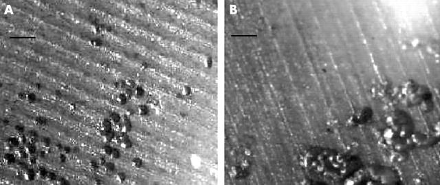
Method: Cultures of lymphocytes and monocytes (osteoclast precursors) or of purified CD14(+) cells alone (osteoclast precursors) were exposed to various combinations of TNFalpha, RANKL, and IL1beta or the inhibitors osteoprotegerin, IL1 receptor antagonist, or neutralising antibodies to RANKL or to IL1. Osteoclastogenesis and resorptive activity were assessed on microscopy of dentine slices.

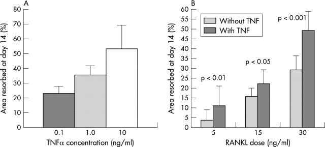
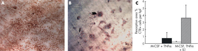
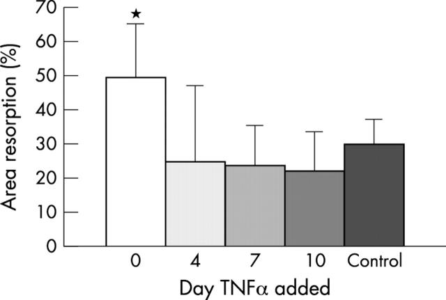
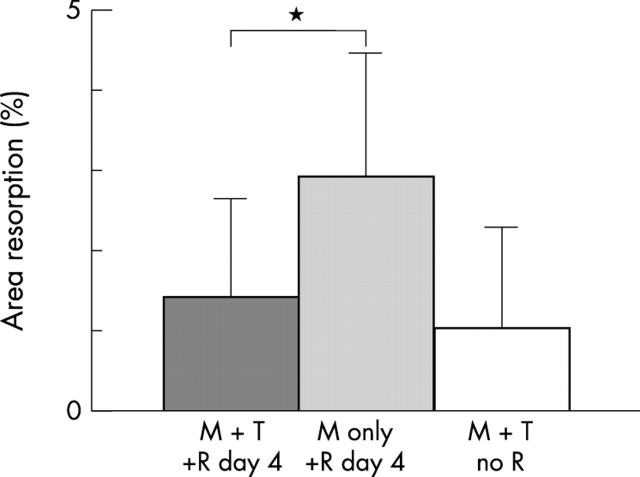
Results: TNFalpha potently increased osteoclast proliferation/differentiation in the presence of RANKL. This effect was greatest when RANKL was present before but not after exposure of osteoclast precursor cells to TNFalpha. The resorptive activity of osteoclasts generated by TNFalpha in the absence of RANKL was critically dependent upon IL1, which was expressed by lymphocyte-monocyte interaction.

Conclusion: TNFalpha potently enhances RANKL mediated osteoclast activity. Interactions between TNFalpha and IL1 also result in osteoclastic activity independently of RANKL. These findings will inform therapeutic approaches to the prevention of joint erosion in rheumatoid arthritis.


Figures

Figure 1
Figure 1
TNFα is synergistic with RANKL in osteoclast regulation. (A) Dose-resorption effect on area of osteoclastic resorption of dentine slices on adding TNFα in a range of doses to PBMCs cultured in RANKL 30 ng/ml and M-CSF 25 ng/ml. (B) Effect on area of osteoclastic resorption of dentine slices of adding TNFα 1 ng/ml to RANKL at 5, 15, or 30 ng/ml. Mean of four to six slices in three experiments; error bars are the standard deviation.
Figure 2
Figure 2
RANKL-independent osteoclast formation in cultures of PBMCs. Reflected light microscopy of dentine slices stained with toluidine blue showing individual and grouped resorption pits. (A) cells cultured in M-CSF and TNFα (1 ng/ml); (B) cells cultured in M-CSF, TNFα (1 ng/ml), and OPG (200 ng/ml). Bar = 50 µm.
Figure 3
Figure 3
Osteoclastogenesis from CD14+ selected cells cultured in (A) M-CSF and TNFα or (B) in M-CSF, TNFα, and IL1. Addition of IL1 resulted in resorptive activity (pits shown by arrows) by CTR+ cells (arrow head), whereas in the absence of IL1, the CTR+ cells did not resorb. Bar (photomicrographs) = 50 µm. Staining by indirect immunoperoxidase method. Data from repeated experiments are shown in the graph (C). The number of CTR+ cells per high power field in the absence (black bar) or presence (hatched bar) of IL1 did not differ significantly. The resorption area was only measurable when IL1 was added (white bar). Error bars are the standard deviation.
Figure 4
Figure 4
Effect of TNFα depends on timing of addition to cultures. PBMCs were cultured in M-CSF and RANKL, with TNF added at various time points. Significantly more resorption occurred when TNFα was added on day 0 (white bar) compared with control to which no TNFα was added (black bar), p<0.001. The resorption area was not increased when TNFα was added on day 4, 7 or 10. Mean of six slices in three experiments; error bars are the standard deviation.
Figure 5
Figure 5
Exposure to TNFα before RANKL reduces osteoclastic resorption activity. PBMCs were cultured in M-CSF (light grey bar) or in M-CSF with TNFα (dark grey bar) before adding RANKL after 4 days, and compared with cells treated with M-CSF and TNFα alone (white bar). Osteoclast resorption was reduced when cells were exposed to TNFα before RANKL (*p = 0.08). Mean of six slices in three experiments; error bars are the standard deviation. M, M-CSF 25 ng/ml; T, TNFα 1 ng/ml; R, RANKL 30 ng/ml.

References

    1. Endocrinology. 1998 Jul;139(7):3157-64 - PubMed
    1. Am J Pathol. 1998 Apr;152(4):943-51 - PubMed
    1. J Immunol. 1999 Mar 1;162(5):2754-60 - PubMed
    1. Arthritis Rheum. 1999 May;42(5):963-70 - PubMed
    1. Semin Arthritis Rheum. 1999 Aug;29(1):27-35 - PubMed

Publication types

MeSH terms