. 2004 May;15(5):2470-83.
doi: 10.1091/mbc.e03-11-0848. Epub 2004 Mar 12.

Cytoplasmic dynein-dynactin complex is required for spermatid growth but not axoneme assembly in Drosophila

Affiliations

Cytoplasmic dynein-dynactin complex is required for spermatid growth but not axoneme assembly in Drosophila

Anindya Ghosh-Roy et al. Mol Biol Cell. 2004 May.

Abstract

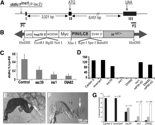
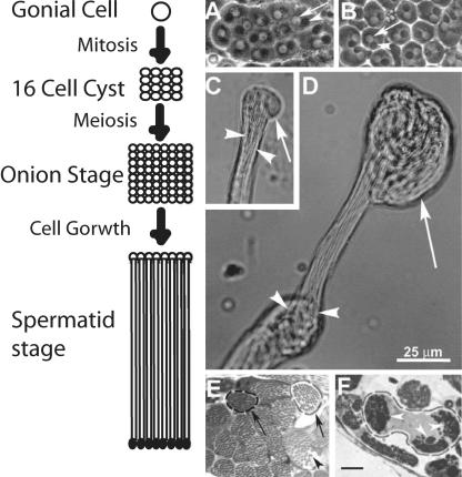
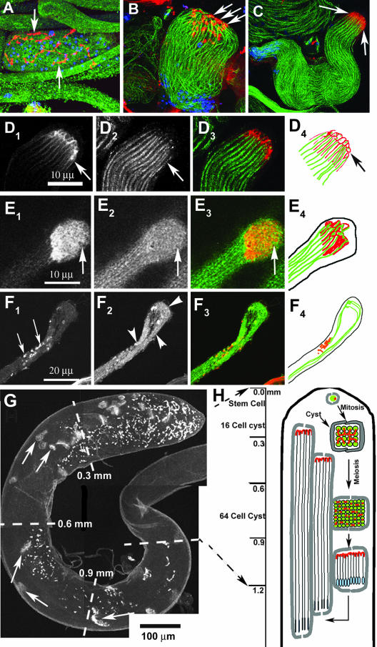
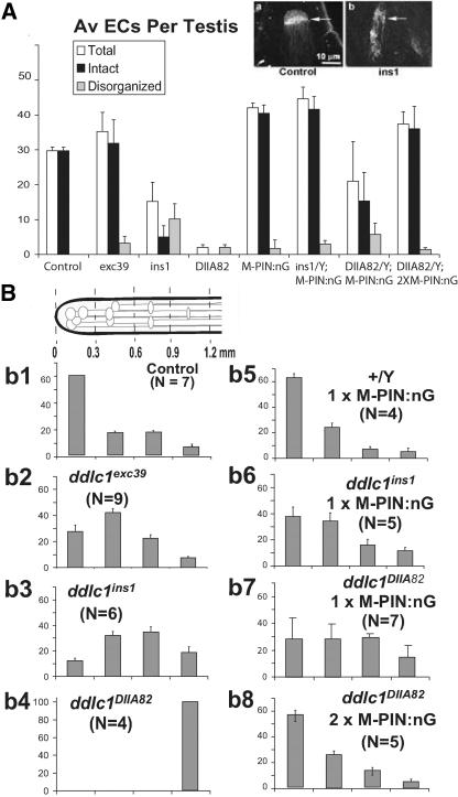
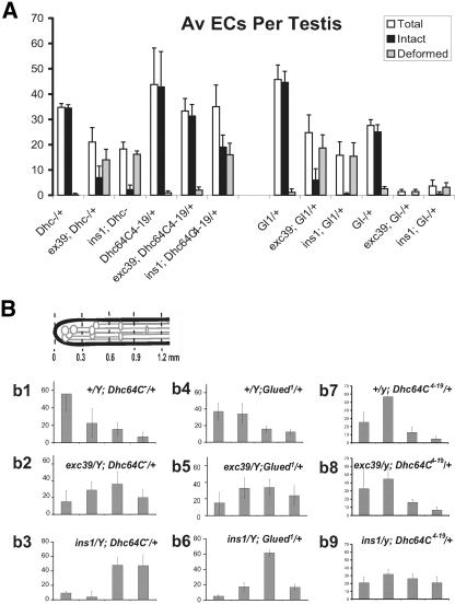
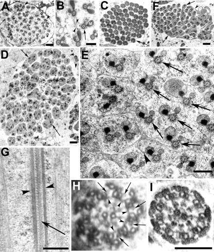
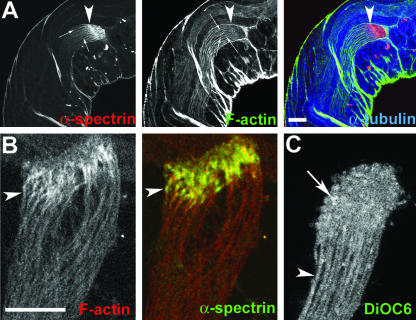
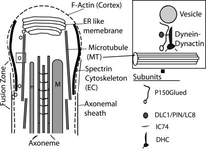
Spermatids derived from a single gonial cell remain interconnected within a cyst and elongate by synchronized growth inside the testis in Drosophila. Cylindrical spectrin-rich elongation cones form at their distal ends during the growth. The mechanism underlying this process is poorly understood. We found that developing sperm tails were abnormally coiled at the growing ends inside the cysts in the Drosophila Dynein light chain 1 (ddlc1) hemizygous mutant testis. A quantitative assay showed that average number of elongation cones was reduced, they were increasingly deformed, and average cyst lengths were shortened in ddlc1 hemizygous testes. These phenotypes were further enhanced by additional partial reduction of Dhc64C and Glued and rescued by Myc-PIN/LC8 expression in the gonial cells in ddlc1 backgrounds. Furthermore, DDLC1, DHC, and GLUED were enriched at the distal ends of growing spermatids. Finally, ultrastructure analysis of ddlc1 testes revealed abnormally formed interspermatid membrane, but the 9 + 2 microtubule organization, the radial spoke structures, and the Dynein arms of the axoneme were normal. Together, these findings suggest that axoneme assembly and spermatid growth involve independent mechanisms in Drosophila and DDLC1 interacts with the Dynein-Dynactin complex at the distal ends of spermatids to maintain the spectrin cytoskeleton assembly and cell growth.


Figures

Figure 1.
Figure 1.
Reduced levels of Ddlc1 cause temperature sensitive recessive lethality and male sterility. (A) Schematic shows the relative positions of exons (I, II, and III), the start (ATG) and stop (UAA) codons in Ddlc1 gene. The positions of the P-lacW insertion in ddlc1ins1 (vertical gray arrow) and the Ddlc1 specific primers (P1 and P2) are also indicated. (B) Schematic map of the Myc-PIN/LC8 transgene cloned in pPUAST. (C) Histogram represents relative fluorescence intensity of the Ddlc1 amplicons with respect to the rp49. Error bar indicates ± SD, and N ≥4 for all data points. (D) Histograms represent average progenies per wild-type and mutant males, respectively. mPIN:nG represents the presence of UAS-MycPIN/LC8 and nos-Gal4-VP16, and N = 20 or more single male crosses for each bar. (E and F) Seminal vesicles isolated from wild-type control (E) and ddlc1DIIA82 hemizygous (F) males. Arrow indicates bundles of active sperms in E and a few motile sperms in F. (G) Histograms indicate percentage of distribution of seminal vesicles (SV) in wild-type controls and in different ddlc1 alleles with a large number of motile sperm (black filled bars), relatively fewer (gray filled bars), and no (white filled bars) motile sperm. All the seminal vesicles from Canton S and ddlc1rev adults were filled with vigorously motile sperm (see Supplemental Movie 1), whereas the ones from ddlc1 hemizygous adults were partly filled with relatively sluggish sperm (see Supplemental Movie 2).
Figure 2.
Figure 2.
Ddlc1 is essential for proper growth of sperm tails in developing cysts. (A–D) Phase contrast images of isolated cysts from wild-type control (A and C) and ddlc1exc39 hemizygous (B and D) testes. (A and B) Onion stage spermatocytes isolated from the wild-type control (A) and mutant testes (B), respectively. The arrows and arrowheads indicate nuclei and onion stage mitochondria, respectively. (C) Distal end of an elongating cyst from wild-type control testis. The spermatid tails (arrowheads) are tightly bundled and have a membranous bulge (arrow) at their distal ends. (D) Spermatids are abnormally knotted (arrowheads) in the middle and coiled (arrow) at their distal ends inside the cysts in ddlc1 testis. Scale is equal for all figures and as indicated in D. (E and F) Thin sections (1 μm) of testis from the wild-type control (E) and ddlc1DIIA82 (F) adults stained with methylene blue. (E) Two typical cyst boundaries (arrows) are marked, and the arrowhead indicates a single spermatid. (F) Spermatids are abnormally bundled (arrowheads) in multiple fascicles even within a single cyst. Bar, 10 μm for E and F.
Figure 3.
Figure 3.
Spectrin scaffold of fusomes is reorganized to form the cylindrical spectrin caps at the growing ends of axoneme. A–D show anti-β-spectrin (colored red) and anti-β-tubulin (colored green) staining in developing spermatids of wild-type testis. The nuclear DNA (colored blue) is stained using Sytox Green. (A) Postmeiotic stage cyst with regular organization of fusomes (arrow) and 64 nuclei inside. (B) Elongating stage cyst with the spermatid heads (blue) bundled together at one end and condensed spectrin staining (arrows) at the other end. The axonemal microtubules (green) are marked by anti-β-tubulin. (C) The β-spectrin staining (arrows) at the growing ends of axoneme seems compact at a later stage, and the spermatids also seemed uniformly bundled. (D1–4) Bundle of cortical spectrin-rich structures (arrows in D1) is visible at the distal end when the cyst is between 1.5 and 2 mm in length, and each one seems to cap individual axoneme (arrows in D2). A red green merge of D1 and D2 is presented in D3, and D4 shows a tracing of individual axoneme (green) and the spectrin caps at this stage. (E1–4) Distal end of a wild-type cyst stained with α-spectrin (E1) and α-tubulin (E2), respectively. Arrows indicate the distal end in E1–3 and E3 shows a red green merge of E1 and E2. A tracing of α-spectrin and α-tubulin staining is shown in E4. (F1–4) Similar staining in ddlc1ins1 cyst shows that the spectrin-rich honeycomb structure is deformed into punctate spots (F1, arrows) and abnormally twisted axonemes (F2, arrowheads). (G) Anti-α-spectrin staining of intact testis depicts a regular distribution of the spectrin scaffolds (arrows) along its length. These are called elongation cone (EC) in this study. (H) Illustration indicates the orientation and position of the spermatid heads (blue) and the spectrin scaffolds (red) within a cyst at different stages of spermatogenesis inside the testis.
Figure 4.
Figure 4.
Mutations in the ddlc1 locus disrupt elongation cone formation and reduce spermatid growth. (A) Histogram depicts average number of recognizable intact and disrupted EC in wild-type control and mutant testes. Error bars indicates ± SD, and N = 4 or more for each data set. The insets show typical examples of an intact (arrow in a) and a disorganized (arrow in b) elongation cones revealed by anti-α-spectrin staining. (B) Schematic at the top left corner indicates the apical one-half of a testis, and gray lines with elliptical heads indicate ECs of individual cysts. The set of histograms (b1–b8) represent percentage of EC distributions ± SD in different length segments of the testis as indicated in the schematic. The scale is set from the apical tip of the testis, and the length segments are indicated in millimeters. MPIN:nG indicates the presence of UAS-Myc-PIN/+; nosGal4-VP16/+ in the background.
Figure 5.
Figure 5.
Additional reduction of DHC and Glued enhanced the EC deformation and spermatid growth defects of ddlc1 alleles. (A) Histogram represents EC morphology parameters counted in different mutant combinations (shown in the x-axis) containing ddlc1 alleles in the background and as described in the previous figure. The error bars indicates ± SD, and N = 4 or more for each data set. (B) EC distribution profile along the length of the testes for different mutant combinations containing ddlc1 alleles in the background and as described in the previous figure. The error bars indicate ± SD, and N = 4 or more for each data set. Df(3L)GN24 is mentioned as DHC and Df(3L)fzGF3b is mentioned as Gl in this figure.
Figure 6.
Figure 6.
DDLC1 is enriched at the EC region along with DHC and GLUED. Isolated cysts were immunostained with anti-DDLC1 (A–C), anti-DHC (D), and anti-GLUED (E), and the same sample were also stained with anti-α-tubulin (A and B), anti-β-spectrin (D), and anti-α-spectrin (C and E) antisera. (A) In wild-type cyst, the DDLC1 and α-tubulin were enriched at the distal ends of individual spermatids (arrowheads). (B) In ddlc1ins1 cyst, the DDLC1 enrichment was absent, and the axonemal microtubules seemed abnormally bent in the middle (arrowhead) and coiled at the distal end (arrow). (C) Higher magnification of the EC region stained with anti-α-spectrin (red), rat-anti-DDLC1 (green), and phalloidin (blue) showed enrichment of DDLC1 along the cortical spectrin network of the EC (arrows). In addition, some cytosolic DDLC1-enriched spots were also visible (fine arrow). (D and E) The DHC (D, green) and the GLUED (E, green) antisera also showed punctate staining along the cortical spectrin cytoskeleton in the EC (arrows). Bar, 20 μm in A and B and 10 μm in C–E.
Figure 7.
Figure 7.
Mutations in ddlc1 affects the cellularization of spermatids. Electron microscopic images of transverse sections of ddlc1rev (A–C), and ddlc1DIIA82 (D–F) testes collected from 2-d-old adults. (A) Section close to the caudal end of a cyst shows 64 axoneme-mitochondria sets mostly wrapped in individual axonemal sheath connected by thin membranous bridges (arrows). But often, more than one axoneme-mitochondria set was found within a single envelope (arrowheads). (B) Axoneme (arrow) and the major (M) and minor (m) mitochondrial derivatives surrounded by cellular microtubules are in syncytium at the caudal most end, and some incompletely formed membrane lamellae with vesicular bodies (arrowheads) attached were seen in this region. (C) Matured cysts showing individually wrapped axoneme-mitochondria pairs (arrow). (D) Mega cyst from ddlc1DIIA82 testis containing 112 axonemes and with multiple axonemes within a single envelope (arrows). In total, 64 such envelopes were visible in this section. (E) Higher magnification image of a part of a different cyst from the same section shows incompletely formed axonemal sheath (arrowhead) and multiple axoneme-mitochondria sets (arrows) within a single cellular envelope. The morphology of the axoneme-mitochondria complex seemed normal. (F) Section of a seemingly mature cyst from ddlc1DIIA82 testis showing incompletely individualized spermatids (arrows) with multiple axoneme-mitochondria sets within one envelope and interconnected by thin membranous bridges (arrowhead). (G) Longitudinal section through the axoneme from an immature (preindividualization) spermatid from ddlc1DIIA82 hemizygous testis revealed normal peripheral (arrowheads) and central (arrow) microtubule assembly. (H) Cross section of an axoneme at the growing end of the spermatid from the same sample revealed 9 + 2 organization of microtubules, the outer (arrows) and inner (arrowheads) Dynein arms, and the radial spoke structures. (I) Section of a relatively matured spermatid (postindividualization) from the mutant testis also showed normal organization. Bar, 1 μm in A, D, and F; 0.5 μm in B, E, and G; and 0.2 μm for H and I as shown in I.
Figure 8.
Figure 8.
Cortical F-actin assembly is formed around developing spermatids at the EC region. (A) Optical section of an intact adult testis stained with anti-α-spectrin (red), rhodamine isothiocyanate: phalloidin (green), and anti-α-tubulin (blue). Arrowheads indicate the distal end (EC) of a cyst and arrows indicate cortical localization of α-spectrin and F-actin around individual spermatids in the cyst. Note that the cortical F-actin as well as the axonemal α-tubulin staining end at the proximal part of the EC structure. (B) Distal end of an isolated cyst stained with phalloidin (red) and anti-α-spectrin (green). Arrowhead indicates cortical enrichment of F-actin and spectrin at the distal end of a single spermatid. (C) Single optical section of an isolated live cyst stained with DiOC6. Arrowhead indicates a single spermatid cell, and arrow indicates enrichment of staining in individual spermatids at the distal ends.
Figure 9.
Figure 9.
An illustration of the proposed role of cytoplasmic Dynein-Dynactin in the membrane deposition process at the caudal end of growing spermatids. The caudal end of a single elongation stage spermatid is shown in this illustration. The smooth “ER-like membrane” lamellae, the cellular “microtubule (MT)”, the major (M) and minor (m) mitochondrial derivatives, “axoneme,” the cortical “F-actin” and “spectrin” cytoskeleton assembly of the EC region, and the “axonemal sheath” is indicated with appropriate labels. Growing spermatids remain in syncytium at the distal most end and individual axoneme-mitochondria assembly as shown in this illustration is found to be enveloped in separate axonemal sheath just below the elongation cone region marked by the spectrin rich cytoskeleton. The cytoplasmic Dynactin–Dynactin complex is proposed to transport membrane bound vesicles generated from the ER lamellae to the fusion zone surrounding the axoneme-mitochondria assembly and these vesicles upon fusion would generate the axonemal sheath. The cellular microtubules are likely to play a role in directing the vesicular traffic toward the basal side. The box at the top right corner illustrates how the cellular Dynein–Dynactin subunits could interact with the vesicle and the microtubule. The subunits indicated are the ones tested for their role in spermatid growth in this study. The membrane organization at the caudal most end of spermatid is not shown in this illustration because the ultrastructure of this region is not clearly understood.

Similar articles

Cited by

References

    1. Abe, T.K., Tanaka, H., Iwanaga, T., Odani, S., and Kuwano, R. (1997). The presence of the 50-kDa subunit of dynactin complex in the nerve growth cone. Biochem. Biophys. Res. Commun. 233, 295–299. - PubMed
    1. Arama, E., Agapite, J., and Steller, H. (2003). Caspase activity and a specific cytochrome C are required for sperm differentiation in Drosophila. Dev. Cell. 4, 687–697. - PubMed
    1. Beckwith, S.M., Roghi, C.H., Liu, B., and Ronald Morris, N. (1998). The “8-kD”cytoplasmic dynein light chain is required for nuclear migration and for dynein heavy chain localization in Aspergillus nidulans. J. Cell Biol. 143, 1239–1247. - PMC - PubMed
    1. Bergeret, E., Pignot-Paintrand, I., Guichard, A., Raymond, K., Fauvarque, M.O., Cazemajor, M, and Griffin-Shea, R. (2001). RotundRacGAP functions with Ras during spermatogenesis and retinal differentiation in Drosophila melanogaster. Mol. Cell. Biol. 21, 6280–6291. - PMC - PubMed
    1. Burkhardt, J.K., Echeverri, C.J., Nilsson, T., and Vallee, R.B. (1997). Overexpression of the dynamitin (p50) subunit of the dynactin complex disrupts dynein-dependent maintenance of membrane organelle distribution. J. Cell Biol. 139, 469–484. - PMC - PubMed

Publication types

MeSH terms

LinkOut - more resources