. 2004 Jun;15(6):2720-8.
doi: 10.1091/mbc.e04-01-0062. Epub 2004 Mar 12.

Modulation of microtubule dynamics by tau in living cells: implications for development and neurodegeneration

Affiliations

Modulation of microtubule dynamics by tau in living cells: implications for development and neurodegeneration

Janis M Bunker et al. Mol Biol Cell. 2004 Jun.

Abstract

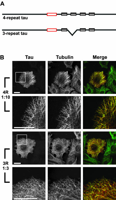
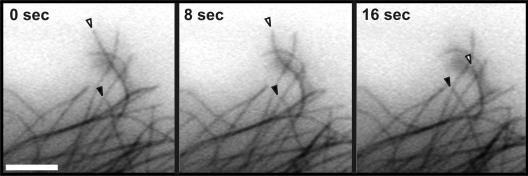
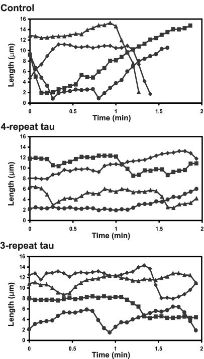
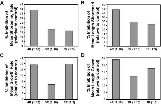
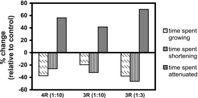
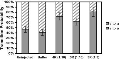
The neural microtubule-associated protein tau binds to and stabilizes microtubules. Because of alternative mRNA splicing, tau is expressed with either 3 or 4 C-terminal repeats. Two observations indicate that differences between these tau isoforms are functionally important. First, the pattern of tau isoform expression is tightly regulated during development. Second, mutation-induced changes in tau RNA splicing cause neuronal cell death and dementia simply by altering the isoform expression ratio. To investigate whether 3- and 4-repeat tau differentially regulate microtubule behavior in cells, we microinjected physiological levels of these two isoforms into EGFP-tubulin-expressing cultured MCF7 cells and measured the effects on the dynamic instability behavior of individual microtubules by time-lapse microscopy. Both isoforms suppressed microtubule dynamics, though to different extents. Specifically, 4-repeat tau reduced the rate and extent of both growing and shortening events. In contrast, 3-repeat tau stabilized most dynamic parameters about threefold less potently than 4-repeat tau and had only a minimal ability to suppress shortening events. These differences provide a mechanistic rationale for the developmental shift in tau isoform expression and are consistent with a loss-of-function model in which abnormal tau isoform expression results in the inability to properly regulate microtubule dynamics, leading to neuronal cell death and dementia.


Figures

Figure 1.
Figure 1.
Immunocytochemical analysis of MCF7 cells injected with 4- or 3-repeat tau. (A) Schematic of 4- and 3-repeat tau proteins. Gray shaded boxes represent the imperfect repeats. Red boxes indicate the epitope of the Tau 5 mAb used in B (LoPresti et al., 1995). (B) MCF7 cells injected with either 4-repeat tau (at a 1:10 tau:tubulin ratio) or 3-repeat tau (at a 1:3 tau:tubulin ratio). Cells were fixed and stained for tau (left column) and tubulin (center column) as described in MATERIALS AND METHODS. In the merged column, the tau staining is shown in red and the tubulin staining is shown in green; yellow indicates colocalization. The boxed areas in the first and third rows are enlarged in the second and fourth rows in order to visualize colocalization on individual microtubules. For both 4- and 3-repeat tau, tau colocalized with microtubules in injected cells, whereas uninjected cells (seen surrounding the injected cell at low magnification) did not stain positively for tau. Scale bars, 20 μm.
Figure 2.
Figure 2.
Dynamic microtubules visualized after microinjection of buffer into an MCF7 cell expressing EGFP-tubulin. Time-lapse images of fluorescent (EGFP) microtubules in buffer-injected cells allow tracking of microtubule growth and shortening over time. One growing microtubule (closed arrowheads) and one shortening microtubule (open arrowheads) are indicated. Although microtubule ends are visible in these still images, it is much easier to discern changes in length in a time-lapse movie (online supplemental material). Scale bar, 5 μm.
Figure 3.
Figure 3.
Life history plots of the growth and shortening dynamics of individual microtubules. The positions of the ends of individual microtubules were tracked in cells injected with buffer or a 1:10 molar ratio of 4- or 3-repeat tau. Changes in the length of the microtubules were plotted vs. time. Each line represents a single microtubule. From these life history plots, individual growth, shortening, and attenuation events were identified and rates and durations of each event was measured.
Figure 4.
Figure 4.
Effects of 4- and 3-repeat tau on microtubule growth and shortening parameters. Microtubule growth and shortening parameters were measured in cells injected with 4- or 3-repeat tau. Presented are the mean rates (A) and lengths (B) of individual shortening events as well as the mean rates (C) and lengths (D) of individual growth events. Values are graphed as percent differences from buffer controls [(experimental value - buffer control value)/buffer control value × 100]. Tau-to-tubulin molar ratios are presented in parentheses.
Figure 5.
Figure 5.
Effects of 4- and 3-repeat tau on time spent growing, shortening and attenuated. The percentage of time microtubules spent growing (diagonal hatches), shortening (vertical stripes), or attenuated (solid gray) was measured in cells injected with 4- or 3-repeat tau. Values are graphed as percent differences from buffer controls [(experimental value - buffer control value)/buffer control value × 100]. Tau-to-tubulin molar ratios are presented in parentheses.
Figure 6.
Figure 6.
Effects of 4- and 3-repeat tau on transitions from shortening to growth or attenuation. The probability that a shortening event would be followed by a growth event vs. an attenuation event was calculated from cells injected with buffer, GST, 4-repeat tau or 3-repeat tau. Probabilities of shortening to attenuation transitions (s to a; solid bars) and shortening to growth transitions (s to g; hatched bars) are presented ± SE. Tau to tubulin molar ratios are presented in parentheses.

References

    1. Bamberger, M.E., and Landreth, G.E. (2002). Inflammation, apoptosis, and Alzheimer's disease. Neuroscientist 8, 276-283. - PubMed
    1. Barghorn, S., and Mandelkow, E. (2002). Toward a unified scheme for the aggregation of tau into Alzheimer paired helical filaments. Biochemistry 41, 14885-14896. - PubMed
    1. Buee, L., Bussiere, T., Buee-Scherrer, V., Delacourte, A., and Hof, P.R. (2000). Tau protein isoforms, phosphorylation and role in neurodegenerative disorders. Brain Res Brain Res Rev 33, 95-130. - PubMed
    1. Butner, K.A., and Kirschner, M.W. (1991). Tau protein binds to microtubules through a flexible array of distributed weak sites. J. Cell Biol. 115, 717-730. - PMC - PubMed
    1. Caceres, A., and Kosik, K.S. (1990). Inhibition of neurite polarity by tau antisense oligonucleotides in primary cerebellar neurons. Nature 343, 461-463. - PubMed

Publication types

LinkOut - more resources