. 2004 Mar 15;164(6):831-42.
doi: 10.1083/jcb.200311121.

In vivo kinetics of Cajal body components

Affiliations

In vivo kinetics of Cajal body components

Miroslav Dundr et al. J Cell Biol. .

Abstract

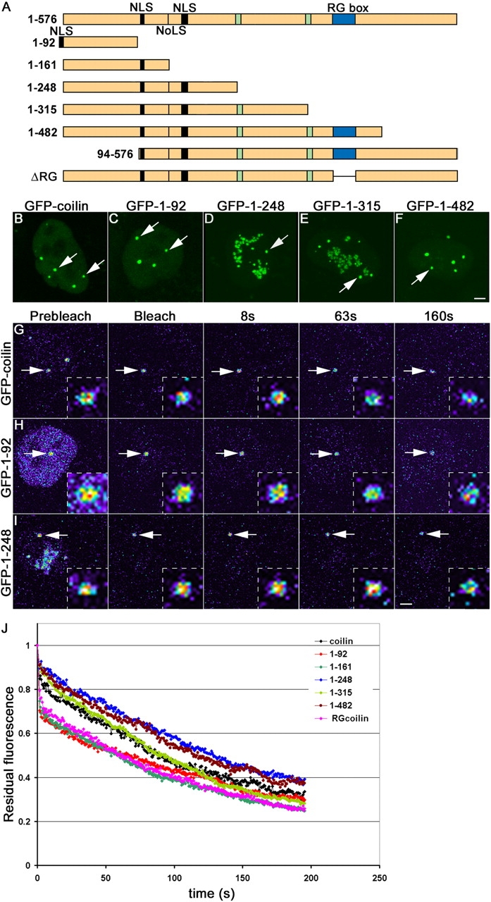
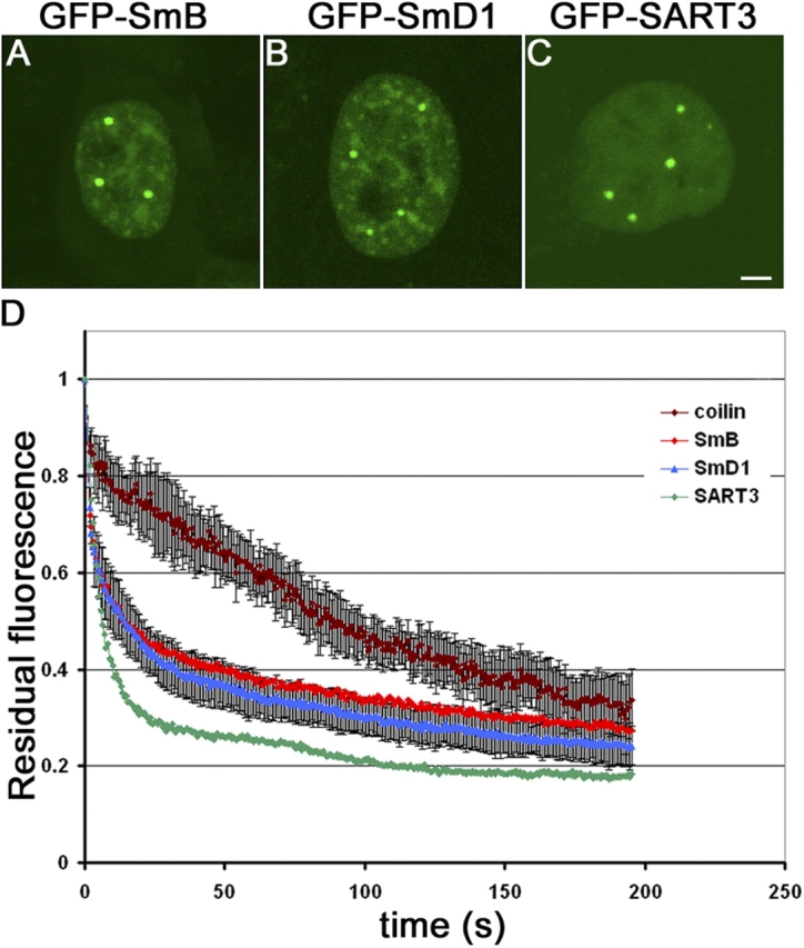
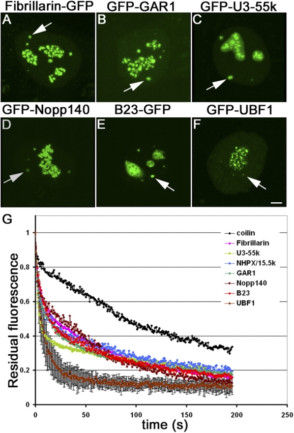
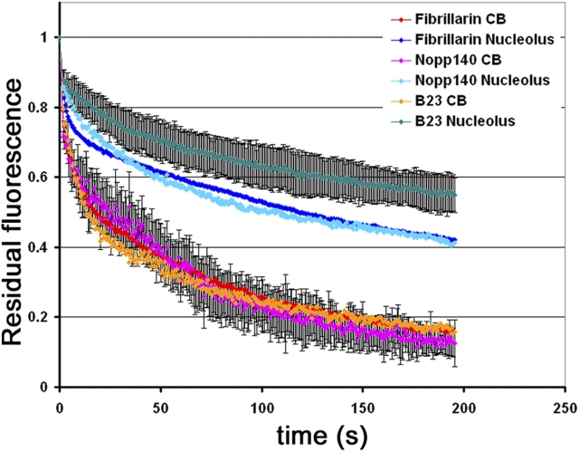
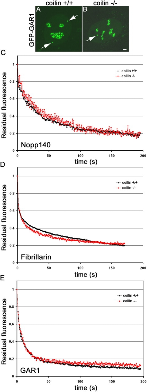
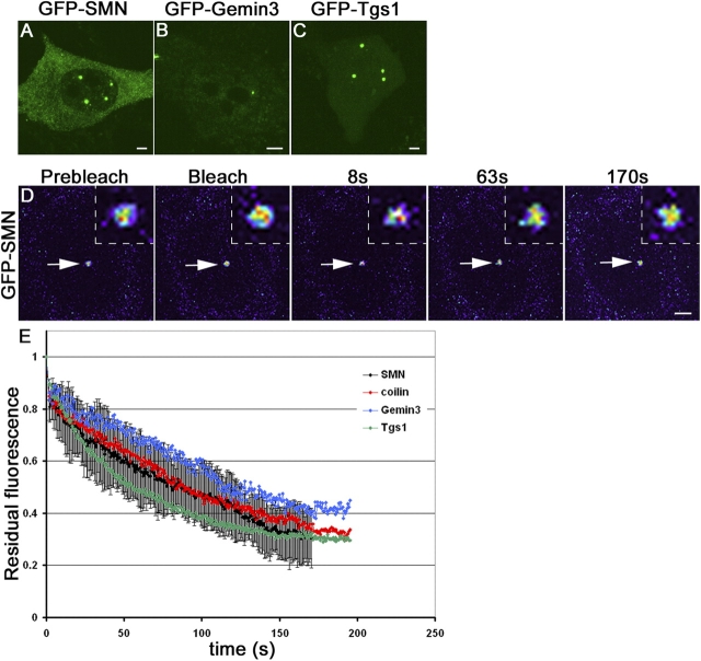
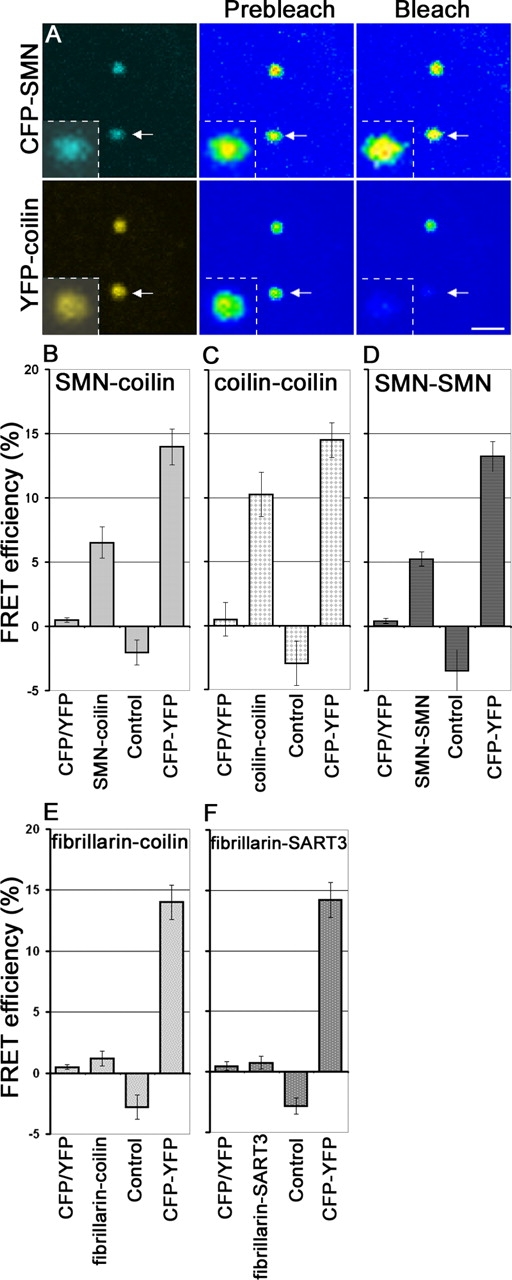
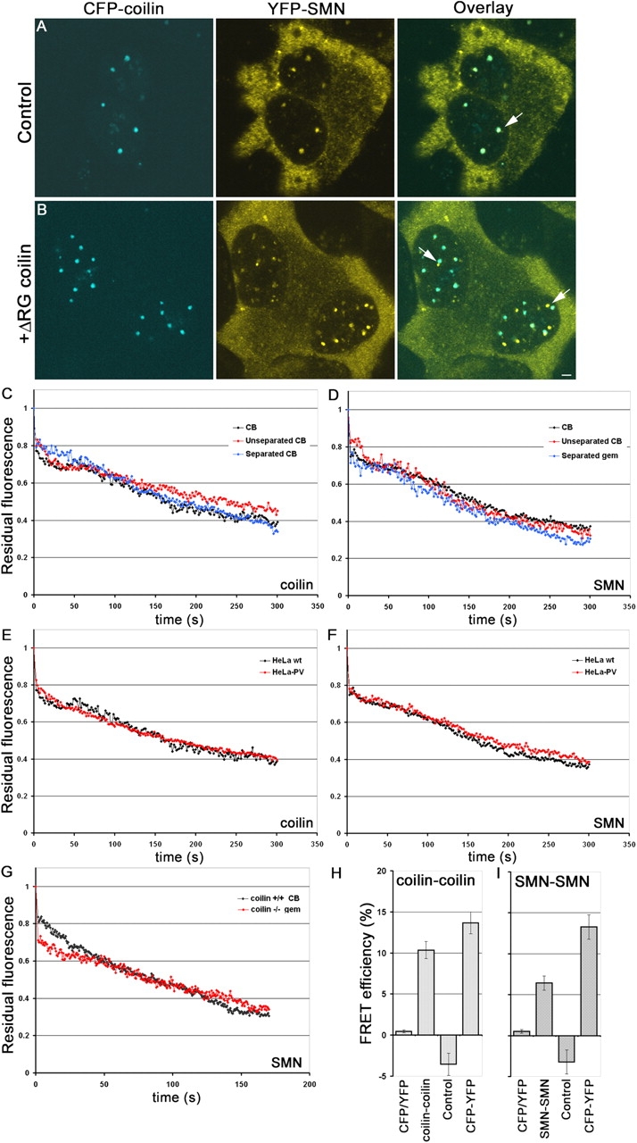
Cajal bodies (CBs) are subnuclear domains implicated in small nuclear ribonucleoprotein (snRNP) biogenesis. In most cell types, CBs coincide with nuclear gems, which contain the survival of motor neurons (SMN) complex, an essential snRNP assembly factor. Here, we analyze the exchange kinetics of multiple components of CBs and gems in living cells using photobleaching microscopy. We demonstrate differences in dissociation kinetics of CB constituents and relate them to their functions. Coilin and SMN complex members exhibit relatively long CB residence times, whereas components of snRNPs, small nucleolar RNPs, and factors shared with the nucleolus have significantly shorter residence times. Comparison of the dissociation kinetics of these shared proteins from either the nucleolus or the CB suggests the existence of compartment-specific retention mechanisms. The dynamic properties of several CB components do not depend on their interaction with coilin because their dissociation kinetics are unaltered in residual nuclear bodies of coilin knockout cells. Photobleaching and fluorescence resonance energy transfer experiments demonstrate that coilin and SMN can interact within CBs, but their interaction is not the major determinant of their residence times. These results suggest that CBs and gems are kinetically independent structures.


Figures

Figure 1.
Figure 1.
iFRAP on coilin and coilin mutants. (A) Full-length coilin (1–576) and coilin mutants fused to GFP at the NH2 terminus. The putative coilin nucleolar localization signal (NoLS), nuclear localization signals (NLS), and serine patches from amino acids 242–259 and 312–325 (green boxes) are indicated. The NLS in the mutant (1–92) was added exogeneously. (B–F) Localization of GFP-coilin (B), GFP-coilin 1–92 (C), GFP-coilin 1–248 (D), GFP-coilin 1–315 (E), and GFP-coilin 1–482 (F) in HeLa cells. CBs are indicated by arrows. Note that mutants 1–248 and 1–315 are also present in nucleoli. (G–I) iFRAP on GFP-coilin (G), GFP-coilin 1–92 (H), and GFP-coilin 1–248 (I). Cells were imaged before and after photobleaching of the entire nucleus with the exception of one CB. The loss of fluorescent signal was monitored using time-lapse microscopy. The unbleached CBs monitored are indicated by arrows and are shown as enlarged insets. Pseudocolored images are shown with high signal levels in red/yellow and low signals in blue. (J) Quantification of iFRAP kinetics. Mutants 1–92, 1–161, and ΔRG showed faster loss than wild-type and the other mutants. Averages from at least 20 cells from two independent experiments are shown. Typical measurement errors were ∼15%. Bars, 2 μm; insets, 0.5 μm.
Figure 2.
Figure 2.
iFRAP of spliceosomal CB components. (A–C) Localization of spliceosomal CB components GFP-SmB (A), GFP-SmD1 (B), and GFP-SART3 (C). (D) Quantification of iFRAP kinetics. GFP-SmB and GFP-SmD1 exhibit similar dissociation kinetics from CBs, which are faster than GFP-coilin loss. In contrast, GFP-SART3 is only transiently associated with CBs. Values represent averages from at least 20 cells ± SDs. Bar, 2 μm.
Figure 3.
Figure 3.
iFRAP of nucleolar CB components. (A–F) Localization of nucleolar CB components in HeLa cells. (A) fibrillarin-GFP, (B) GFP-GAR1, (C) GFP-U3–55k, (D) GFP-Nopp140, (E) B23-GFP, and (F) GFP-UBF1. (G) Quantification of iFRAP kinetics. All nucleolar CB components followed similar dissociation kinetics from CBs with the exception of GFP-UBF1. GFP-coilin dissociation kinetics are show for comparison. Values represent averages from at least 20 cells ± SDs. Bar, 2 μm.
Figure 4.
Figure 4.
Compartment-specific retention mechanism in CBs and nucleoli. Quantification of iFRAP kinetics. The nucleolar CB components, fibrillarin-GFP, GFP-Nopp140, and B23-GFP exhibit significantly faster dissociation kinetics from CBs compared with nucleoli, indicating that they are retained in both nuclear compartments by specific retention mechanisms. Values represent averages from at least 20 cells ± SDs.
Figure 5.
Figure 5.
Coilin-independent dissociation kinetics. (A and B) GFP-GAR1 was expressed in coilin+/+ MEFs (A) or in coilin−/− MEFs (B). In contrast to the normal localization of GFP-GAR1 in coilin+/+ cells (A, arrow), in the absence of coilin GFP-GAR1 localized in residual CBs (B, arrow). (C–E) Quantification of iFRAP kinetics. The dissociation kinetics of GFP-Nopp140 (C), which directly interacts with coilin, fibrillarin-GFP (D), and GFP-GAR1 (E) from CBs is unaffected in the absence of coilin. Averages from at least 20 cells are shown. Typical measurement errors were ∼15%. Bar, 2 μm.
Figure 6.
Figure 6.
iFRAP on RNP assembly machinery. (A–C) Localization of expressed gem components in HeLa cells. (A) GFP-SMN, (B) Gemin3-GFP, and (C) GFP-Tgs1. (D) Cells expressing GFP-SMN were imaged before and after photobleaching of the entire nucleus with the exception of one CB/gem as shown in pseudocolor. The unbleached CB/gem monitored is indicated by arrows and is shown as enlarged insets. (E) Quantification of iFRAP kinetics. The components of SMN protein complex, GFP-SMN, Gemin3-GFP, and GFP-Tgs1 exhibit similar dissociation kinetics from CBs. Values represent averages from at least 20 cells ± SDs. Bars, 2 μm; insets, 0.5 μm.
Figure 7.
Figure 7.
Coilin and SMN interact in CBs in vivo. Acceptor bleaching FRET was performed on living HeLa cells coexpressing (A) CFP-SMN and YFP-coilin. The acceptor (YFP-coilin) was irreversibly bleached, resulting in an increase in donor (CFP-SMN) fluorescence, suggestive of physical interaction between coilin and SMN. Note the increase of fluorescence intensity of CFP-SMN in pseudocolored CB after the bleach (arrow, inset). (B–F) Quantification of FRET. A fusion protein between CFP and YFP was used as a positive control. Coexpressed monomeric CFP and YFP and a CB in the same nucleus to which no acceptor bleaching had been applied were used as negative controls. A significant increase of CB donor fluorescence in CBs was detected for CFP- SMN/YFP-coilin (B), CFP-coilin/YFP-coilin (C), CFP-SMN/YFP-SMN self-interaction (D), fibrillarin-CFP/YFP-coilin (E), and fibrillarin-CFP/YFP-SART3 (F). Averages from at least 20 cells are shown. Error bars represent SEM. Bar, 2 μm.
Figure 8.
Figure 8.
CB and gems are kinetically independent domains. (A) CFP-coilin and YFP-SMN colocalize in CBs in HeLa cells (arrow). (B) When CFP-coilin and YFP-SMN are coexpressed with an untagged dominant ΔRG coilin mutant, CBs physically separate from gems (arrows). (C–G) Quantification of iFRAP kinetics. (C and D) YFP-coilin and YFP-SMN exhibit indistinguishable dissociation kinetics in their respective separated organelles as in unseparated CB/gems in control cells. (E and F) YFP-coilin and YFP-SMN exhibited indistinguishable dissociation kinetics from CBs and gems, respectively, in HeLa wild-type and HeLa-PV cells where CBs and gems are naturally separated. (G) GFP-SMN also shows similar dissociation kinetics in coilin−/− cells where gems are naturally separated from residual CBs. Averages from at least 20 cells are shown. (H and I) Self-interaction of coilin and SMN in separated CBs and gems detected by FRET in living cells. Acceptor bleaching FRET was performed on CFP and YFP pairs of coilin or SMN coexpressed with untagged ΔRG coilin mutant. Upon bleaching of separated CBs or gems, significant increases in donor fluorescence of CFP-coilin (H) and CFP-SMN (I) were observed, indicating that both coilin in CBs and SMN in gems self-interact even when CBs and gems are physically separated. Averages from at least 20 cells are shown. Typical measurement errors in iFRAP experiments were ∼15%. Bar, 2 μm.

References

    1. Bell, M., S. Schreiner, A. Damianov, R. Reddy, and A. Bindereif. 2002. p110, a novel human U6 snRNP protein and U4/U6 snRNP recycling factor. EMBO J. 21:2724–2735. - PMC - PubMed
    1. Boisvert, F.M., M.J. Kruhlak, A.K. Box, M.J. Hendzel, and D.P. Bazett-Jones. 2001. The transcription coactivator CBP is a dynamic component of the promyelocytic leukemia nuclear body. J. Cell Biol. 152:1099–1106. - PMC - PubMed
    1. Boisvert, F.M., J. Cote, M.C. Boulanger, P. Cleroux, F. Bachand, C. Autexier, and S. Richard. 2002. Symmetrical dimethylarginine methylation is required for the localization of SMN in Cajal bodies and pre-mRNA splicing. J. Cell Biol. 159:957–969. - PMC - PubMed
    1. Boudonck, K., L. Dolan, and P.J. Shaw. 1999. The movement of coiled bodies visualized in living plant cells by the green fluorescent protein. Mol. Biol. Cell. 10:2297–2307. - PMC - PubMed
    1. Carmo-Fonseca, M. 2002. New clues to the function of the Cajal body. EMBO Rep. 3:726–727. - PMC - PubMed

Publication types

MeSH terms