. 2004 Mar 15;164(6):887-97.
doi: 10.1083/jcb.200310055.

An actin molecular treadmill and myosins maintain stereocilia functional architecture and self-renewal

Affiliations

An actin molecular treadmill and myosins maintain stereocilia functional architecture and self-renewal

Agnieszka K Rzadzinska et al. J Cell Biol. .

Abstract

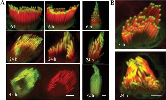
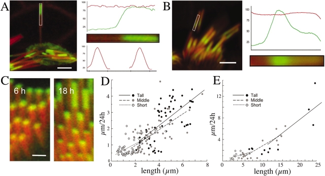
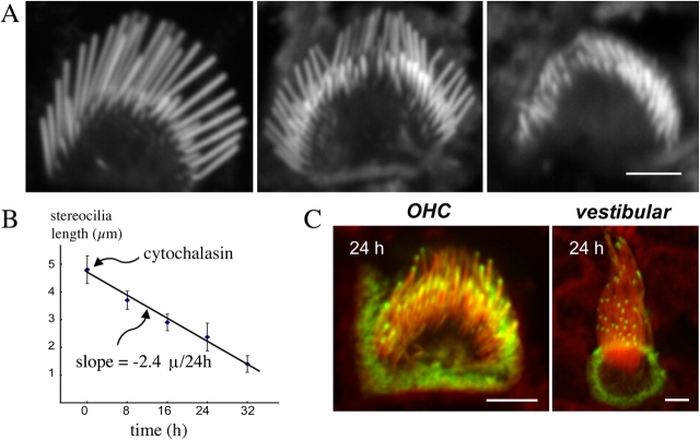
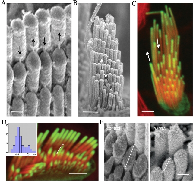
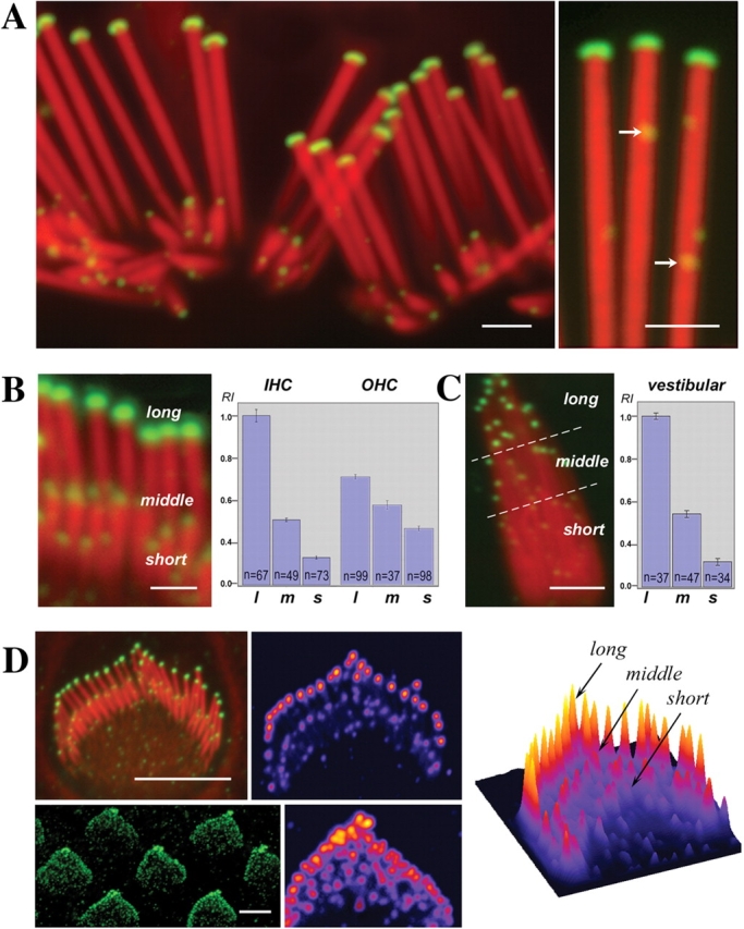
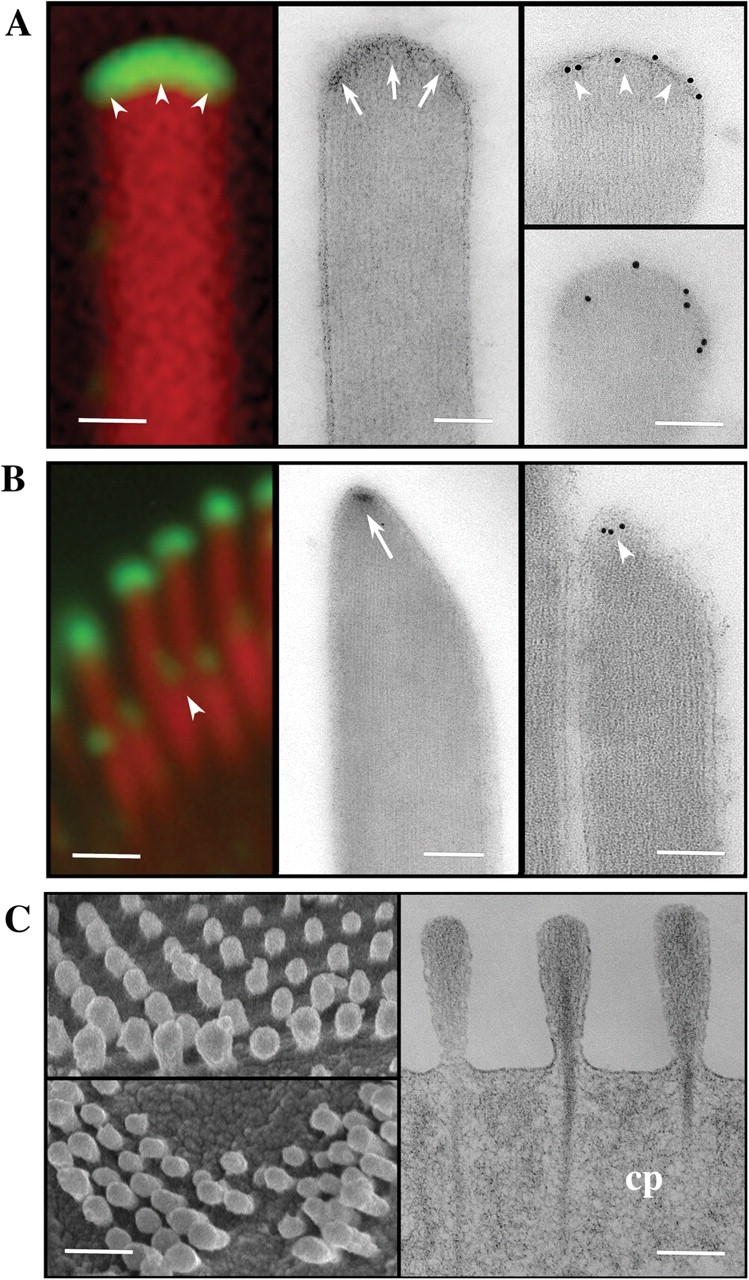
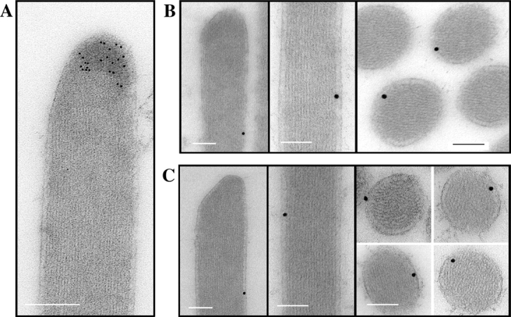
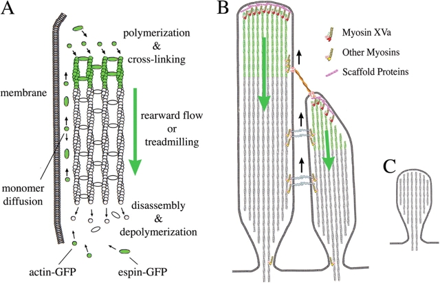
We have previously shown that the seemingly static paracrystalline actin core of hair cell stereocilia undergoes continuous turnover. Here, we used the same approach of transfecting hair cells with actin-green fluorescent protein (GFP) and espin-GFP to characterize the turnover process. Actin and espin are incorporated at the paracrystal tip and flow rearwards at the same rate. The flux rates (approximately 0.002-0.04 actin subunits s(-1)) were proportional to the stereocilia length so that the entire staircase stereocilia bundle was turned over synchronously. Cytochalasin D caused stereocilia to shorten at rates matching paracrystal turnover. Myosins VI and VIIa were localized alongside the actin paracrystal, whereas myosin XVa was observed at the tips at levels proportional to stereocilia lengths. Electron microscopy analysis of the abnormally short stereocilia in the shaker 2 mice did not show the characteristic tip density. We argue that actin renewal in the paracrystal follows a treadmill mechanism, which, together with the myosins, dynamically shapes the functional architecture of the stereocilia bundle.


Figures

Figure 1.
Figure 1.
Incorporation of β actin and espin in stereocilia. (A) β actin–GFP incorporation into the stereocilia of the hair cells of organ of Corti (left) and vestibular organs (right) after transfection. Confocal microscopy revealed that β actin–GFP (green) appeared at the tips of stereocilia counterstained with rhodamine/phalloidin (red) 6 h after transfection. β actin–GFP was progressively incorporated from the stereocilia tips to their bases within 48 h in the organ of Corti hair cells and within 72 h in the vestibular hair cells. Stereocilia maintained their lengths as evident in the bottom left panel where actin-GFP fluorescence had reached the base of the stereocilia, yet the stereocilia length is similar to the neighboring nontransfected cell. Bars, 2.5 μm. (B) Espin-GFP incorporation into the stereocilia of the hair cells of organ of Corti. Espin-GFP incorporation began at the tips of stereocilia and was progressively incorporated into stereocilia at similar rates as the β actin–GFP shown in A. Bar, 2 μm.
Figure 2.
Figure 2.
Characterization of the actin incorporation and flux. (A) Characterization of actin-GFP incorporation into the stereocilia actin paracrystal. The pixel intensity profile along an individual stereocilium (rectangular area) shows the sharp demarcation of the β actin–GFP incorporation (green), whereas the pixel intensity of the rhodamine/phalloidin counterstained actin filaments (red) shows a constant profile. The pixel intensity from the rhodamine/phalloidin shows the same profile when measured across the region of β actin–GFP incorporation or across the region without actin-GFP, excluding the possibility that incorporation was due to nucleation of additional actin filaments. Bar, 2 μm. (B) Transient β actin–GFP expression in an auditory hair cell. The serendipitous pulse of actin-GFP (green) expression was arrested midway along the stereocilia (left). The incorporation appears as a band that has progressed halfway along the stereocilia showing a sharp front facing the stereocilia base (similar to the stereocilia in C) and a diffuse attenuated trailing edge facing the tip. It also shows no change in the total actin fluorescence along the stereocilia, confirming that no anomalous actin filaments are added laterally to the actin paracrystal. Bar, 2 μm. (C) The extent of actin incorporation and treadmill rates is proportional to the length of the stereocilia in a bundle. Even at the earliest times of transfection, we could detect in the fluorescence confocal images that the actin-GFP incorporation starts at the same time in all stereocilia in a bundle but the rate of incorporation and subsequent actin treadmilling are proportional to the stereocilia length in both organ of Corti (left, 6 h after transfection) and vestibular (right, 18 h after transfection) hair bundles. Bar, 600 nm. (D and E) Relationship of the treadmill rates to the length of the stereocilia. The overall actin-GFP treadmill rates calculated by measuring lengths of incorporation 12–24 h after actin-GFP transfection and expressed as μm/24 h for the organ of Corti (D) and vestibular (E) stereocilia. The rate of actin treadmill is proportional to the length of the stereocilium irrespective of its rank (tall, middle, or short) within the staircase bundle.
Figure 3.
Figure 3.
Effects of cytochalasin D on stereocilia. (A) Cytochalasin D shortens stereocilia as it blocks actin polymerization. Confocal images of phalloidin-stained stereocilia of representative hair cells after 1-μM cytochalasin D treatment of middle turn cochlear cultures (n = 10) indicate that stereocilia progressively decrease in length. All cells in the culture demonstrated stereocilia shortening over time without apparent change in the staircase pattern of the hair bundle. Bar, 2.5 μm. (B) The rate of stereocilia shortening is linear and matches the rate of actin polymerization. The lengths of the tall row of stereocilia were measured at 8-h intervals after 1 μM cytochalasin treatment, and the average values ± SD (n = 50) were plotted in the graph. The slope of the best-fit curve indicates an actin shortening rate of 2.4 μm/24 h. (C) Cytochalasin inhibits actin-GFP incorporation. Representative confocal images (n = 30 cells) of stereocilia from the apical turn of the organ of Corti (left) and vestibular (right) hair cells showing actin-GFP marks that have been incorporated for 8 h into the paracrystal. Actin-GFP marks remained at the tips and did not increase in length or progress down the stereocilia over the ensuing 16 h after the incubation with cytochalasin. Bars: (left) 2.4 μm; (right) 1.5 μm.
Figure 4.
Figure 4.
Dynamic regulation of stereocilia structure. (A–C) SEM images of organ of Corti (A) and vestibular (B) hair bundles demonstrate slight irregularities in stereocilia lengths within the characteristically packed rows of the hair bundle. Arrows pointing down show slightly shorter stereocilia, whereas arrows pointing up show longer stereocilia in the same row. In the actin-GFP–transfected vestibular hair cell, the incorporation and treadmill rates (C) reflect the slight length variations within stereocilia of the same row in the bundle. The actin treadmill rates are faster in stereocilia that are taller than their neighbors in the same row (upward arrow) and slower in stereocilia that are shorter than their peers of the same row (downward arrow). Bars: (A) 250 μm; (B) 1 μm; (C) 2 μm. (D) Natural variability in stereocilia tip shape and rate of actin-GFP incorporation. The stereocilia of the tallest row in a bundle have uniform oblate tips, whereas the stereocilia from the second and lower rows exhibit prolate tips that can be slightly pointed to very elongated. The rate of actin-GFP incorporation is increased in the more elongated tips. Bar, 2 μm. Inset shows the frequency distribution of tip length measured as indicated by the measuring bar in the figure. (E) Remodeling of stereocilia tips exposed to BAPTA. SEM images of the stereocilia after 5-min incubation with 5 mM BAPTA in calcium-free L-15 media and 30-min recovery (right) reveal that tip links are disrupted and stereocilia tips become rounded when compared with control (left). The length of the tip defined as shown on the figure was 0.51 ± 0.05 μm for control and 0.34 ± 0.02 μm for BAPTA-treated stereocilia (n = 50). Bar, 250 nm.
Figure 5.
Figure 5.
Intensity of myosin XVa labeling in the tips of stereocilia is graded with length. (A–C) Myosin XVa is localized to the stereocilia tips of auditory (A and B) and vestibular (C) hair cells. Confocal images revealed myosin XVa immunofluorescence in all stereocilia tips of the rat auditory hair cells (A) and at higher magnification also exhibited small fluorescence puncta (A, right, arrows) along the entire stereocilia. Levels of myosin XVa labeling are highest in the longest stereocilia within the auditory (B) and vestibular (C) hair bundle. The relationship between myosin XVa and stereocilia length is visible in fully developed bundles (A) as well as in cultured auditory (B) and vestibular hair cells (C). Quantitative analysis of average pixel intensity confirms myosin XVa gradation within hair bundles of inner (IHC) and outer (OHC) auditory hair cells (B) as well as vestibular hair cells (C). Error bars equal mean ± standard error of the mean. l, long; m, middle; s, short stereocilia. The gradient of myosin XVa expression is noticeable at the earliest stages of the staircase formation in inner (D, top) and outer (D, bottom) hair cells from postnatal day 1 rat neonates whose shorter stereocilia are indistinguishable from supernumerary microvilli. Scaling of myosin XVa levels to the stereocilia length and their distribution within a bundle is clearly visible on the pseudo-colorized images and surface plot of pixel intensities (D); the highest intensities are shown in red. Bars: (A and B) 1 μm; (C) 2 μm; (D) 4 μm.
Figure 6.
Figure 6.
Myosin XVa is a component of the stereocilia tip complex. (A and B) Comparison of high magnification confocal immunofluorescence (left), TEM of uranyl acetate–stained sections (middle), and immunogold-labeled unstained sections (right) of the oblate (A) and prolate (B) stereocilia tips. Stereocilia tips of the tallest row of the bundle are prolate in shape with an electron-dense region just below the stereocilia membrane (A, arrows), where intense myosin XVa labeling is visualized by immunofluorescence or by immunogold EM (A, arrowheads). The second row stereocilia in the bundle are prolate and pointed (B) with a smaller discrete electron-dense region at the end of their longest actin filaments (B, arrow), which also coincides to where myosin XVa is visualized by either fluorescence or by immunogold EM (B, arrowheads). (C) SEM images of shaker 2J mutants confirm previous observations that stereocilia, despite being very short, maintain a slight length gradation reminiscent of the staircase pattern of a normal hair bundle. TEM of osmium and uranyl acetate–stained thin sections (right) show that these very short stereocilia maintain the overall organization of the actin filaments including the rootlets that insert into the cuticular plate (cp) but their tips lack the electron-dense material seen in normal stereocilia (A and B). Bars: (A) 100 nm; (B) 1 μm (left) and 100 nm (right); (C) 1 μm (left) and 100 nm (right).
Figure 7.
Figure 7.
Comparative localization of myosins XVa, VI, and VIIa in vestibular hair cell stereocilia. Immunogold labeling of vestibular hair cell stereocilia showing myosin XVa at the stereocilia tip region (A) while myosins VI (B) and VIIa (C) were localized between the actin core and the lateral membrane as seen in longitudinal and cross-sections. Bars, 100 nm.
Figure 8.
Figure 8.
An actin molecular treadmill model for the dynamic regulation of stereocilia functional architecture. (A) Diagram illustrating the actin paracrystal treadmill model. Actin polymerization and espin cross-linking into a paracrystal occur at the barbed (plus) end of actin filaments near the stereocilia tip complex. Paracrystal disassembly and actin filament depolymerization occurs at the pointed (minus) end of actin filaments at the base of the stereocilia. When the rate of assembly at the tips is equivalent to the rate of disassembly at the base, the paracrystal undergoes a rearward flow or treadmilling and the paracrystal length is dynamically maintained constant. (B) Diagram of a longitudinal section view of two neighboring stereocilia linked by tip and lateral links illustrating the localization and possible relationship of myosins to the actin paracrystal treadmill. Actin flux or treadmill in each stereocilia is independently regulated. Myosin XVa localized to the stereocilia tip complex could regulate or coordinate actin polymerization and/or paracrystal assembly. Tip link tension may regulate the stereocilia tip complex and actin incorporation in the shorter stereocilia. Other myosins localized lateral to the paracrystal may have a role in retrograde flow or dynamic positioning the stereocilia links along the paracrystal treadmill. (C) Diagram illustrating the shaker 2J mutant mice stereocilia, which are abnormally very short, have a rounded tip, and lack myosin XVa and the electron-dense structure or tip complex.

References

    1. Anderson, D.W., F.J. Probst, I.A. Belyantseva, R.A. Fridell, L. Beyer, D.M. Martin, D. Wu, B. Kachar, T.B. Friedman, Y. Raphael, and S.A. Camper. 2000. The motor and tail regions of myosin XV are critical for normal structure and function of auditory and vestibular hair cells. Hum. Mol. Genet. 9:1729–1738. - PubMed
    1. Assad, J.A., G.M. Shepherd, and D.P. Corey. 1991. Tip-link integrity and mechanical transduction in vertebrate hair cells. Neuron. 7:985–994. - PubMed
    1. Bartles, J.R. 2000. Parallel actin bundles and their multiple actin-bundling proteins. Curr. Opin. Cell Biol. 12:72–78. - PMC - PubMed
    1. Boeda, B., A. El-Amraoui, A. Bahloul, R. Goodyear, L. Daviet, S. Blanchard, I. Perfettini, K.R. Fath, S. Shorte, J. Reiners, et al. 2002. Myosin VIIa, harmonin and cadherin 23, three Usher I gene products that cooperate to shape the sensory hair cell bundle. EMBO J. 21:6689–6699. - PMC - PubMed
    1. Chen, B., A. Li, D. Wang, M. Wang, L. Zheng, and J.R. Bartles. 1999. Espin contains an additional actin-binding site in its N terminus and is a major actin-bundling protein of the Sertoli cell-spermatid ectoplasmic specialization junctional plaque. Mol. Biol. Cell. 10:4327–4339. - PMC - PubMed

MeSH terms