Comparative Study
. 2004 Mar 15;199(6):805-14.
doi: 10.1084/jem.20031454.

Sclerostin is an osteocyte-expressed negative regulator of bone formation, but not a classical BMP antagonist

Affiliations
Comparative Study

Sclerostin is an osteocyte-expressed negative regulator of bone formation, but not a classical BMP antagonist

Rutger L van Bezooijen et al. J Exp Med. .

Abstract

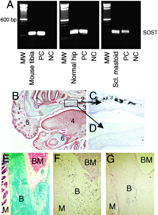
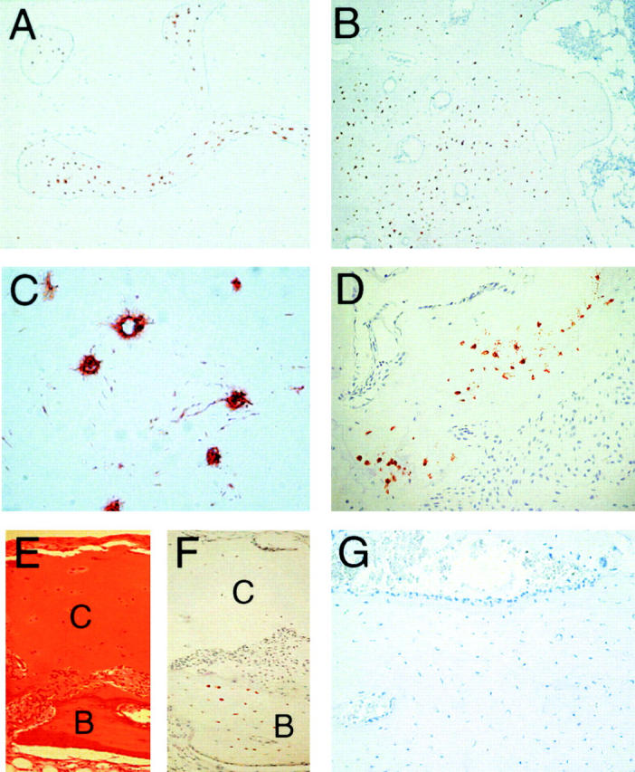
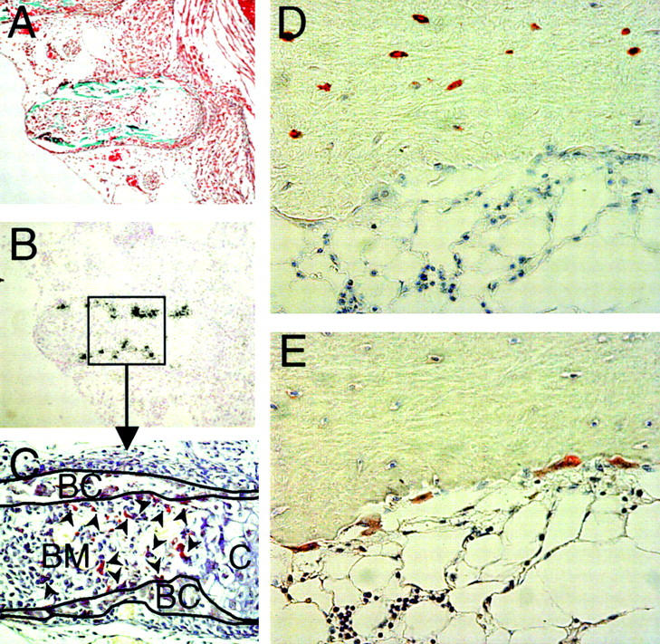
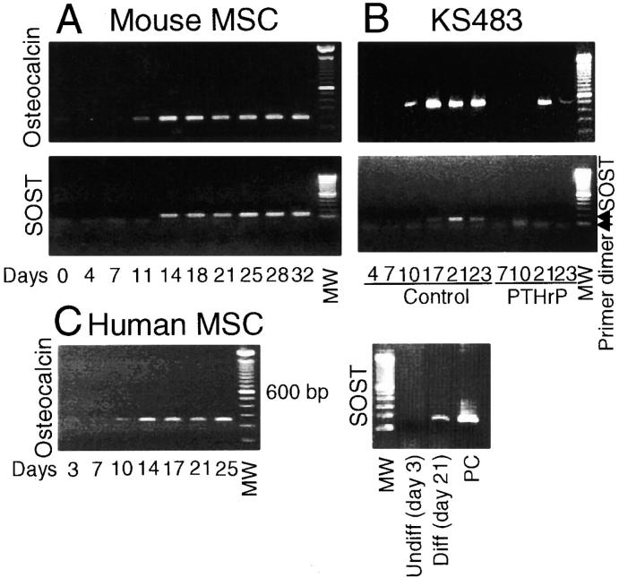
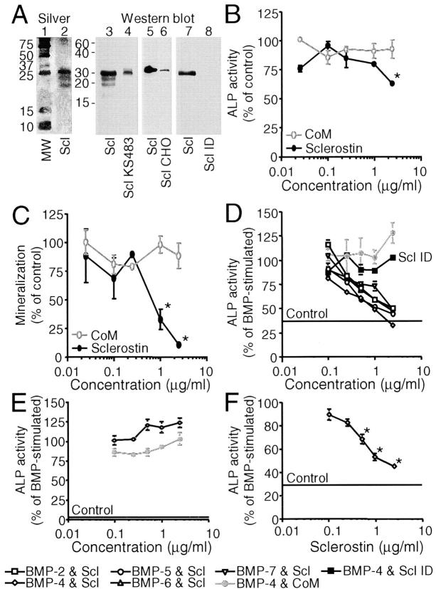
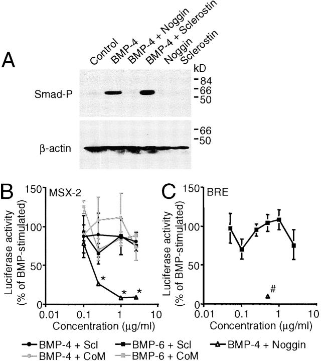
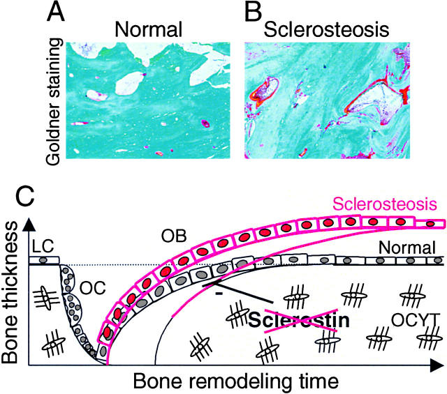
Sclerosteosis, a skeletal disorder characterized by high bone mass due to increased osteoblast activity, is caused by loss of the SOST gene product, sclerostin. The localization in bone and the mechanism of action of sclerostin are not yet known, but it has been hypothesized that it may act as a bone morphogenetic protein (BMP) antagonist. We show here that SOST/sclerostin is expressed exclusively by osteocytes in mouse and human bone and inhibits the differentiation and mineralization of murine preosteoblastic cells (KS483). Although sclerostin shares some of the actions of the BMP antagonist noggin, we show here that it also has actions distinctly different from it. In contrast to noggin, sclerostin did not inhibit basal alkaline phosphatase (ALP) activity in KS483 cells, nor did it antagonize BMP-stimulated ALP activity in mouse C2C12 cells. In addition, sclerostin had no effect on BMP-stimulated Smad phosphorylation and direct transcriptional activation of MSX-2 and BMP response element reporter constructs in KS483 cells. Its unique localization and action on osteoblasts suggest that sclerostin may be the previously proposed osteocyte-derived factor that is transported to osteoblasts at the bone surface and inhibits bone formation.


Figures

Figure 1.
Figure 1.
Localization of SOST mRNA expression in vivo. RT-PCR analysis of SOST mRNA expression in tibia of a young adult mouse, human hipbone, and mastoid of a patient with sclerosteosis (A). Amplicons were amplified over 35 cycles and had the predicted size of 185 bp. In situ hybridization analysis of SOST mRNA expression in 17.5-d-old fetal mouse embryos (B–D) and adult mouse tibia (E–G). Goldner staining of the upper and lower jaw to visualize mineralized bone (B, green/blue). Details of the palatal shelf in the upper jaw showing specific SOST mRNA expression in mineralized bone using antisense probe against SOST (C) and sense probe as control (D). Goldner staining of adult mouse tibia (E). Specific SOST mRNA expression in osteocytes using antisense probe against SOST (F) and sense probe as control (G). Counterstaining was hematoxylin and eosin (C, D, F, and G). Magnification is (B) 40, (C and D) 200, and (E–G) 100. (1) Nasal cavity. (2) Nasopharynx. (3) Oropharynx. (4) Tongue. (5) Nose. (6) Mandible. B, Bone. BM, bone marrow. M, muscle. MW, 100-bp molecular weight marker. NC, negative control. PC, positive control.
Figure 2.
Figure 2.
Sclerostin protein is expressed by osteocytes in human bone biopsies. Sclerostin protein expression was analyzed in control and sclerosteosis human bone biopsies using mouse monoclonal antibodies generated against human sclerostin. Staining with clone H11 showed sclerostin expression in osteocytes of both trabecular (A) and cortical (B) normal femur. Canaliculi and/or lacunae of osteocytes were positive for sclerostin (C). Sclerostin staining of a toe bone biopsy of a 1-yr-old boy showed clear restricted expression of sclerostin to osteocytes (D). No sclerostin expression was found in articular cartilage (E and F) of a 12-yr-old girl. No sclerostin staining was found in mastoid of a patient with sclerosteosis (G). Note the presence of active bone-forming osteoblasts on the bone surface that is characteristic for a site of active bone formation. Counterstaining was Mayers Hematoxylin. C, cartilage. B, bone. Magnification is (A and B) 40, (C) 400, and (D–G) 100.
Figure 3.
Figure 3.
Sclerostin protein is not expressed by osteoclasts in mouse and human bone. Goldner staining of a rib of a 17.5-d-old fetal mouse embryo (A). Specific SOST mRNA expression in mineralized bone of the rib using antisense probe against SOST (B). TRAcP staining of the rib showing that osteoclasts do not colocalize with SOST mRNA expression. TRAcP+ osteoclasts (arrowheads) are present in the bone marrow and on the surface of the bone collars, whereas SOST mRNA is located within the bone collar (C). Sclerostin staining of human bone biopsy of a 12-yr-old girl shows that sclerostin is expressed by osteocytes, but not by multinuclear osteoclasts on the bone surface (D). TRAcP staining to identify multinuclear osteoclasts on the bone surface (E). Magnification is (A and B) 100 and (C–E) 200.
Figure 4.
Figure 4.
SOST mRNA is expressed during the mineralization phase of osteoblastic cultures. Mouse MSCs (A), KS483 cells (B), and human MSCs (C) were cultured under osteogenic conditions. SOST mRNA expression during osteoblastic differentiation is shown in relation to the late osteoblast differentiation marker osteocalcin. Osteocalcin was amplified over 30, 30, and 35 cycles for mouse MSCs, KS483 cells, and human MSCs, respectively. The amplicons had the predicted size of 199 bp (mouse primer set 1), 430 bp (mouse primer set 2), and 242 bp (human primer set). SOST was amplified over 35, 35, and 40 cycles, respectively, and the amplicons had the predicted size of 137 bp (mouse primer set) and 185 bp (human primer set). KS483 cells were cultured in the absence or presence of 10−8 M PTHrP with continuous treatment from day 4 onwards. MW, 100-bp molecular weight marker. PC, positive control.
Figure 5.
Figure 5.
Sclerostin inhibits unstimulated and BMP-stimulated osteoblastic differentiation of KS483 and primary human MSCs, but not of C2C12 cells. Silver staining (lanes 1 and 2) and Western blotting with a rabbit anti–human sclerostin antibody (lanes 3–8) of human sclerostin (lanes 2 [100 ng/ml], 3 [500 ng/ml], 5 [100 ng/ml], and 7 [100 ng/ml]), sclerostin containing medium of KS483 (lane 4), CHO cells (lane 6), and immunodepleted sclerostin preparation (lane 8, equal volume as 100 ng/ml sclerostin preparation) run under reducing conditions (A). Long-term (18 d) osteogenic KS483 cell cultures were treated with sclerostin (2.5 μg/ml or concentrations indicated) or CoM (equal volume) from day 4 onwards and analyzed for ALP activity (B) and mineralization (C). Confluent KS483 were stimulated with BMPs (50 ng/ml BMP-2, 50 ng/ml BMP-4, 300 ng/ml BMP-5, 100 ng/ml BMP-6, and 300 ng/ml BMP-7) in the absence or presence of the dose range of sclerostin or CoM. ALP activity was measured kinetically 4 d after stimulation (D). Confluent C2C12 cells were stimulated with 50 ng/ml BMP-4 in the absence or presence of the dose range of sclerostin or CoM. ALP activity was measured kinetically 4 d after stimulation (E). Confluent hMSCs were stimulated with 100 ng/ml BMP-4 in the absence or presence of the dose range of sclerostin. ALP activity was measured kinetically 4 d after stimulation (F). CoM, Control medium. hMSCs, human mesenchymal stromal cells. MW, molecular weight marker. Scl, sclerostin. Scl CHO, sclerostin containing medium of CHO cells. Scl ID, Immunodepleted sclerostin preparation. Scl KS483, sclerostin containing medium of KS483 cells. Results are expressed as mean ± SEM of triplicate (B and C) or octagonal (D–F) experiments relative to control (B and C) and BMP stimulation (D–F) that was set at 100%. *, Significant versus CoM (P < 0.05). Statistical differences were omitted from C for clarity.
Figure 6.
Figure 6.
Sclerostin does not antagonize early BMP responses in KS483 cells. Western analysis of Smad-1, -5, and/or -8 phosphorylation in confluent KS483 cells that were stimulated for 45 min with 50 ng/ml BMP-4 in the absence or presence of 2.5 μg/ml sclerostin or 500 ng/ml noggin (A). Subconfluent KS483 cells were transfected with MSX-2 luciferase reporter construct (B) or BRE luciferase reporter construct (C) and stimulated for 24 h with 100 ng/ml BMP-6 or 50 ng/ml BMP-4 in the absence or presence of the dose range of sclerostin, CoM, or noggin (500 ng/ml), respectively. CoM, Control medium. Scl, sclerostin. Smad-P, phosphorylated Smad-1, -5, and/or -8. Results are expressed as mean ± SEM of triplicate experiments relative to BMP stimulation that was set at 100%. *, Significant versus CoM. #, Significant versus BMP-4. P < 0.05.
Figure 7.
Figure 7.
Time-dependent effects of sclerostin and BMPs on ALP activity. Sclerostin antagonizes BMP responses in KS483 cells when added as long as 24 h after BMP stimulation (A). Confluent KS483 cells were stimulated with 100 ng/ml BMP-6 in the absence or presence of 1.0 μg/ml sclerostin that was added at the indicated time points after BMP stimulation. ALP activity was measured kinetically 3 d after stimulation. BMPs need to be present for >24 h to stimulate ALP activity (B). Confluent KS483 cells were stimulated with 50 ng/ml BMP-4 for indicated time periods. BMP-4 was removed from the medium or its activity antagonized by changing the medium (light gray bars) or adding soluble truncated BMP receptor-1A (250 ng/ml; dark gray bars), respectively. ALP activity was measured kinetically 4 d after stimulation. Results are expressed as mean ± SEM of octagonal experiments. *, Significant versus BMP-6. #, Significant versus control (P < 0.05). strBMPR-1A, soluble truncated BMP receptor-1A.
Figure 8.
Figure 8.
Histological pictures and a schematic model of increased bone formation in sclerosteosis due to sclerostin deficiency. Representative Goldner staining of calvarial bones of a control (A) and a patient with sclerosteosis (B). Schematic model of sclerostin's mechanism of action in bone remodeling (C). Sclerostin produced by osteocytes may be transported through lacunae to the bone surface, where it inhibits osteoblastic bone formation. In sclerosteosis, loss of sclerostin may prolong the active bone-forming phase of osteoblasts and thereby increase the amount of bone formed by osteoblasts. The increased osteoid levels in sclerosteosis reflect this. In the absence of increased osteoclastic bone resorption, the increased bone formation results in a positive bone balance and, subsequently, in the excess bone mass of normal architecture and increased strength found in sclerosteosis. LC, lining cell. OB, osteoblast. OC, osteoclast. OCYT, osteocyte.
Figure 9.
Figure 9.
Two schematic models that may explain the mechanism of action of sclerostin. Both models provide a mechanism by which sclerostin may antagonize the late BMP response ALP activity, but not the early BMP responses of Smad phosphorylation and direct gene transcription (BRE and MSX-2 reporter construct activation). (Option I) Sclerostin functions as a BMP antagonist when a BMP-inducible factor is coexpressed. The cofactor is necessary for sclerostin to prevent BMP binding to its receptor. The BMP-inducible cofactor may, for example, increase sclerostin's binding affinity for BMPs. (Option II) Sclerostin prevents receptor binding of an yet unknown BMP-inducible factor that stimulates ALP activity. BMPR, BMP receptor. OB, osteoblast. Smad-P, phosphorylated Smad.

References

    1. Truswell, A.S. 1958. Osteopetrosis with syndactyly, a morphologic variant of Albers-Schonberg disease. J. Bone Joint Surg. Br. 40:208–218. - PubMed
    1. Hansen, H. 1967. Sklerosteose. Handbuch der Kinderheilkunde. H. Opitz and F. Schmid, editors. Springer-Verlag, Berlin. 351–355.
    1. Beighton, P., A. Barnard, H. Hamersma, and A. Van Der Wouden. 1984. The syndromic status of sclerosteosis and van Buchem disease. Clin. Genet. 25:175–181. - PubMed
    1. Beighton, P. 1988. Sclerosteosis. J. Med. Genet. 25:200–203. - PMC - PubMed
    1. Hamersma, H., J. Gardner, and P. Beighton. 2003. The natural history of sclerosteosis. Clin. Genet. 63:192–197. - PubMed

Publication types

MeSH terms