. 2004 Apr;24(7):2720-33.
doi: 10.1128/MCB.24.7.2720-2733.2004.

Securin is a target of the UV response pathway in mammalian cells

Affiliations

Securin is a target of the UV response pathway in mammalian cells

Francisco Romero et al. Mol Cell Biol. 2004 Apr.

Abstract

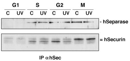
All eukaryotic cells possess elaborate mechanisms to protect genome integrity and ensure survival after DNA damage, ceasing proliferation and granting time for DNA repair. Securin is an inhibitory protein that is bound to a protease called Separase to inhibit sister chromatid separation until the onset of anaphase. At the metaphase-to-anaphase transition, Securin is degraded by the anaphase-promoting complex or cyclosome, and Separase contributes to the release of cohesins from the chromosome, allowing for the segregation of sister chromatids to opposite spindle poles. Here we provide evidence that human Securin (hSecurin) has a novel role in cell cycle arrest after exposure to UV light or ionizing radiation. In fact, irradiation downregulated the level of hSecurin protein, accelerating its degradation via the proteasome and reducing hSecurin mRNA translation, but the presence of hSecurin is necessary for cell proliferation arrest following UV treatment. Moreover, an alteration of UV-induced hSecurin downregulation could lead directly to the accumulation of DNA damage and the subsequent development of malignant tumors.


Figures

FIG. 1.
FIG. 1.
Effect of UV-C on hSecurin expression. HeLa cells were exposed to UV-C radiation (A, 100 J/m2; B, 50 J/m2; C, as indicated) and harvested at the indicated times. Lanes C, untreated cells. Equal amounts of NP-40 extracts were resolved by SDS-12% PAGE. Immunoblotting was performed with polyclonal anti-hSecurin antibodies. Anti-Grb2 antibody was used as a control for equal loading of proteins.
FIG. 2.
FIG. 2.
UV-mediated depletion of hSecurin is independent of p53 and hSecurin mRNA expression but is dependent on the cell cycle. (A) Securin expression from different cell lines that were left untreated (lanes C), after UV irradiation (100 J/m2, harvested at 1 h 30 min after irradiation) (lanes UV), or after X-ray irradiation (25 Gy, harvested at 1 h after irradiation) (lane X-r) was analyzed by Western blotting. Extracts of HeLa cells (T.Ex.) were whole-cell extracts prepared as described in Materials and Methods. wt, wild type. (B) Northern blot analysis of hSecurin mRNA expression from control (lane C) or UV-treated (100 J/m2) HeLa cells harvested at the indicated times. Hybridization to a β-actin probe is also shown as a control for RNA loading. (C) HeLa cells were arrested in the indicated phases of the cell cycle and harvested (lanes C) or UV irradiated (100 J/m2, collected 1 h 30 min later). Equal amounts of NP-40 extracts were resolved by SDS-12% PAGE and blotted with anti-hSecurin antibody.
FIG. 3.
FIG. 3.
Caffeine inhibits the effect of UV light on hSecurin expression. HeLa cells were left untreated (lanes C) or UV irradiated (100 J/m2, harvested at 1 h 30 min after irradiation) in the presence or absence of various compounds to inhibit the effect of UV light on hSecurin expression. Equal amounts of NP-40 extracts were resolved by SDS-12% PAGE. Immunoblotting was performed with polyclonal anti-hSecurin antibodies. (A) The caspase inhibitors Ac-DEVD-CHO (100 μM) (DEVD) and Z-VAD-FMK (20 μM) (VAD) were added 1 h before to irradiate HeLa cells. (B) The proteasome inhibitors Ac-LLnL-CHO (100 μM) (LLnL) and Z-LLnD (50 μM) (LLnV) were added 30 min before to irradiate HeLa cells. (C) Wortmannin (5 μM) (Wortm), UCN-01 (1 μM), PD98059 (50 μM), SB202190 (2 μM), and SP600125 (25 μM) were added between 30 min and 1 h before to irradiate cells. (D) Caffeine (10 mM) (Caf) was added 1 h before UV treatment, and cells were collected after 1 h 30 min or 3 h.
FIG. 4.
FIG. 4.
UV light accelerates hSecurin degradation by the ubiquitin-proteasome pathway. (A) Half-life of hSecurin protein in Cos-7 cells. Cells were transiently transfected with hSec-VSV, and expression was induced for 18 h. Cycloheximide (CHX, 25 μg/ml) was added to the medium at 1 h after irradiation (100 J/m2), and cells were collected at the indicated times. hSec-VSV was detected by immunoblotting with anti-VSV monoclonal antibody. (B) Ectopic hSec-VSV is degraded in a proteasome-dependent manner in UV-irradiated Cos-7 cells. Cells were transfected as described for panel A. Ac-LLnL-CHO (100 μM) (LLnL) was added to the culture medium of control (lanes C) or irradiated (100 J/m2) cells immediately after treatment. Cells were harvested at 1 h 30 min after treatment. Samples were allowed to migrate and were immunoblotted. (C) A nondegradable hSecurin mutant is completely stable after UV irradiation. Cos-7 cells were transfected with hSecurin or hSecurin KAA-DM and treated as described for panel B. Western blotting was performed with anti-Securin antibodies. (D) The effect of UV light on endogenous hSecurin expression is inhibited by proteasome inhibitors in HeLa cells with a block in protein synthesis. Cells were preincubated with LLnL (100 μM) or CHX (25 μg/ml) or both for 1 h and irradiated as described above. Immunoblotting were performed with anti-hSecurin antibodies.
FIG. 5.
FIG. 5.
UV irradiation provokes specific hSecurin protein synthesis inhibition. (A) Cos-7 cells were transfected with hSec-VSV or 5UTR-hSec-VSV and treated as described in the legend to Fig. 4B. (B) Cos-7 cells were cotransfected with pXP2 (including the firefly luciferase cDNA without a promoter) and pRL-Renilla (columns labeled 1) or with pXP2-0.7kb (including the hSecurin promoter, the entire hSecurin 5′ UTR, and the firefly luciferase cDNA) and pRL-Renilla (columns labeled 2). After 48 h, cells were irradiated (100 J/m2) or not and then were collected. Equal amounts of proteins were used for the determination of luciferase activities, and after normalization, the results were expressed as a percentage of the expression of pXP2-0.7kb in nonirradiated cells. These results are representative of three independent experiments.
FIG. 6.
FIG. 6.
UV irradiation actives APC/C-dependent hSecurin degradation in S and G2 phases. HeLa cells were synchronized in S (A) and G2 (B) phases, and the expression of hSecurin was studied. hSecurin expression was also analyzed at 1 h 30 min after synchronization in cells that were left untreated (lane −), cells in the presence of Ac-LLnL-CHO (100 μM) (LLnL) or UV irradiation (100 J/m2), or cells in the presence of both LLnL and UV irradiation. Western blots were developed with anti-Securin antibodies.
FIG. 7.
FIG. 7.
Effect of UV irradiation on hSecurin-hSeparase complexes. Extracts from HeLa cells synchronized in various phases of the cell cycle (lanes C) or synchronized and UV irradiated (100 J/m2, harvested at 1 h 30 min after irradiation) were used to immunoprecipitate (IP) hSecurin. Coimmunoprecipitated proteins were transferred to nitrocellulose filters, and Western blotting was performed with anti-hSeparase antibodies. The same filters were decorated with anti-Securin antibodies.
FIG. 8.
FIG. 8.
Cell proliferation studies of wild-type and hSecurin−/− HCT116 cells after UV irradiation. (A) Representative growth curves for wild-type (filled circles) and hSecurin−/− (empty squares) HCT116 cells under control conditions. (B) HCT116 cells were exposed to UV irradiation (30 J/m2), and their proliferation was compared with that of nonirradiated cells. Viable cells were measured as described in Materials and Methods. (C) hSecurin−/− HCT116 cells were treated as described for panel B. (D) Comparison of the cell proliferation of wild-type and hSecurin−/− HCT116 cells after UV irradiation (30 J/m2).
FIG. 9.
FIG. 9.
Effect of UV light on the cell cycle of HCT116 cells and hSecurin−/− HCT116 cells. (A) Cells were synchronized by treatment with aphidicolin (APH) for 18 h. At the indicated time points after release from the APH block, cells were harvested and analyzed by FACS. (B) Cells were synchronized as described for panel A and, at 1 h after release from the APH block, were UV irradiated (30 J/m2), harvested at the indicated times, and analyzed by FACS. (C) Percentages of cells in each cell cycle phase. Async, asynchronous cells.
FIG. 10.
FIG. 10.
BrdU pulse-chase analysis of the cell cycle kinetics of wild-type and hSecurin−/− HCT116 cells exposed to UV radiation. (A) After UV irradiation of asynchronous cell cultures, S-phase cells were labeled with a 30-min pulse of BrdU. The transit of labeled cells through the cell cycle was monitored by measuring the percentage of cells in S phase at intervals after BrdU pulse-labeling (time zero). The relative percentages of cells were calculated as (percentage of cells in S phase at each time interval/percentage of cells in S phase at time zero) × 100. (B) Histograms showing the DNA content of cells used in the BrdU pulse-chase experiment described for panel A. Areas filled in black correspond to BrdU-labeled cells and overlay global areas, i.e., labeled plus unlabeled cells (unfilled areas). The percentages represent increments of BrdU-labeled cells in G1 at each time interval relative to the percentage of labeled cells under the G1 peak at time zero. PI, propidium iodide.
FIG. 11.
FIG. 11.
UV light-mediated apoptosis in wild-type and hSecurin−/− HCT116 cells. (A) Cells were synchronized and irradiated as described in the legend to Fig. 9B. Control cells were not irradiated. After 6 days, cells were harvested and assayed for apoptotic events. Histogram overlays show FITC-annexin V binding of control and irradiated wild-type (grey line) and hSecurin−/− (black line) HCT116 cells. (B) In situ detection of apoptosis in control and irradiated wild-type and hSecurin−/− HCT116 cells at day 6 after UV irradiation. Apoptotic cells (brown) were detected after monoclonal antibody M30 immunostaining. Original magnification, ×70.
FIG. 11.
FIG. 11.
UV light-mediated apoptosis in wild-type and hSecurin−/− HCT116 cells. (A) Cells were synchronized and irradiated as described in the legend to Fig. 9B. Control cells were not irradiated. After 6 days, cells were harvested and assayed for apoptotic events. Histogram overlays show FITC-annexin V binding of control and irradiated wild-type (grey line) and hSecurin−/− (black line) HCT116 cells. (B) In situ detection of apoptosis in control and irradiated wild-type and hSecurin−/− HCT116 cells at day 6 after UV irradiation. Apoptotic cells (brown) were detected after monoclonal antibody M30 immunostaining. Original magnification, ×70.

Similar articles

Cited by

References

    1. Agami, R., and R. Bernards. 2000. Distinct initiation and maintenance mechanisms cooperate to induce G1 cell cycle arrest in response to DNA damage. Cell 102:55-66. - PubMed
    1. Alexandru, G., F. Uhlmann, K. Mechtler, M. A. Poupart, and K. Nasmyth. 2001. Phosphorylation of the cohesin subunit Scc1 by Polo/Cdc5 kinase regulates sister chromatid separation in yeast. Cell 105:459-472. - PubMed
    1. Alexandru, G., W. Zachariae, A. Schleiffer, and K. Nasmyth. 1999. Sister chromatid separation and chromosome re-duplication are regulated by different mechanisms in response to spindle damage. EMBO J. 18:2707-2721. - PMC - PubMed
    1. Bartek, J., and J. Lukas. 2001. Mammalian G1- and S-phase checkpoints in response to DNA damage. Curr. Opin. Cell Biol. 13:738-747. - PubMed
    1. Bernal, J. A., R. Luna, A. Espina, I. Lazaro, F. Ramos-Morales, F. Romero, C. Arias, A. Silva, M. Tortolero, and J. A. Pintor-Toro. 2002. Human securin interacts with p53 and modulates p53-mediated transcriptional activity and apoptosis. Nat. Genet. 32:306-311. - PubMed

Publication types

MeSH terms

LinkOut - more resources