. 2004 Apr;24(7):2734-46.
doi: 10.1128/MCB.24.7.2734-2746.2004.

Palm mutants in DNA polymerases alpha and eta alter DNA replication fidelity and translesion activity

Affiliations

Palm mutants in DNA polymerases alpha and eta alter DNA replication fidelity and translesion activity

Atsuko Niimi et al. Mol Cell Biol. 2004 Apr.

Abstract

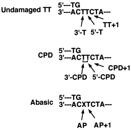
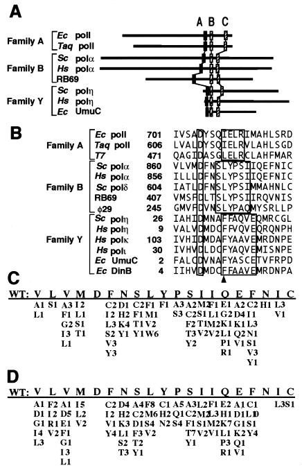
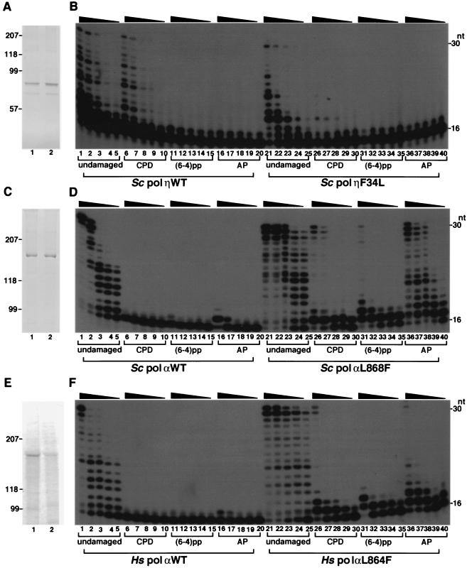
We isolated active mutants in Saccharomyces cerevisiae DNA polymerase alpha that were associated with a defect in error discrimination. Among them, L868F DNA polymerase alpha has a spontaneous error frequency of 3 in 100 nucleotides and 570-fold lower replication fidelity than wild-type (WT) polymerase alpha. In vivo, mutant DNA polymerases confer a mutator phenotype and are synergistic with msh2 or msh6, suggesting that DNA polymerase alpha-dependent replication errors are recognized and repaired by mismatch repair. In vitro, L868F DNA polymerase alpha catalyzes efficient bypass of a cis-syn cyclobutane pyrimidine dimer, extending the 3' T 26000-fold more efficiently than the WT. Phe34 is equivalent to residue Leu868 in translesion DNA polymerase eta, and the F34L mutant of S. cerevisiae DNA polymerase eta has reduced translesion DNA synthesis activity in vitro. These data suggest that high-fidelity DNA synthesis by DNA polymerase alpha is required for genomic stability in yeast. The data also suggest that the phenylalanine and leucine residues in translesion and replicative DNA polymerases, respectively, might have played a role in the functional evolution of these enzyme classes.


Figures

FIG. 1.
FIG. 1.
Each template site used for the kinetic analysis is schematically indicated.
FIG. 2.
FIG. 2.
DNA polymerase motif A. (A) Conserved motifs A, B, and C are illustrated schematically. (B) Amino acid sequences of family A, family B, and family Y DNA polymerases are compared. Conserved amino acids are boxed. Phe34 in S. cerevisiae pol η is indicated by an arrowhead. Sc, S. cerevisiae; Hs, Homo sapiens; RB69, bacteriophage RB69; φ29, bacteriophage φ29; Ec, E. coli; Taq, Thermus aquaticus; T7, bacteriophage T7. (C) Mutant pol α with single substitutions, which are shown under the WT sequence. Amino acid substitutions in active mutants are shown, and the number of isolates is indicated. Amino acid positions of the WT sequence are 860 to 879. Mutations at catalytically essential Asp864 were not obtained. (D) Mutant pol α with multiple substitutions is indicated.
FIG. 3.
FIG. 3.
S. cerevisiae L868F DNA pol α mutation spectrum in M13mp2 lacZα. Mutation spectra were determined by sequencing 135 (WT pol α) and 137 (L868F pol α) mutant plaques. The M13mp2 DNA sequence is shown. Base substitutions (A) and frameshifts (B) produced by S. cerevisiae L868F are indicated above the M13mp2 sequence, and those produced by WT pol α are indicated below the sequence. The +1 and −1 frameshift mutations are indicated by Δ (deletion) and + (insertion), respectively. For a frameshift in a run sequence, the symbol is centered under the run. Endpoints of large deletions are indicated by one arrow pointing down and one arrow pointing up. The nucleotide position at the corresponding other endpoint is indicated at the arrow.
FIG. 3.
FIG. 3.
S. cerevisiae L868F DNA pol α mutation spectrum in M13mp2 lacZα. Mutation spectra were determined by sequencing 135 (WT pol α) and 137 (L868F pol α) mutant plaques. The M13mp2 DNA sequence is shown. Base substitutions (A) and frameshifts (B) produced by S. cerevisiae L868F are indicated above the M13mp2 sequence, and those produced by WT pol α are indicated below the sequence. The +1 and −1 frameshift mutations are indicated by Δ (deletion) and + (insertion), respectively. For a frameshift in a run sequence, the symbol is centered under the run. Endpoints of large deletions are indicated by one arrow pointing down and one arrow pointing up. The nucleotide position at the corresponding other endpoint is indicated at the arrow.
FIG. 4.
FIG. 4.
Translesion synthesis by pol η and α. Sodium dodecyl sulfate gel electrophoresis profiles for each enzyme preparation are shown in panels A (S. cerevisiae pol η WT and F34L), C (S. cerevisiae pol α WT and L868F), and E (human pol α WT and L864F). WT and mutant proteins were loaded onto lanes 1 and 2, respectively. Positions of protein standards (in kilodaltons) are indicated in the left. The second major bands shown in panel A were considered to be the degradation products of the major ones, because they were also stained by Western blotting (data not shown). For translesion DNA synthesis, enzymes were incubated with undamaged or damaged template as indicated. The primer terminus is properly base paired to the nucleotide adjacent to the first damaged base such that the damaged base is the first template site during primer extension. Enzymes were titrated to result in similar levels of primer extension efficiency for the WT and mutant strains on the undamaged template primer. (B) Reaction mixtures included pol η at the following concentrations in the indicated lanes: pol η WT at 3.5 (lanes 1, 6, 11, and 16), 1.8 (lanes 2, 7, 12, and 17), 0.9 (lanes 3, 8, 13, and 18), 0.44 (lanes 4, 9, 14, and 19), and 0.22 (lanes 5, 10, 15, and 20) nM; and pol η F34L at 440 (lanes 21, 26, 31, and 36), 220 (lanes 22, 27, 32, and 37), 110 (lanes 23, 28, 33, and 38), 52 (lanes 24, 29, 34, and 39), and 26 (lanes 25, 30, 35, and 40) nM. (D) Reactions were carried out in the presence of S. cerevisiae pol α at the following concentrations in the indicated lanes: WT pol α at 560 (lanes 1, 6, 11, and 16), 280 (lanes 2, 7, 12, and 17), 140 (lanes 3, 8, 13, and 18), 70 (lanes 4, 9, 14, and 19), and 35 (lanes 5, 10, 15, and 20) nM; and L868F pol α at 56 (lanes 21, 26, 31, and 36), 28 (lanes 22, 27, 32, and 37), 14 (lanes 23, 28, 33, and 38), 7 (lanes 24, 29, 34, and 39), and 3.5 (lanes 25, 30, 35, and 40) nM. (F) Reactions were carried out in the presence of human pol α at the following concentrations in the indicated lanes: WT and L864F pol α at 110 (lanes 1, 6, 11, 16, 21, 26, 31, and 36), 56 (lanes 2, 7, 12, 17, 22, 27, 32, and 37), 28 (lanes 3, 8, 13, 18, 23, 28, 33, and 38), 14 (lanes 4, 9, 14, 19, 24, 29, 34, and 39), and 7 (lanes 5, 10, 15, 20, 25, 30, 35, and 40) nM. Positions of primers and the full-length products are indicated as 16 and 30, respectively. Some reactions resulted in a 31-mer product that was 1 nucleotide longer than the template. This template-independent extra nucleotide forms a single-stranded DNA 3′ overhang, as observed for other proofreading-deficient polymerases.
FIG. 5.
FIG. 5.
Structural models of S. cerevisiae pol η and RB69 pol. (A) The representations of the crystal structures of S. cerevisiae pol η (blue) and RB69 pol (dark yellow) are superimposed. Side chains are indicated as F/Y (Phe35 in pol η and Tyr416 in RB69 are indicated with blue and dark yellow colors), F/L (Phe34 in pol η and Leu415 in RB69), and D (Asp30 and Asp155 in pol η and Asp623 and Asp411 in RB69). The α helix C, F, J, and β sheets 8 and 10 in pol η are also indicated. (B) Van der Waals contacts of Phe34 in S. cerevisiae pol η are shown in purple. A putative interaction with the Leu substitution (dark yellow) is also simulated. Distances between atoms are shown in parentheses. (C) Van der Waals contacts of Leu415 in RB69 pol are shown in blue. A putative interaction with the Phe substitution (blue) is also simulated. These structure models are also relevant to the recently crystallized Dpo4 pol catalytic complex with CPD (27).

Similar articles

Cited by

References

    1. Ayyagari, R., K. J. Impellizzeri, B. L. Yoder, S. L. Gary, and P. M. Burgers. 1995. A mutational analysis of the yeast proliferating cell nuclear antigen indicates distinct roles in DNA replication and DNA repair. Mol. Cell. Biol. 15:4420-4429. - PMC - PubMed
    1. Blasco, M. A., J. M. Lazaro, L. Blanco, and M. Salas. 1993. Phi 29 DNA polymerase active site. The conserved amino acid motif “Kx3NSxYG” is involved in template-primer binding and dNTP selection. J. Biol. Chem. 268:16763-16770. - PubMed
    1. Boudsocq, F., H. Ling, W. Yang, and R. Woodgate. 2002. Structure-based interpretation of missense mutations in Y-family DNA polymerases and their implications for polymerase function and lesion bypass. DNA Repair (Amsterdam) 1:343-358. - PubMed
    1. Braithwaite, D. K., and J. Ito. 1993. Compilation, alignment, and phylogenetic relationships of DNA polymerases. Nucleic Acids Res. 21:787-802. - PMC - PubMed
    1. Capizzi, R. L., and J. W. Jameson. 1973. A table for the estimation of the spontaneous mutation rate of cells in culture. Mutat. Res. 17:147-148. - PubMed

Publication types

MeSH terms