. 2004 Apr;24(7):2905-14.
doi: 10.1128/MCB.24.7.2905-2914.2004.

Growth factor-mediated induction of HDM2 positively regulates hypoxia-inducible factor 1alpha expression

Affiliations

Growth factor-mediated induction of HDM2 positively regulates hypoxia-inducible factor 1alpha expression

Julia I Bárdos et al. Mol Cell Biol. 2004 Apr.

Abstract

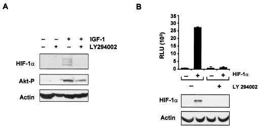
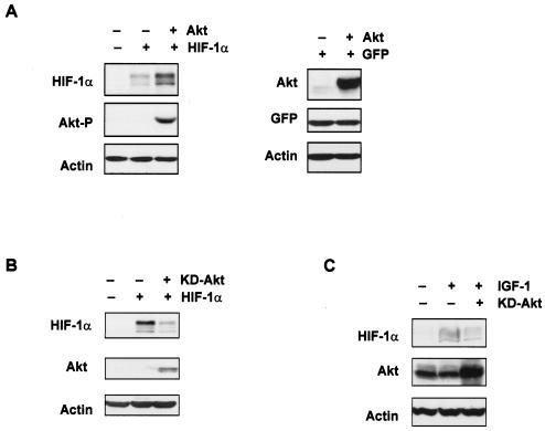
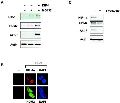
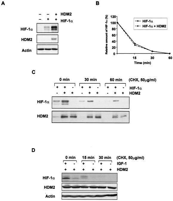
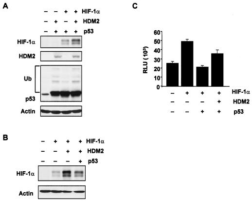
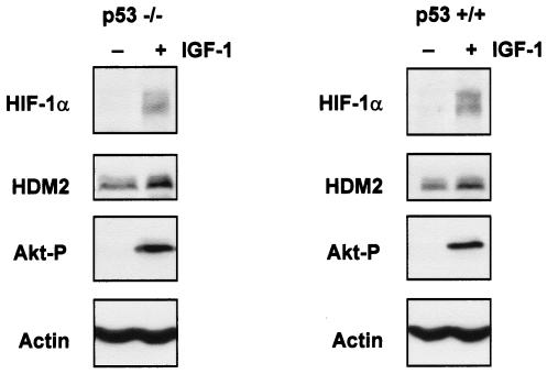
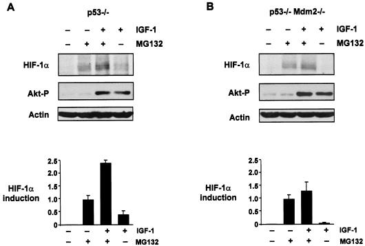
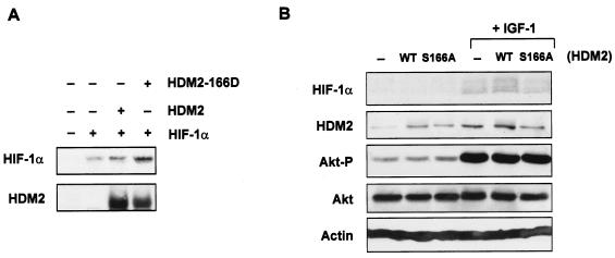
The hypoxia-inducible factor 1 (HIF-1) transcriptional complex is regulated by cellular oxygen levels and growth factors. The phosphoinosotide 3-kinase (PI-3K)-Akt/protein kinase B (PKB) pathway has been shown to regulate HIF-1 activity in response to oncogenic signals and growth factors. We assessed whether the HDM2 oncoprotein, a direct target of Akt/PKB, could regulate HIF-1alpha expression and HIF-1 activity under normoxic conditions. We found that growth factor stimulation, overexpression of Akt/PKB, or loss of PTEN resulted in enhanced expression of both HIF-1alpha and HDM2. Growth factor-mediated induction of HIF-1alpha was ablated by transient expression of a dominant negative form of Akt/PKB or by treatment with LY294002. Transient expression of HDM2 led to increased expression of HIF-1alpha. Pulse-chase and cycloheximide experiments revealed that HDM2 did not significantly affect the half-life of HIF-1alpha. Growth factor-induced HIF-1alpha and HDM2 proteins were localized to the nucleus, and induction of both proteins was observed in both p53(+/+) and p53(-/-) HCT116 cells to comparable levels. Importantly, insulin-like growth factor 1-induced HIF-1alpha expression was observed in p53-null mouse embryo fibroblasts (MEFs) but was significantly impaired in p53 Mdm2 double-null MEFs, indicating a requirement for Mdm2 in this process. Finally, we showed that phosphorylation at Ser166 in HDM2 contributed in part to growth factor-mediated induction of HIF-1alpha. Our study has important implications for the role of the PI-3K-Akt/PKB-HDM2 pathway in tumor progression and angiogenesis.


Figures

FIG. 1.
FIG. 1.
HIF-1α expression is regulated by growth factor stimulation and the PI-3K-Akt/PKB signaling pathway. (A) Western blot analysis of HIF-1α and phosphorylated Akt (Akt-P) (Ser473) proteins from HCT116 cells treated with LY294002 (10 μM) for 30 min prior to stimulation with IGF-1 (100 ng/ml) for 6 h. β-Actin expression was used as a load control in all experiments. (B) Inhibition of PI-3K ablates HIF-1 activity. U2OS cells stably expressing pGL-HRE (U2OS-HRE-luc) were transiently transfected with either 5 μg of the pCMV control vector (−) or 5 μg of HIF-1α (pCMVβ-HA-HIF-1α) (+) expression constructs. pCMV-β-Gal (50 ng) was cotransfected in all experiments as an internal transfection control. LY294002 (10 μM) was added for 6 h prior to harvest, and HIF-1 activity was determined by luciferase assay. Experiments were performed in triplicate, with the standard deviation indicated. Activity is expressed as RLU standardized to β-Gal expression. The lower panel shows Western blot analysis of HIF-1α expression. β-Actin expression was used as the load control.
FIG. 2.
FIG. 2.
Growth factor-mediated induction of HIF-1α requires activation by Akt/PKB. (A) Western blot analysis of HIF-1α and phosphorylated Akt (Akt-P) (Ser473) or total Akt proteins from U2OS cells which were transiently cotransfected with 10 μg of the pCMV vector control (−), HIF-1α expression constructs, and/or 5 μg of Akt expression constructs. Green fluorescent protein (GFP) (1 μg) was used as a transfection control as previously described (4). (B) Western blot analysis of HIF-1α or total Akt proteins from U2OS cells transiently cotransfected with either the pCMV vector control (−), 10 μg of HIF-1α, and/or 5 μg of kinase-dead (KD)-Akt expression constructs. (C) Western blot analysis of HIF-1α and Akt proteins from U2OS cells transiently cotransfected with either 5 μg of the pCMV vector control (−) or 5 μg of KD-Akt expression constructs. Cells were starved for 36 h posttransfection and then stimulated with IGF-1 (100 ng/ml) for 6 h. β-Actin expression was used as the load control in all experiments.
FIG. 3.
FIG. 3.
HDM2 expression is regulated by the PI-3K-Akt/PKB signaling pathway. (A) Western blot analysis of HIF-1α, HDM2, and phosphorylated Akt (Akt-P) (Ser473) proteins from primary human fibroblasts (MRC-5 cells). Cells were starved overnight and then treated with MG132 (50 μM) for 3 h, followed by incubation with or without IGF-1 (100 ng/ml) for 6 h. (B) Immunofluorescence staining of HIF-1α (upper panels) and HDM2 (lower panels) proteins in HCT116 cells before treatment (−) and after treatment with IGF-1 (+IGF-1). Cells were counterstained with DAPI to localize the nucleus. (C) Western blot analysis of HIF-1α, HDM2, and phosphorylated Akt (Akt-P) (Ser473) proteins from U87MG cells treated or not treated with LY294002 (10 μM) 30 min prior to lysis. β-Actin expression was used as the load control in all experiments.
FIG. 4.
FIG. 4.
The HDM2 oncoprotein enhances HIF-1α expression. (A) Western blot analysis of HIF-1α and HDM2 proteins from U2OS cells that were transiently cotransfected with either the pCMV vector control (−), 10 μg of HIF-1α, and/or 5 μg of HDM2 expression constructs. (B) Graphical representation of radioactively labeled HIF-1α protein transfected as described in panel A. Transiently transfected U2OS cells were pulse-labeled with methionine-cysteine-free medium containing [35S]methionine/cysteine and chased with unlabeled complete medium for the times indicated. (C) Western blot analysis of HIF-1α and HDM2 proteins from U2OS cells which were transiently cotransfected as described in panel A and then treated with 50 μg of cycloheximide (CHX)/ml for the times indicated prior to harvesting. (D) Western blot analysis of HIF-1α and HDM2 proteins from HCT116 cells, which were transiently transfected with 10 μg of HDM2 expression plasmid, starved for 36 h, stimulated with IGF-1 (100 ng/ml) for 6 h, and then treated with 50 μg of cycloheximide/ml for the times indicated prior to harvesting.
FIG. 5.
FIG. 5.
HDM2-mediated effects in the presence of p53. (A and B) U2OS cells were transiently cotransfected with the vector control (−), 10 μg of HIF-1α, 5 μg of HDM2, and/or 5 μg of p53-FLAG expression constructs. Western blot analysis was carried out using specific antibodies against HIF-1α, p53, and HDM2. Ub, ubiquitinated p53. (C) Saos-2 HIF-1α TetOn-inducible cells were transiently cotransfected with the pGL-HRE plasmid and 2.5 μg of either the vector control (−), p53-FLAG, or HDM2 expression constructs, followed by doxycycline treatment (6 μg/ml) for 24 h. pCMV-β-Gal (50 ng) was cotransfected in all experiments as an internal transfection control. HIF-1 activity was determined by luciferase assay. Experiments were performed in duplicate, with the standard deviation indicated. Activity is expressed as RLU standardized to β-Gal expression.
FIG. 6.
FIG. 6.
Induction of HIF-1α protein expression by growth factors is p53 independent. (A) Western blot analysis of HIF-1α, HDM2, and phosphorylated Akt (Akt-P) (Ser473) proteins from p53−/− HCT116 (left panel) or p53+/+ HCT116 cells (right panel) starved for 36 h prior to IGF-1 treatment (100 ng/ml) for 6 h.
FIG. 7.
FIG. 7.
Induction of HIF-1α protein expression by growth factors requires HDM2. Western blot analysis of HIF-1α and phosphorylated Akt proteins from p53−/− (A) or p53−/− Mdm2−/− (B) MEFs starved overnight and then treated with MG132 (50 μM) for 3 h, followed by incubation with or without IGF-1 (100 ng/ml) for 6 h. β-Actin expression was used as the load control. Graphs are representative of two independent experiments and show the relative amounts of HIF-1α induced for each treatment as outlined. HIF-1α levels from Western blot analyses were measured by densitometric analysis and quantified relative to the actin load control.
FIG. 8.
FIG. 8.
Role of HDM2 phosphorylation in growth factor-mediated induction of HIF-1α. (A) Western blot analysis of HIF-1α and HDM2 proteins from Saos-2 cells (p53-null) after transient cotransfection with the pCMV vector control, 10 μg of HIF-1α, 5 μg of wild-type HDM2 (HDM2), and/or 5 μg of mutant HDM2-S166D expression constructs. (B) Western blot analysis of HIF-1α, HDM2, phosphorylated Akt (Akt-P) (Ser473), and total Akt proteins from HCT116 cells transiently transfected with either 10 μg of the pCMV vector control (−), wild-type (WT) HDM2, or mutant HDM-S166A (S166A) expression constructs. Cells were then starved for 36 h and treated with or without IGF-1 (100 ng/ml) for 6 h prior to harvest. β-Actin expression was used as the load control in all experiments.

References

    1. Alvarez-Tejado, M., A. Alfranca, J. Aragones, A. Vara, M. O. Landazuri, and L. del Peso. 2002. Lack of evidence for the involvement of the phosphoinositide 3-kinase/Akt pathway in the activation of hypoxia-inducible factors by low oxygen tension. J. Biol. Chem. 277:13508-13517. - PubMed
    1. Arany, Z., L. E. Huang, R. Eckner, S. Bhattacharya, C. Jiang, M. A. Goldberg, H. F. Bunn, and D. M. Livingston. 1996. An essential role for p300/CBP in the cellular response to hypoxia. Proc. Natl. Acad. Sci. USA 93:12969-12973. - PMC - PubMed
    1. Arsham, A. M., D. R. Plas, C. B. Thompson, and M. C. Simon. 2002. Phosphatidylinositol 3-kinase/Akt signaling is neither required for hypoxic stabilization of HIF-1 alpha nor sufficient for HIF-1-dependent target gene transcription. J. Biol. Chem. 277:15162-15170. - PubMed
    1. Ashcroft, M., R. L. Ludwig, D. B. Woods, T. D. Copeland, H. O. Weber, E. J. MacRae, and K. H. Vousden. 2002. Phosphorylation of HDM2 by Akt. Oncogene 21:1955-1962. - PubMed
    1. Ashcroft, M., Y. Taya, and K. H. Vousden. 2000. Stress signals utilize multiple pathways to stabilize p53. Mol. Cell. Biol. 20:3224-3233. - PMC - PubMed

Publication types

MeSH terms