. 2004 Apr;24(7):2915-22.
doi: 10.1128/MCB.24.7.2915-2922.2004.

Brn-2 expression controls melanoma proliferation and is directly regulated by beta-catenin

Affiliations

Brn-2 expression controls melanoma proliferation and is directly regulated by beta-catenin

Jane Goodall et al. Mol Cell Biol. 2004 Apr.

Abstract

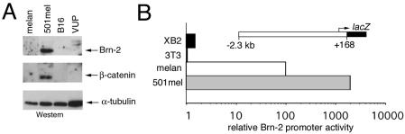
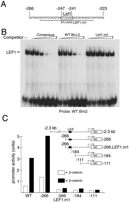
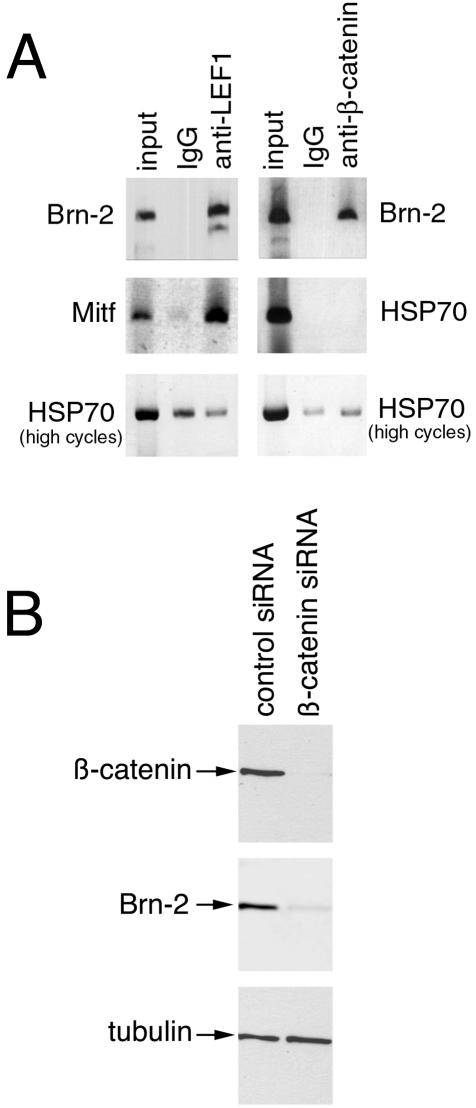
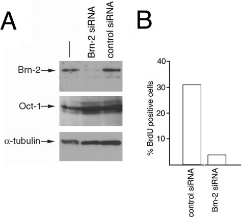
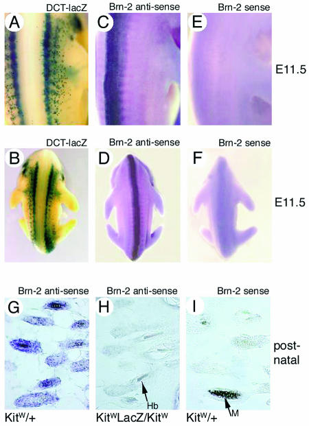
Constitutive activation of the Wnt/beta-catenin signaling pathway is a notable feature of a large minority of cases of malignant melanoma, an aggressive and increasingly common cancer. The identification of target genes downstream from this pathway is therefore crucial to our understanding of the disease. The POU domain transcription factor Brn-2 has been implicated in control of proliferation and melanoma survival, and its expression is strongly upregulated in melanoma. We show here that in vivo Brn-2 is expressed in melanocytes but not in embryonic day 11.5 melanoblasts and that its expression is directly controlled by the Wnt/beta-catenin signaling pathway in melanoma cell lines and in transgenic mice. Moreover, silent interfering RNA-mediated inhibition of Brn-2 expression in melanoma cells overexpressing beta-catenin results in significantly decreased proliferation. These results, together with the observation that BRAF signaling also induces Brn-2 expression, reveal that Brn-2 is a focus for the convergence of two key melanoma-associated signaling pathways that are linked to cell proliferation.


Figures

FIG. 1.
FIG. 1.
Brn-2 and β-catenin expression in melanocyte and melanoma cell lines. (A) Extracts from the indicated melanocyte (melan-a) and melanoma (B16, 501 mel, and VUP) cell lines were subjected to Western blotting with the indicated antibodies. Note that a low level of β-catenin could be detected in all cell lines on longer exposure. (B) The Brn-2 promoter is highly active in the 501 mel cell line. The melanocyte cell line melan-c, the human melanoma cell line 501 mel, NIH 3T3 cells, and the keratinocyte cell line XB2 were transfected with the indicated promoter construct driving expression of a lacZ reporter, and β-galactosidase activity was assayed 48 h posttransfection. All results are presented relative to those obtained with a cotransfected simian virus 40 promoter-luciferase reporter.
FIG. 2.
FIG. 2.
Brn-2 expression is activated by β-catenin in vitro and in vivo. (A) Sequence of a region of the Brn-2 promoter required for activity in melanocytes and melanoma, with the consensus Lef1/Tcf factor binding site indicated (−241 to −247). The sequence of the mutation in the binding site is indicated, and the underlined region was used as a probe for the DNA-binding assays presented below. (B) Lef1 binds the Brn-2 promoter in vitro. Band shift assay with a probe corresponding to the sequence underlined in panel A together with bacterially expressed Lef1. The indicated competitor oligonucleotides were used at 2, 5, 10, 20, 50, and 250 ng. The sequence of the Lef1 consensus oligonucleotide was 5′-CTAGAAGGGCACCCTTTGAAGCTCT-3′. (C) β-Catenin activates the Brn-2 promoter. The 501 mel cells were transfected with a Brn-2 promoter-luciferase reporter construct extending to either −2.3 kb (wild type) or deleted to −266, −184, or −111 or a reporter, −266/Lef.m1, in which the Lef1/Tcf site has been mutated, as illustrated in the inset. The indicated reporters were transfected alone or together with a β-catenin expression vector, and luciferase activity was determined.
FIG. 3.
FIG. 3.
Brn-2 promoter is a target for Lef1 and β-catenin in vivo. (A) Chromatin immunoprecipitated from 501 mel cells with either nonspecific IgG or anti-β-catenin or anti-Lef1 antibodies was subjected to PCR with primers specific for the Brn-2 and Mitf promoters. Primers for the HSP70 promoter were used as a negative control. For the HSP70 promoter, PCR for the same number of cycles (25) used to generate a signal for the Brn-2 or Mitf promoter immunoprecipitated with anti-Lef1 or anti-β-catenin antibodies failed to reveal a signal. The PCR cycles were therefore increased to 30 so that a product was evident (high cycles). (B) siRNA-mediated downregulation of β-catenin results in decreased Brn-2 expression; 501 mel cells were transfected with either a control siRNA or an siRNA specific for β-catenin (47), harvested after 3 days, and subjected to Western blotting with the indicated antibodies.
FIG. 4.
FIG. 4.
Brn-2 expression is required for proliferation in 501 mel cells. 501 mel cells were transfected with siRNA specific for Brn-2 or a control siRNA. After 3 days, cells were grown in the presence of bromodeoxyuridine for 1 h before being subjected to either Western blotting or immunofluorescence. (A) Results of Western blotting with the indicated antibodies after Brn-2 or control siRNA treatment. (B) Quantification of the immunofluorescence results obtained by counting 300 control or Brn-2 siRNA-transfected cells stained for DNA with DAPI and for bromodeoxyuridine (BrdU) incorporation.
FIG. 5.
FIG. 5.
Brn-2 is not expressed in melanoblasts in vivo but is expressed in hair follicle melanocytes. (A and B) E11.5 mouse embryo bearing a DCT-lacZ reporter transgene stained for lacZ expression as a marker of melanoblasts. (C and D) In situ hybridization of an E11.5 embryo with a Brn-2 antisense probe. (E and F) In situ hybridization of an E11.5 embryo with a Brn-2 sense probe. Note that Brn-2 expression is not detected in melanoblasts. (G to I) In situ hybridization of a cryosection of newborn mouse skin with the antisense (G and H) and sense (I) Brn-2 probes with wild-type and KitW-lacZ/KitW mice. The KitW-lacZ/KitW mice lack neural crest-derived melanocytes. The arrow in panel I indicates melanin (M), and the arrow in panel H indicates a hair bulb (hb).
FIG. 6.
FIG. 6.
Brn-2 is upregulated by β-catenin in vivo. β-Catenin-GFP was overexpressed in the melanocyte lineage from a tyrosinase promoter including an upstream tyrosinase locus control region (Martinozzi et al., unpublished data). RNA derived from the skin of transgenic (Tg) and nontransgenic wild-type (WT) littermates were subjected to RT-PCR with primers for the indicated genes. Primers specific for GFP were used to detect the transgene, and cyclin D1 and hypoxanthine phosphoribosyltransferase (HPRT) were used as controls.

References

    1. Behrens, J., J. P. von Kries, M. Kuhl, L. Bruhn, D. Wedlich, R. Grosschedl, and W. Birchmeier. 1996. Functional interaction of beta-catenin with the transcription factor LEF-1. Nature 382:638-642. - PubMed
    1. Bert, A. G., J. Burrows, A. Hawwari, M. A. Vadas, and P. N. Cockerill. 2000. Reconstitution of T cell-specific transcription directed by composite NFAT/Oct elements. J. Immunol. 165:5646-5655. - PubMed
    1. Cadigan, K. M., and R. Nusse. 1997. Wnt signaling: a common theme in animal development. Genes Dev. 11:3286-3305. - PubMed
    1. Caelles, C., H. Hennemann, and M. Karin. 1995. M-phase-specific phosphorylation of the POU transcription factor GHF-1 by a cell cycle-regulated protein kinase inhibits DNA binding. Mol. Cell. Biol. 15:6694-6701. - PMC - PubMed
    1. Chin, L., G. Merlino, and R. A. Depinho. 1998. Malignant melanoma: modern black plague and genetic black box. Genes Dev. 12:3467-3481. - PubMed

Publication types

MeSH terms