. 2004 Apr;24(7):2923-31.
doi: 10.1128/MCB.24.7.2923-2931.2004.

The Brn-2 transcription factor links activated BRAF to melanoma proliferation

Affiliations

The Brn-2 transcription factor links activated BRAF to melanoma proliferation

Jane Goodall et al. Mol Cell Biol. 2004 Apr.

Abstract

Malignant melanoma, an aggressive and increasingly common cancer, is characterized by a strikingly high rate (70%) of mutations in BRAF, a key component of the mitogen-activated protein (MAP) kinase signaling pathway. How signaling events downstream from BRAF affect the underlying program of gene expression is poorly understood. We show that the Brn-2 POU domain transcription factor is highly expressed in melanoma cell lines but not in melanocytes or melanoblasts and that overexpression of Brn-2 in melanocytes results in increased proliferation. Expression of Brn-2 is strongly upregulated by Ras and MAP kinase signaling. Importantly, the Brn-2 promoter is stimulated by kinase-activating BRAF mutants and endogenous Brn-2 expression is inhibited by RNA interference-mediated downregulation of BRAF. Moreover, silent interfering RNA-mediated depletion of Brn-2 in melanoma cells expressing activated BRAF leads to decreased proliferation. The results suggest that the high levels of Brn-2 expression observed in melanomas link BRAF signaling to increased proliferation.


Figures
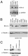
FIG. 1.
FIG. 1.
Brn-2 is expressed at high levels in melanoma cell lines. (A) Reverse transcription-PCR showing relative levels of Brn-2 mRNA in the indicated cell lines compared to a glyceraldehyde-3-phosphate dehydrogenase (G3PDH) control. 501 mel and HMB2 are human melanoma cell lines, melan-c is a mouse melanocyte cell line, and K1735 is an undifferentiated mouse melanoma cell line. Cos7 cells were used as a negative control. (B and C) Western blots with an anti-Brn-2 monoclonal antibody, showing relative levels of Brn-2 protein expressed in the indicated cell lines compared to the expression of α-tubulin. Melan-c is a mouse melanocyte cell line, melb is a mouse melanoblast line, 1° melanocytes are primary mouse melanocytes, and B16 and K1735 represent relatively differentiated and undifferentiated mouse melanomas, respectively. Cos7 cells were used as a negative control. The remaining cell lines were derived from human melanomas, VUP representing a uveal melanoma.
FIG. 2.
FIG. 2.
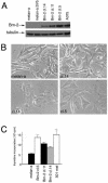
Brn-2 overexpression results in increased [3H]thymidine incorporation into melanocytes. (A) Western blot with anti-Brn-2 antibody of melan-a cells, melan-a cells infected with an empty retrovirus (SV5), or three independent clones of melan-a cells infected with a retrovirus expressing SV5 epitope-tagged Brn-2. The A375 melanoma cell line was used a positive control. The same blot was also probed for α-tubulin as a loading control. (B) Phase contrast image of melan-a cells and the indicated clones of melan-a cells stably overexpressing Brn-2 from the pBabePuro retrovirus. (C) [3H]thymidine incorporation into the indicated cell lines.
FIG. 3.
FIG. 3.
Robust activation of Brn-2 expression in response to a melanoma-associated RTK. (A) Schematic showing the MAP kinase signaling pathway downstream from the HER-mrk chimeric receptor in which the ligand binding domain of the EGFR is fused to the intracellular signaling domain of the Xmrk RTK. U0126 is a specific inhibitor of the MAP kinase kinase MEK. (B) Western blot showing expression of ERK in cells expressing the HER-mrk fusion protein (melan-a Hm) (36) grown in the presence of EGF (50 ng/ml). The smaller panel indicates that EGF-mediated stimulation of MAP kinase was inhibited by treatment with the MEK inhibitor U0126. (C) Western blot with anti-Brn-2 antibody, showing that Brn-2 expression is induced by treatment of cells expressing the HER-mrk fusion protein (melan-a Hm) with EGF (50 ng/ml) for 24 h. The location of Brn-2 is indicated, and the asterisk indicates a nonspecific band that cross-reacted with the antibody. This band was apparent when bovine serum albumin rather then skim milk was used as a blocking agent for the Western blot. Parental melan-a cells and melan-a Hm cells grown in the absence of EGF are shown as controls. The blot was reprobed with anti-ERK2 polyclonal antibody as a loading control. No expression of Brn-2 was observed in the absence of treatment. ERK2 was used as a loading control. (D) Western blot with anti-Brn-2 monoclonal antibody of melan-a Hm cells grown in either the absence or presence of EGF (50 ng/ml) for 24 or 48 h, as indicated. The blot was probed with anti-Brn-2 monoclonal or anti-ERK2 polyclonal antibodies. (E) Induction of Brn-2 expression by Xmrk requires MAP kinase signaling. Melan-a Hm cells were stimulated with EGF for 48 h in the presence or absence of 20 μM U0126. Cell extracts were Western blotted with anti-Brn-2 or anti-ERK2 antibody, as indicated.
FIG. 4.
FIG. 4.
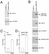
Ras- and MAP kinase-induced expression of the Brn-2 promoter. (A) Western blot with anti-Brn-2 or anti-α-tubulin antibodies and either melan-a cells or Ras-transformed melan-a cells, as indicated. (B) Western blot of melan-a cells and a melan-a cell line stably expressing constitutively activated MEK (MEK.EE). MEK.EE expression was determined with an anti-His tag antibody. Antibodies against Brn-2, Rsk, phospho-Rsk, and α-tubulin were used where indicated. (C and D) Regulation of the Brn-2 promoter by Ras. The wild-type Brn-2 promoter fused to a luciferase reporter extending to −2.3 kb or to −266 (ΔNhe) was transfected into the melan-c melanocyte cell line together with either an empty expression vector or a vector expressing activated Ras (RasN12), as indicated. The results presented are the averages of four experiments.
FIG. 5.
FIG. 5.
Brn-2 is a target for BRAF signaling. (A) Western blot of human melanoma cell lines A375M and WM266.4, which express constitutively active forms of BRAF (11), with either an anti-Brn-2 antibody or anti-ERK2 antibody. Where indicated, cells were treated for 24 h with the MEK inhibitor U0126 at a concentration of 10 μM. (B) BRAF activates the Brn-2 promoter. B16 melanoma cells were transfected with the Brn-2 promoter (−266)-luciferase reporter (500 ng) and either an empty vector or a vector expressing wild-type BRAF or the indicated BRAF mutants (200 ng). (C) BRAF and β-catenin can cooperate for activation of the Brn-2 promoter. B16 melanoma cells were transfected with the Brn-2 promoter (−266)-luciferase reporter (100 ng) and either an empty vector or a vector expressing wild-type BRAF, β-catenin, or both, as indicated. Luciferase activity was determined 48 h posttransfection. (D) siRNA-mediated inhibition of BRAF leads to loss of MAP kinase activity and Brn-2 expression. Shown is a Western blot of WM266.4 cells either untreated or transfected for 60 h with BRAF-specific siRNA or a scrambled oligonucleotide control with either anti-Brn2 monoclonal, anti-ERK2, or anti-phospho-ERK2 antibodies, where indicated.
FIG. 6.
FIG. 6.
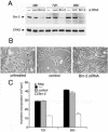
siRNA-mediated downregulation of Brn-2 leads to decreased [3H]thymidine incorporation in melanoma cells. (A) Western blot with anti-Brn-2 antibody of WM266.4 cell cells transfected with a Brn-2-specific siRNA or a control nonsilencing siRNA for the indicated times. Untransfected cells were used as an additional control. (B) Phase contrast image of WM266.4 cells transfected with a Brn-2-specific siRNA or a control nonsilencing siRNA, as indicated. (C) [3H]thymidine incorporation into untransfected WM266.4 cells or cells transfected with a Brn-2-specific siRNA or a control nonsilencing siRNA, as indicated, either 72 or 96 h posttransfection.

References

    1. Angel, P., M. Imagawa, R. Chiu, B. Stein, R. J. Imbra, H. J. Rahmsdorf, C. Jonat, P. Herrlich, and M. Karin. 1987. Phorbol ester-inducible genes contain a common cis element recognized by a TPA-modulated trans-acting factor. Cell 49:729-739. - PubMed
    1. Bennett, D. C., P. J. Cooper, T. J. Dexter, L. M. Devlin, J. Heasman, and B. Nester. 1989. Cloned mouse melanocyte lines carrying the germline mutations albino and brown: complementation in culture. Development 105:379-385. - PubMed
    1. Bert, A. G., J. Burrows, A. Hawwari, M. A. Vadas, and P. N. Cockerill. 2000. Reconstitution of T cell-specific transcription directed by composite NFAT/Oct elements. J. Immunol. 165:5646-5655. - PubMed
    1. Carreira, S., T. J. Dexter, U. Yavuzer, D. J. Easty, and C. R. Goding. 1998. Brachyury-related transcription factor Tbx2 and repression of the melanocyte-specific TRP-1 promoter. Mol. Cell. Biol. 18:5099-5108. - PMC - PubMed
    1. Carreira, S., B. Liu, and C. R. Goding. 2000. The gene encoding the T-box transcription factor Tbx2 is a target for the microphthalmia-associated transcription factor in melanocytes. J. Biol. Chem. 275:21920-21927. - PubMed

Publication types

MeSH terms