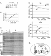
. 2004 Apr;24(7):3036-47.
doi: 10.1128/MCB.24.7.3036-3047.2004.

Chromatin-mediated restriction of nuclear factor 1/CTF binding in a repressed and hormone-activated promoter in vivo

Affiliations

Chromatin-mediated restriction of nuclear factor 1/CTF binding in a repressed and hormone-activated promoter in vivo

Sergey Belikov et al. Mol Cell Biol. 2004 Apr.

Erratum in

  • Mol Cell Biol. 2004 Jun;24(12):5636

Abstract

Mouse mammary tumor virus (MMTV) promoter-driven transcription is induced by glucocorticoid hormone via binding of the glucocorticoid receptor (GR). The MMTV promoter also harbors a binding site for nuclear factor 1 (NF1). NF1 and GR were expressed in Xenopus oocytes; this revealed GR-NF1 cooperativity both in terms of DNA binding and chromatin remodeling but not transcription. A fraction of NF1 sites were occupied in a hormone-dependent fashion, but a significant and NF1 concentration-dependent fraction were constitutively bound. Activation of the MMTV promoter resulted in an approximately 50-fold increase in the NF1 accessibility for its DNA site. The hormone-dependent component of NF1 binding was dissociated by addition of a GR antagonist; however, the antagonist RU486, which supports partial GR-DNA binding, also maintained partial NF1 binding. Hence GR-NF1 cooperativity is independent of agonist-driven chromatin remodeling. NF1 induced the formation of a micrococcal-nuclease-resistant protein-DNA complex containing the DNA segment from -185 to -55, the MMTV enhanceosome. Coexpression of NF1 and Oct1 resulted in a significant stimulation of hormone-induced MMTV transcription and also in increased basal transcription. We propose that hormone-independent NF1 binding may be involved in maintaining transcriptional competence and establishment of tissue-specific gene networks.


Figures
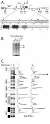
FIG. 1.
FIG. 1.
(A) The reporter DNA construct pMMTV:M13 used for injection. Solid black arrows, primers used for primer extension analysis of SacI in situ accessibility and DMS methylation protection. The restriction enzyme sites shown are referred to in the text. Arrow (+1), transcription start site. GRE I to IV (white boxes), the NF1 binding site (light gray), Oct1-binding sites (black), and the TATA box (dark gray) and their indicated cognate DNA binding segments are displayed with the same shading. Black dots, protected guanines in DMS in vivo footprinting. (B) Autoradiography of Xenopus oocyte extract 24 h after injection of 6 ng of GR mRNA alone or together with 12 ng of NF1 mRNA followed by incubation in [35S]methionine (see Materials and Methods). (C) DMS methylation protection analysis of the MMTV DNA segment −200 to −50 for oocytes injected with 3 ng of pMMTV:M13 DNA and GR-NF1 mRNA as for panel B and the next day not treated (−) or treated (+) with 1 μM synthetic glucocorticoid TA. Radioactivity scans show the two lanes with highlighted hormone-dependent effects: methylation-protected bands (white circles) and a hypermethylated band (black dot).
FIG. 2.
FIG. 2.
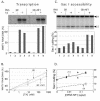
Effects on MMTV transcription (A and B) and SacI in situ cutting (C and D) by GR and NF1. MMTV reporter DNA and the indicated mRNA mixtures injected into oocytes were kept for 19 h and then exposed to hormone (TA) for 9 h. Shown are an S1 nuclease analysis (A) and a chromatin-remodeling assay by SacI in situ cutting (C) of double samples from the same oocyte homogenate, a pool of eight oocytes in each sample. (B) Oocytes injected as in panel A were incubated with the indicated hormone concentrations. Double samples of five oocytes each were analyzed for MMTV transcription. (D) SacI in situ cutting of double samples of seven oocytes each injected as in panel A but with the indicated amounts of mRNA for NF1 per oocyte and 3 ng of GR mRNA. A subsaturating dose of 8 nM hormone was added (+TA) or not added (−TA).
FIG. 3.
FIG. 3.
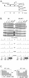
GR-dependent NF1 binding is reversible. (A) Experimental design. (B) Autoradiogram of DMS in vivo footprinting developed by primer extension. Lanes: A, no hormone; B1 and B2, corticosterone agonist at time 13 h (B1) and 23 h (B2); C, oocytes exposed to the indicated antagonist at time 13 h and analyzed as double samples. Grey arrows, right, protected bands; black arrowheads, reference bands.Scans are averages of each double lane, with indicated binding sites below. (C) Quantified DMS methylation of the two guanosines in the NF1 site, with double samples for the indicated lanes. (Right) The level of NF1 binding without hormone (bars A) is used as reference, i.e., 100% (double arrow).
FIG. 4.
FIG. 4.
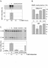
NF1-DNA binding depends on ligand-induced GR-GRE binding also in the presence of the antagonist RU486. MMTV reporter DNA- and GR-NF1 mRNA-injected oocytes were treated with the indicated ligands for 10 h. Two pools of 10 oocytes each were analyzed for transcription. (A) Diagram displaying the amount of mRNA expressed as a percentage of the agonist response. (B) DMS in vivo footprinting to quantify GR-GRE binding, an average of GRE II to IV (top), and NF1-DNA binding (bottom) from two pools of five oocytes each. (C) SacI in situ access analysis, showing the increase in cutting in relation to the mock-treated control. SacI bands (S) and HinfI bands (H) are indicated on the left. Error bars signify the two values of the double samples.
FIG. 5.
FIG. 5.
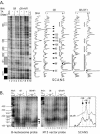
MMTV chromatin structure analysis using DNase I and MNase. Oocytes injected with the MMTV DNA reporter followed by GR mRNA with or without NF1 mRNA were either not treated (−) or treated (+) with hormone (TA). Pools of 10 oocytes were homogenized and divided in three aliquots for digestion with the indicated enzyme in increasing concentrations. (A) Primer extension of DNase I-digested samples with scans of one of the two displayed lanes for each oocyte sample. Lane DNA, naked DNA control; lane G>A, DMS sequence ladder. Black arrowheads, 10- or 11-bp pattern of chromatin-specific hypercutting; black dots, hypersensitive sites; arrowheads with numbers, positions of chromatin-specific hypersensitivity relative to transcription start (+1); boxes, protected areas. (B) MNase-digested samples analyzed on a 3.6% agarose gel followed by electroblotting to a filter and probing first with a B nucleosome probe (−218 to −54; left) and then with the M13 vector probe (right). Size markers (base pairs) are given to the left. Black diamond, mononucleosome band of ∼146 bp; gray arrowhead, ∼120-bp subnucleosome band (lanes 4 to 6); black arrowhead, ∼10-bp larger subnucleosome band induced by NF1 (lanes 10 to 12). Scans to the right are of lanes 5 (bottom) and 11 (top) of the B nucleosome-probed filter.
FIG. 6.
FIG. 6.
Quantification of intranuclear GR and NF1 protein and specific DNA binding as a function of increasing concentrations of injected NF1 mRNA. Injection of MMTV DNA reporter was followed by injection of 5.8 ng of GR mRNA also containing 0 to 5.8 ng of NF1 mRNA prepared as a serial threefold dilution in five steps. Two pools of oocytes were analyzed for each NF1 mRNA concentration. (A) Western blot of purified rat GR and of nuclear GR from one nuclear equivalent of GR and NF1 mRNA-injected and hormone-treated oocytes. (B) Standard curve for GR quantification. (C) DMS in vivo footprinting of oocytes containing increasing intracellular NF1 protein not treated (−) or treated (+) with hormone (TA). Boxes and circles (left), protein binding sites and protected guanines, respectively. (D) Quantification of DMS methylation (from panel C) at the GREs, an average of GRE II to IV (circles, individual samples; dashed line, average), and at the NF1 binding site (triangles and solid line) as a function of NF1 protein concentrations. The intranuclear GR concentration was 7.8 μM in the presence of hormone; the intranuclear NF1 concentrations were 0 nM, 240 nM, and 6.5 μM without hormone (lanes 1 to 6) and 0 nM, 80 nM, 240 nM, 720 nM, 2.2 μM, and 6.5 μM with hormone (lanes 7 to 18). (E) Hypothetical model of NF1 homodimer formation and DNA binding.
FIG. 7.
FIG. 7.
NF1 stimulates MMTV transcription in the presence of Oct1. Oocytes were injected with 2 ng of pMMTV:M13 mixed with 0.4 ng of AdML reporter construct 18 h before hormone treatment and then with 3.8 ng of GR and/or Oct1 mRNA and/or 0.4 ng of NF1 mRNA as indicated 14 h before hormone treatment. Double samples without hormone and triple samples with hormone, 10 oocytes in each pool, were taken for RNA and DNA analysis. Hormone-treated oocytes containing GR only were used as reference (100%). Error bars signify the 95% confidence interval. Student's t test of the hormone-treated oocytes containing GR and Oct1 in the absence (258% ± 5% [mean ± standard deviation]; n = 3) and presence (325% ± 3%; n = 3) of NF1 rendered a P value of 0.105. The AdML RNA was not used as reference due to hormone- and protein context-dependent systematic variation.

References

    1. Adams, C. C., and J. L. Workman. 1995. Binding of disparate transcriptional activators to nucleosomal DNA is inherently cooperative. Mol. Cell. Biol. 15:1405-1421. - PMC - PubMed
    1. Almouzni, G., and A. P. Wolffe. 1993. Replication-coupled chromatin assembly is required for repression of basal transcription in vivo. Genes Dev. 7:2033-2047. - PubMed
    1. Anderson, J. D., A. Thastrom, and J. Widom. 2002. Spontaneous access of proteins to buried nucleosomal DNA target sites occurs via a mechanism that is distinct from nucleosome translocation. Mol. Cell. Biol. 22:7147-7157. - PMC - PubMed
    1. Archer, T. K., M. G. Cordingley, R. G. Wolford, and G. L. Hager. 1991. Transcription factor access is mediated by accurately positioned nucleosomes on the mouse mammary tumor virus promoter. Mol. Cell. Biol. 11:688-698. - PMC - PubMed
    1. Belikov, S., B. Gelius, G. Almouzni, and Ö. Wrange. 2000. Hormone activation induces nucleosome positioning in vivo. EMBO J. 19:1023-1033. - PMC - PubMed

Publication types

MeSH terms