. 2004 Apr;74(4):752-60.
doi: 10.1086/383252. Epub 2004 Mar 11.

High proportion of mutant osteoblasts is compatible with normal skeletal function in mosaic carriers of osteogenesis imperfecta

Affiliations

High proportion of mutant osteoblasts is compatible with normal skeletal function in mosaic carriers of osteogenesis imperfecta

Wayne A Cabral et al. Am J Hum Genet. 2004 Apr.

Abstract

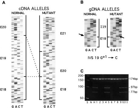
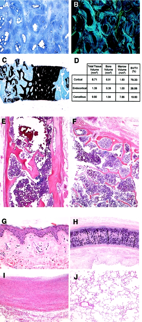
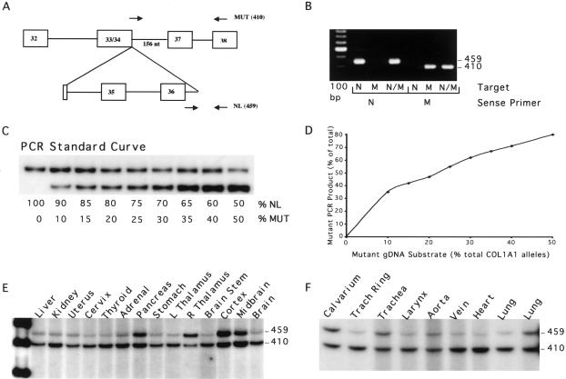
Individuals with mosaicism for the autosomal dominant bone dysplasia osteogenesis imperfecta (OI) are generally identified by having more than one affected child. The mosaic carriers have both normal and mutant cell populations in somatic and germline tissues but are unaffected or minimally affected by the type I collagen mutation that manifests clinically in their heterozygous offspring. We determined the proportion of mutant osteoblasts in skeletal tissue of two mosaic carriers who each have a COL1A1 mutation in a high proportion of dermal fibroblasts. Both carriers had normal height and bone histology; the first carrier had normal lumbar spine measurements (L1-L4), as determined by dual-energy x-ray absorptiometry (Z = +1.17). In cultured cells from the first carrier, studied by labeled PCR and single-cell PCR over successive passages, the collagen mutation was present in 85% of fibroblasts and 50% and 75% of osteoblasts from her right iliac crest and left patella, respectively, with minimal selection. The second carrier was studied by PCR amplification of DNA from autopsy paraffin blocks. The proportion of heterozygous cells was 40% in calvarium, 65% in tracheal ring, and 70% in aorta. Thus, in OI, substantially normal skeletal growth, density, and histology are compatible with a 40%-75% burden of osteoblasts heterozygous for a COL1A1 mutation. These data are encouraging for mesenchymal stem-cell transplantation, since mosaic carriers are a naturally occurring model for cell therapy.


Figures

Figure  1
Figure 1
Collagen mutation and clonal lines of the mosaic carrier of OI type IV. A, Sequence of the normal and mutant cDNA of son of the mosaic carrier, showing skipping of COL1A1 exon 19 sequences. B, Sequence of COL1A1 gene, showing the splice-donor–site mutation IVS 19 G+1→C. C, Demonstration that the mosaic carrier has two distinct cell lines. The PCR fragment containing the exon 19/intron 19 junction was amplified from DNA of dermal fibroblast clonal lines by PCR, by use of a forward primer in exon 19 (5′-TGCTCCTGGTATTGCTGGTGCTCCTGGCTTC-3′) and a reverse primer in intron 19 (5′-gcgtcttcctgctccccagatgagagccgc-3′) (lowercase letters denote intron nucleotides; uppercase letters denote exon nucleotides). PCR conditions were 94°C for 5 min; 30 cycles of 1 min at 94°C, 30 s at 67°C, and 30 s at 72°C; and, finally, 7 min at 72°C. Amplification products were digested with DdeI and electrophoresed on a 6% acrylamide gel. Five clonal lines (2, 3, 6, 7, 8) have undigested product (174 nt) from normal allele sequence and 6 clonal lines (1, 4, 5, 9, 10, 11) are heterozygous for normal and mutant allele products (DdeI digestion products, 97 nt and 77 nt). S is the 50-bp ladder; N and H are normal and heterozygous control cells.
Figure  2
Figure 2
Histology of tissues from both mosaic carriers. AC, Biopsy of the iliac crest sample from the mosaic carrier of OI type IV. A and B, Toluidine-blue–stained section under normal and polarized light. C, von Kossa stained section, with normal bone volume. D, Quantitative histomorphometry of sample in panel C. E and F, Sections of calvarial and rib bone, respectively, of the mosaic carrier of OI type III, stained with hematoxylin-eosin. GJ, Sections of dermis, tracheal ring, aorta wall, and lung, respectively, of the mosaic carrier of OI type III, stained with hematoxylin-eosin.
Figure  3
Figure 3
Multiplex PCR of autopsy tissues of the mosaic carrier of OI type III. A, Diagram of multiplex PCR amplification of DNA from autopsy tissues of the mosaic carrier. The normal allele sense primer spanned the exon 36/intron 36 junction (5′-TGGCCCCCCTgtgagtaccaagacccccat-3′ [lowercase letters denote intron nucleotides; uppercase letters denote exon nucleotides]), and the mutant allele sense primer spanned the novel junction between exon 34 and the retained portion of intron 36 (5′-CCTGCTGGTGCCCCTGGTGACggaa-3′).The common antisense primer (5′-CGAGCACCTTTGGCTCCAGGAGCACCAACA-3′) is in exon 38. B, Allele specificity of PCR primers, verified using subcloned mutant (M) and normal (N) proband DNA. Only the specific normal COL1A1 allele (459 bp) and mutant allele (410 bp) products were detected by the respective sense primers. C, A 32P-labeled standard curve (200 ng DNA/sample), prepared by mixing genomic DNA from normal leukocytes with DNA from the heterozygous leukocytes of the carrier’s affected son. PCR reactions used 200 ng genomic DNA, 1 U Platinum Taq DNA Polymerase High Fidelity (Invitrogen), and 5 μCi of 111 TBq/mmol [α-32P] dCTP. Normal and mutant allele products have similar numbers of C nucleotides, 132 and 120, respectively; preferential amplification of the mutant allele product is presumably due to differences in secondary structure, as well as smaller size. D, The PCR standard curve, quantitated by densitometry, and the proportion of mutant amplification product for each allele ratio in the standard curve. E and F, PCR amplification products of DNA from various autopsy tissues of the mosaic carrier. The normal allele product (459 bp) and the mutant allele product (410 bp) are indicated. Allele ratios were quantitated by densitometry and normalized to the standard curve.

References

Electronic-Database Information

    1. Database of Type I and III Collagen Mutations, http://www.le.ac.uk/genetics/collagen/
    1. Online Mendelian Inheritance in Man (OMIM), http://www.ncbi.nlm.nih.gov/Omim/ (for OI type I, OI type II, OI type III, and OI type IV)

References

    1. Byers PH, Cole WG (2002) Osteogenesis imperfecta. In: Royce P, Steinmann B (ed) Connective tissue and its heritable disorders, 2nd ed. Wiley-Liss, New York, pp 385–430
    1. Byers PH, Tsipouras P, Bonadio JF, Starman BJ, Schwartz RC (1988) Perinatal lethal osteogenesis imperfecta (OI type II): a biochemically heterogeneous disorder usually due to new mutations in the genes for type I collagen. Am J Hum Genet 42:237–248 - PMC - PubMed
    1. Byers PH, Wallis GA, Willing MC (1991) Osteogenesis imperfecta: translation of mutation to phenotype. J Med Genet 28:433–442. - PMC - PubMed
    1. Chomczynski P, Sacchi N (1987) Single-step method of RNA isolation by acid guanidinium thiocyanate-phenol-chloroform extraction. Anal Biochem 162:156–15910.1006/abio.1987.9999 - DOI - PubMed
    1. Cohen-Solal L, Zolezzi F, Pignatti PF, Mottes M (1996) Intrafamilial variable expressivity of osteogenesis imperfecta due to mosaicism for lethal G382R substitution in the COL1A1 gene. Mol Cell Probes 10:219–22510.1006/mcpr.1996.0030 - DOI - PubMed

Substances