. 2004 Jul 15;381(Pt 2):437-46.
doi: 10.1042/BJ20031375.

The vaccinia virus-stimulated mitogen-activated protein kinase (MAPK) pathway is required for virus multiplication

Affiliations

The vaccinia virus-stimulated mitogen-activated protein kinase (MAPK) pathway is required for virus multiplication

Anderson A Andrade et al. Biochem J. .

Abstract

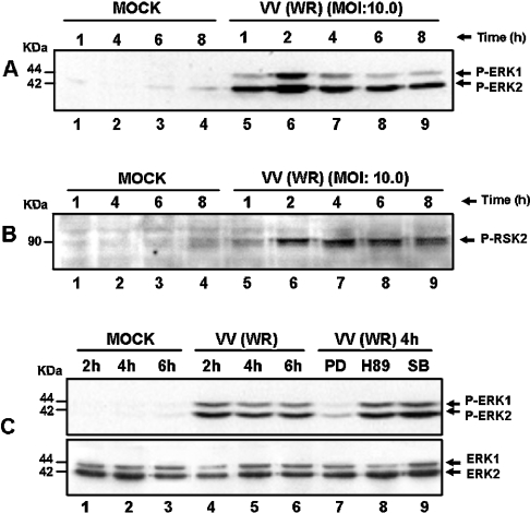
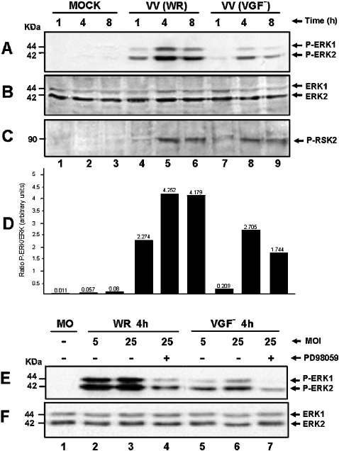
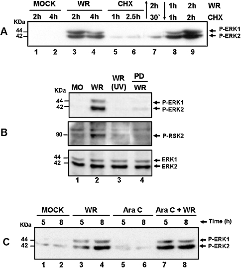
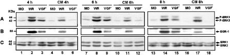
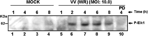
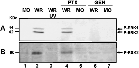
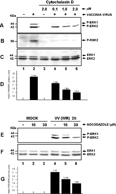
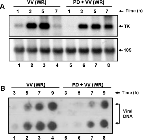
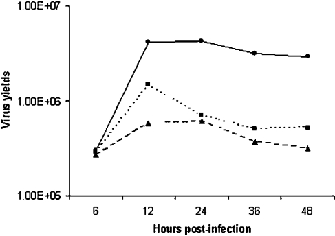
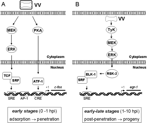
Early events play a decisive role in virus multiplication. We have shown previously that activation of MAPK/ERK1/2 (mitogen-activated protein kinase/extracellular-signal-regulated kinase 1/2) and protein kinase A are pivotal for vaccinia virus (VV) multiplication [de Magalhães, Andrade, Silva, Sousa, Ropert, Ferreira, Kroon, Gazzinelli and Bonjardim (2001) J. Biol. Chem. 276, 38353-38360]. In the present study, we show that VV infection provoked a sustained activation of both ERK1/2 and RSK2 (ribosomal S6 kinase 2). Our results also provide evidence that this pattern of kinase activation depends on virus multiplication and ongoing protein synthesis and is maintained independently of virus DNA synthesis. It is noteworthy that the VGF (VV growth factor), although involved, is not essential for prolonged ERK1/2 activation. Furthermore, our findings suggest that the VV-stimulated ERK1/2 activation also seems to require actin dynamics, microtubule polymerization and tyrosine kinase phosphorylation. The VV-stimulated pathway MEK/ERK1/2/RSK2 (where MEK stands for MAPK/ERK kinase) leads to phosphorylation of the ternary complex factor Elk-1 and expression of the early growth response (egr-1) gene, which kinetically paralleled the kinase activation. The recruitment of this pathway is biologically relevant, since its disruption caused a profound effect on viral thymidine kinase gene expression, viral DNA replication and VV multiplication. This pattern of sustained kinase activation after VV infection is unique. In addition, by connecting upstream signals generated at the cytoskeleton and by tyrosine kinase, the MEK/ERK1/2/RSK2 cascade seems to play a decisive role not only at early stages of the infection, i.e. post-penetration, but is also crucial to define the fate of virus progeny.


Figures

Figure 1
Figure 1. Sustained kinase activation after VV infection
Cells were serum-starved and then infected with VV at the MOI and times indicated. Western-blot analysis was performed with cell lysates, and 30 μg of protein/lane were fractionated by SDS/PAGE, transferred on to nitrocellulose and then probed with the specific anti-phospho-antibody. Cells were either mock- or VV-infected. (A, B) ERK1/2- and RSK2-activation respectively. (C) Upper panel: PD98059 caused a specific inhibition of ERK1/2 phosphorylation. Cells were either left untreated (lanes 1–6), or preincubated for 30 min with protein kinase inhibitors: 50 μM MEK (PD98059), lane 7; 20 μM PKA (H89), lane 8 or 10 μM p38MAPK (SB203580), lane 9; then mock-infected (lanes 1–3) or VV-infected (WR) at an MOI of 10.0 as indicated. Lower panel: the same blot was re-probed with total ERK as an internal control for protein loading. The position of the phosphoproteins or the molecular-mass standard (kDa) is indicated on the right or on the left respectively. The results were consistently repeated in at least two independent experiments.
Figure 2
Figure 2. VGF is partially required for MAPK activation
Western-blot analysis was performed as described in Figure 1. Cells were VV-infected at an MOI of 10.0 for the times shown. (A) Cells were either mock-infected (lanes 1–3) or infected with WR (lanes 4–6) or with VGF virus (lanes 7–9), blotted and then probed with anti-phospho-ERK1/2. (B) Same as in (A), except that the blot was probed with anti-ERK1/2 for loading control. (C) Same as in (A), except that the blot was probed with anti-phospho-RSK2. Blots are representative of at least two independent experiments with identical results. (D) Represents the quantification of two independent experiments taken from Figure 2, showing the ratio between phospho-ERK1/2 (A) and total ERK1/2 (B). Blots were quantified by phosphoimager. (E) Cells were either mock-infected (lane 1) or infected for 4 h with the WR (lanes 2–4) at an MOI of 5.0 (lane 2) or 25.0 (lanes 3 and 4). Lanes 5–7: infected with the VGF at an MOI of 5.0 (lane 5) or 25.0 (lanes 6–7), blotted and then probed with anti-phospho-ERK1/2. (F) Same as in (E), except that the blot was probed with total ERK1/2 as a control of protein loading.
Figure 3
Figure 3. VV multiplication and ongoing protein synthesis is a prerequisite for ERK/RSK activation
Western-blot analysis was performed as described in Figure 1. Cells were either mock-infected (MO) or VV-infected (WR) at an MOI of 10.0, for the indicated times. (A) Effect of CHX (100 μg/ml) on ERK1/2 activation. Lanes: 1 and 2, mock-infected; 3 and 4, VV-infected; 5 and 6, incubated with CHX alone; 7, incubated with CHX for 30 min. Before VV infection for 2 h, lanes 8 and 9 were infected with VV for 1 and 2 h and then treated with CHX for 1 and 2 h respectively. (B) Top and middle panels: ERK1/2 and RSK2 activations respectively rely on virus multiplication. Cells were infected with the WR, or with UV-inactivated virus (UV), or preincubated with PD98059 (50 μM) before VV infection for 4 h as shown and then probed with anti-phospho-ERK or anti-phospho-RSK2. Bottom panel: the same blot as above probed with anti-total ERK1/2 for loading control. (C) Prolonged ERK activation occurs independent of virus DNA synthesis. Lanes 1 and 2, mock-infected; lanes 3 and 4, VV-infected; lanes 5 and 6, incubated with Ara C (40 μg/ml) alone; lanes 7 and 8, incubated with Ara C for 30 min before virus infection. The results were consistently reproduced in at least two independent experiments.
Figure 4
Figure 4. VGF is necessary for both full ERK1/2 activation and EGR-1 expression
Western-blot analysis was performed as described in Figure 1. Infections were performed at an MOI of 10.0. Cells were mock-infected (lanes 1, 7 and 13), or infected with WR (lanes 2, 8 and 14), or infected with VGF (lanes 3, 9 and 15), for 4, 6 or 8 h respectively. Alternatively, the supernatants from WR- or VGF-infected cells were taken after 4, 6 and 8 hpi, collected (conditioned medium, CM) and used to stimulate starved cells for an additional 4, 6 or 8 h respectively. Blots were then probed with (A) anti-phospho-ERK1/2, (B) anti-EGR-1, (C) same blot as in (A) except that the blot was probed with anti-total ERK1/2 for protein loading. Molecular-mass standards (kDa) are indicated on the left. Arrows on the right indicate the investigated proteins. The experiments were repeated at least twice with identical results.
Figure 5
Figure 5. Sustained MAPK activation leads to Elk-1 phosphorylation
Western-blot analysis of VV-infected cells as described in Figure 1. Cells were either mock-infected (lanes 1–4) or infected with WR at an MOI of 10.0 for 1, 2, 4, 6 or 8 h (lanes 5–9). Blots were then probed with anti-phospho-Elk-1. Preincubation with PD98059 (50 μM) blocked the VV-stimulated Elk-1 phosphorylation (lane 10). The blot is representative of two independent experiments with identical results.
Figure 6
Figure 6. VV stimulates a tyrosine-kinase-sensitive pathway
(A, B) Effects of G-protein-coupled receptors and tyrosine kinase inhibitors on VV-stimulated ERK1/2 and RSK2 activation respectively. Western-blot analysis was performed as described above. Cells were mock-infected (lane 1), or were incubated either with 500 μg/ml PTX (lanes 4–5) or with 100 μM GEN (lanes 6–7) for 30 min before VV infection at an MOI of 10.0 for 2 h. As a control for kinase activation, cells were either infected with VV (WR) or with UV-inactivated VV (UV) (lanes 2 and 3) respectively. Blots are representative of at least two independent experiments with identical results.
Figure 7
Figure 7. VV-induced actin polymerization is necessary for both ERK and RSK activations
Western-blot analysis of MO-infected (lanes 1 and 3) or VV-infected cells at an MOI of 10.0 for 2 h (lanes 2 and 4–6), performed with different concentrations of cytochalasin D as shown. Activation of (A) ERK1/2 or (B) RSK2. (C) Blot (A) was re-probed with total ERK for loading control. (E) Effect of nocodazole on VV-stimulated ERK1/2 activation. Cells were either left untreated (lanes 1 and 4) or incubated with the indicated nocodazole concentration before Mock infection (lanes 1–3) or VV infection (lanes 4 –6) and then probed with anti-phospho-ERK1/2. The results were confirmed by at least two independent experiments. (F) Blot (E) was re-probed with anti-total ERK for loading control. (D, G) Quantification of two independent experiments taken from Figure 7, showing the ratio between phospho-ERK1/2 and total ERK1/2 (A/C) or (E/F) respectively. Blots were quantified by phosphoimager.
Figure 8
Figure 8. A delayed/prolonged expression of viral TK gene and viral DNA replication after ERK blockade
(A) Northern-blot assay performed as described in the Experimental section. Upper panel: cells were left untreated (lanes 1–4) or incubated with 50 μM PD for 30 min (lanes 5–8), before VV (WR) infection at an MOI of 10.0 for the times shown and then hybridized with labelled TK probe. Lower panel: blot was stripped of the probe and re-probed with 18 S rRNA as a control for RNA loading. (B) Dot-blot assay performed in duplicates as described in the Experimental section. Cells were either left untreated (lanes 1– 4) or treated with PD98059 (50 μM) (lanes 5–8) for 30 min before and during VV infection at an MOI of 10.0. Infections were performed for 3, 5, 7 or 9 h. Samples were prepared, transferred under vacuum to nylon membrane and then probed with labelled VV DNA.
Figure 9
Figure 9. PD98059 and GEN inhibit VV growth
A time course of VV growth was performed either in the absence or in the presence of PD98059 or GEN (50 μM). Cells were infected with VV at MOI of 10 and 6, 12, 24, 36 or 48 hpi, virus was collected and assayed for infectivity. Results are representative of at least two independent experiments. –•–, VV; --▴--, Gen + VV; ···▪···, PD98059 + VV.
Figure 10
Figure 10. Signalling pathways triggered after VV infection
(A) Early events after VV infection, up to 1 hpi, rely on the activation of MEK/ERK. Both kinase activation and transient c-fos expression are verified very early during virus–host interaction and are independent of the viral protein VGF. A structural component of the virus seems to be sufficient for c-fos stimulation, since no virus gene expression is required. PKA is also recruited to trigger the early signals. (B) Events that occur between 1 hpi until late stages of VV multiplication. Sustained ERK1/2 and RSK2 activation lead to Elk-1 phosphorylation, which in turn binds, possibly in association with SRF, to the cis-acting element SRE found at the 5′-untranslated region of EGR-1 (P. N. G. Silva and C. A. Bonjardim, unpublished work) to promote a sustained egr-1 expression. In connection with this pathway, upstream events are also required, which include tyrosine phosphorylation and actin dynamics. Viral progeny results from an orchestrated programme, which is initiated soon after virus–host interaction and requires, at least partially, sustained MEK/ERK/RSK2 activation.

References

    1. Moss B. Poxviridae viruses and their replication. In: Fields B. N., Knipe D. M., Howley P. M., editors. Fields Virology. 3rd edn. vol. 2. Publishers, NY: Lippincott-Raven; 1996. pp. 2637–2672.
    1. Symons J. A., Alcami A., Smith G. L. Vaccinia virus encodes a soluble type I interferon receptor of novel structure and broad species specificity. Cell (Cambridge, Mass.) 1995;81:551–560. - PubMed
    1. Upton C., Mossman K., McFadden G. Encoding of a homolog of the IFN-γ receptor by myxoma virus. Science. 1992;258:1369–1372. - PubMed
    1. Bowie A., Kiss-Toth E., Symons J. A., Smith G. L., Dower S. K., O'Neill L. A. A46R and A52R from vaccinia virus are antagonists of host IL-1 and toll-like receptor signaling. Proc. Natl. Acad. Sci. U.S.A. 2000;97:10162–10167. - PMC - PubMed
    1. Alcami A. Viral mimicry of cytokines, chemokines and their receptors. Nat. Rev. Immunol. 2003;3:36–50. - PubMed

Publication types

MeSH terms

Substances