. 2004 Mar 16;101(11):3851-6.
doi: 10.1073/pnas.0400611101. Epub 2004 Mar 16.

Coordinate enhancers share common organizational features in the Drosophila genome

Affiliations

Coordinate enhancers share common organizational features in the Drosophila genome

Albert Erives et al. Proc Natl Acad Sci U S A. .

Abstract

The evolution of animal diversity depends on changes in the regulation of a relatively fixed set of protein-coding genes. To understand how these changes might arise, we examined the organization of shared sequence motifs in four coordinately regulated neurogenic enhancers that direct similar patterns of gene expression in the early Drosophila embryo. All four enhancers possess similar arrangements of a subset of putative regulatory elements. These shared features were used to identify a neurogenic enhancer in the distantly related Anopheles genome. We suggest that the constrained organization of metazoan enhancers may be essential for their ability to produce precise patterns of gene expression during development. Organized binding sites should facilitate the identification of regulatory codes that link primary DNA sequence information with predicted patterns of gene activity.


Figures
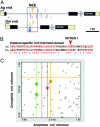
Fig. 3.
Fig. 3.
The Anopheles vnd locus contains an intronic cluster of NEE motifs. (A) There are only several composite clusters of neurogenic motifs in the Anopheles genome (Table 3). One of these clusters maps within the first intron of the vnd ortholog, as determined by reciprocal blast homology to the homeodomain and an NK-2 specific domain located in the third exon (HD-NK). Motifs described in the text, as well as degenerate Dorsal motifs (lower rows of blue clusters), are only shown for intron 1. Intron 2 is not to scale. (B) We find that the Drosophila and Anopheles vnd loci share only one other region of significant conservation, an N-terminal protein-coding sequence spanning intron 1 in Drosophila. (C) Specific comparison of the 1-kb regions encompassing the cluster of sites for the tested enhancers shows that the two sequences lack any extensive sequence alignment spanning the enhancers. Each line in the graph represents a window of sequence alignment containing at least a perfect 7-bp core alignment. Substituting the vnd enhancer sequence with its reverse-complement gives similar results (data not shown). Thus, the actual points of correspondence between sites for Twist (green), Dorsal-like (blue line), Su(H) (red line), and μ (orange lines) do not rise appreciably above background levels of random matches (nonhighlighted dashes). The Anopheles vnd cluster is no more closely related by primary sequence alignment to the Drosophila vnd NEE than to any of the other Drosophila NEEs (data not shown).
Fig. 1.
Fig. 1.
Shared organization of Drosophila enhancers. (Left) Each diagram shows a 640-bp region encompassing the minimal rho (A), vnd (B), brk (C), and vn (D) NEE. Different sequences from these enhancers were attached to a lacZ reporter gene and expressed in transgenic embryos (photomicrographs, Right). Embryos are undergoing cellularization and are oriented with anterior to the left and dorsal up. The staining patterns were determined by in situ hybridization using an antisense RNA probe against lacZ. Red boxes indicate the occurrences of the extended Su(H) site (YGTGRGAAM), whereas the red arrows indicate the orientation of the motif within each enhancer. The specialized μ motif and Dorsal-like sites are shown in yellow and blue, respectively. All CACATGT sequences are shown in green, as well as the related Twist-binding site CATATGT (light green). The distance between linked Twist and Dorsal-like sites is shown directly above each pair. Also indicated are the Twist/μ distances starting from the 3′ position of the palindromic ACATGT core of the Twist site. The centerline at 320 bp bisects the μ core CTGRCC in each enhancer.
Fig. 2.
Fig. 2.
Organized sequence motifs in the rho, vnd, brk, and vn NEEs. All four enhancers contain three organizational features: Twist is linked to a Dorsal-like site (A), the extended Su(H) motif shares 3′ sequences related to the Dorsal-binding site and is located on the same strand of the double helix (B), and there is a Twist site located at a characteristic position upstream of μ (C). A consensus sequence for each signature is shown, as well as an example from a specific enhancer. For example, the Dorsal-like motif in the rho NEE is a perfect match to an optimal Dorsal-binding site (GGGW4-5CCC). The extended Su(H) motif in the vnd enhancer matches overlapping Su(H) and Dorsal-binding sites. The extended μ motif is related to the previously identified Dip3 consensus sequence (14). The genetic organization of the neurogenic genes and their enhancers are summarized in D. The vnd and brk genes are linked on the X chromosome, whereas the vein and rho genes are linked on the left arm of chromosome 3 (D).
Fig. 4.
Fig. 4.
Anopheles vnd enhancer retains conserved NEE structure and function. (A) The mosquito vnd enhancer displays many of the organizational features seen for all of the Drosophila neurogenic enhancers. Comparison with the Drosophila pseudoobscura NEE sequence reveals an interesting intermediate compared to the former two. Both the Twist/Dorsal-like and Twist/μ distances are indicated for each sequence. For discussion of salient aspects of this organization, see text. (B) Alignments of Anopheles vnd sites to Drosophila NEE consensus sequences are depicted with mismatches underlined. The E2 sequence (asterisk) shown here overlaps a CACTTGT E-box in the opposite orientation. Lateral (C) and ventral (D) views of transgenic Drosophila embryos carrying the Anopheles vnd enhancer attached to the lacZ reporter gene are shown. Staining is detected in ventral regions of the neurogenic ectoderm.

References

    1. Thanos, D. & Maniatis, T. (1995) Cell 83, 1091-1100. - PubMed
    1. Belvin, M. P. & Anderson, K. V. (1996) Annu. Rev. Cell Dev. Biol. 12, 393-416. - PubMed
    1. Rusch, J. & Levine, M. (1996) Curr. Opin. Genet. Dev. 6, 416-423. - PubMed
    1. Drier, E. A. & Steward, R. (1997) Semin. Cancer Biol. 8, 83-92. - PubMed
    1. Stathopoulos, A. & Levine, M. (2002) Dev. Biol. 246, 57-67. - PubMed

Publication types

LinkOut - more resources