. 2004 Mar 17;24(11):2593-601.
doi: 10.1523/JNEUROSCI.4461-03.2004.

Aberrant cellular behavior of mutant torsinA implicates nuclear envelope dysfunction in DYT1 dystonia

Affiliations

Aberrant cellular behavior of mutant torsinA implicates nuclear envelope dysfunction in DYT1 dystonia

Pedro Gonzalez-Alegre et al. J Neurosci. .

Abstract

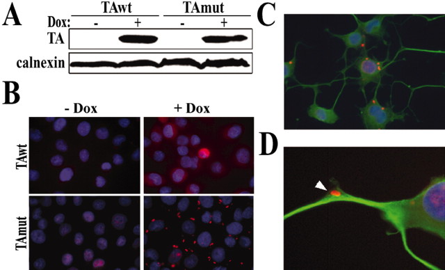
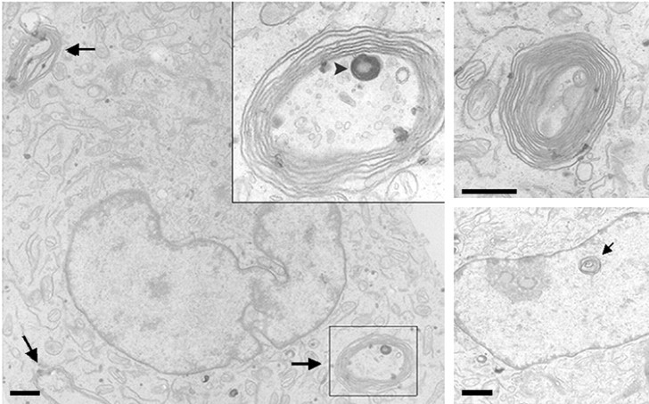
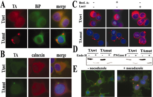
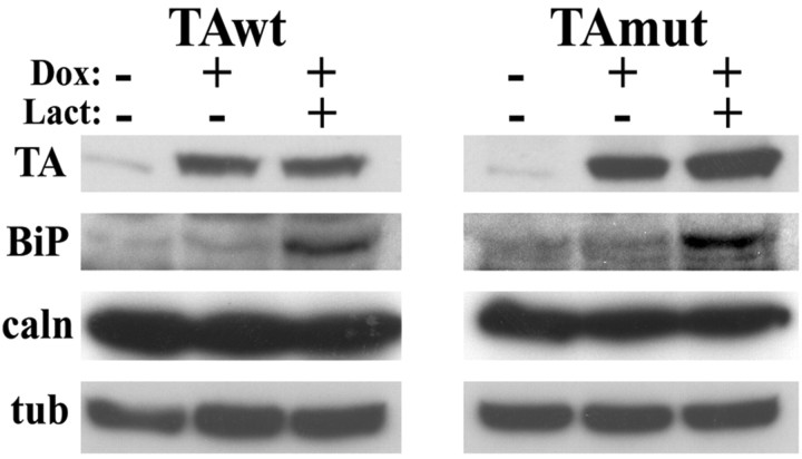
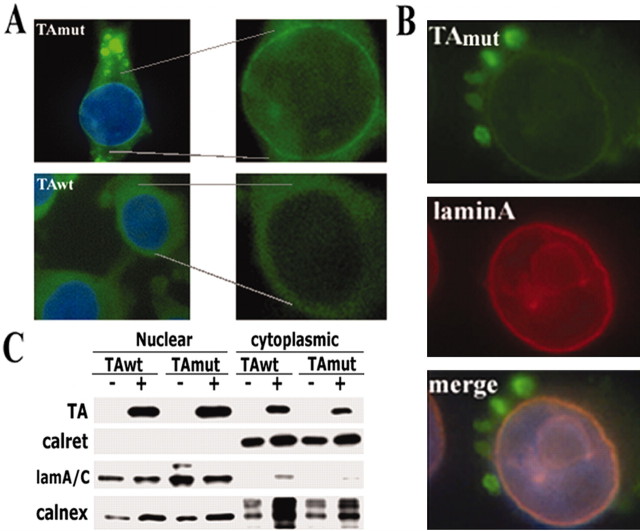
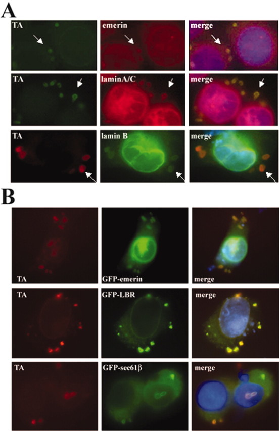
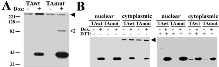
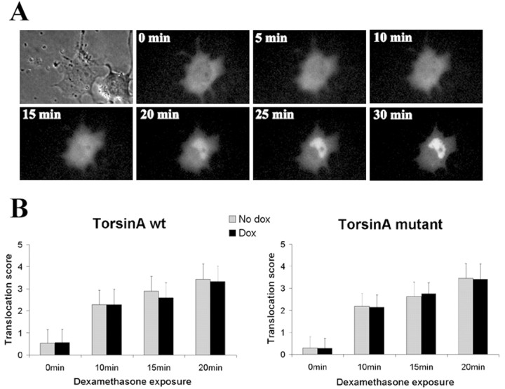
Torsion dystonia-1 (DYT1) dystonia, the most common inherited form of dystonia, is caused by a three base pair deletion that eliminates a single amino acid from the disease protein, torsinA. TorsinA is an "AAA" protein thought to reside in the endoplasmic reticulum (ER), yet both its cellular function and the basis for neuronal dysfunction in DYT1 remain unknown. A clue to disease pathogenesis is the fact that mutant, but not wild-type, torsinA forms membranous inclusions in cell culture. To explore the pathobiology of DYT1 dystonia, we generated PC12 neural cell lines that inducibly express wild-type or mutant torsinA. Although in this model torsinA displays some properties consistent with ER localization, mutant torsinA also accumulates in the nuclear envelope (NE), a structure contiguous with cytoplasmic ER. Consistent with this, membranous inclusions formed by mutant torsinA are shown to derive not from the ER, as thought previously, but from the NE. We demonstrate further that torsinA forms different disulfide-linked complexes that may be linked functionally to subcellular localization in the NE versus cytoplasmic ER. Despite mutant TA accumulation in NE structures, nucleocytoplasmic transport of a reporter protein was unaffected. These findings, together with parallel studies failing to demonstrate perturbation of ER function, implicate the NE as a primary site of dysfunction in DYT1. DYT1 dystonia can be added to the growing list of inherited neurological disorders involving the NE.


Figures

Figure 1.
Figure 1.
An inducible neural cell model of DYT1. Shown are stably transfected PC6-3 clonal cells displaying doxycycline-inducible expression of TAwt or TAmut by WB (A) [anti-TA (MBP) 1:1000], where calnexin is shown as a loading control, and by IF (B), where TA is seen in red and nuclear DAPI staining is in blue. TAwt distributes as a diffuse cytoplasmic pattern, whereas TAmut forms multiple cytoplasmic inclusions. C, A similar IF pattern is seen after TAmut induction in differentiated cells, where TA is seen in red, α-tubulin is in green, and DAPI nuclear staining is in blue. D, Inclusions are sometimes found in neuritic processes.
Figure 2.
Figure 2.
TAmut inclusions are multilamellar bimembrane structures. Shown are representative transmission electron microscopy views of TAmut-expressing cells. The left panel shows a single cell with three different inclusions (arrows). Inset is a higher magnification of one of the inclusions showing loosely arranged concentric membranes. The inclusion also contains a darker, smaller inclusion formed by tightly arranged concentric membranes (arrowhead). The top right panel shows another typical cytoplasmic inclusion, and the right bottom picture shows a possible intranuclear lamellar inclusion (arrow). Scale bars, 1 μm.
Figure 3.
Figure 3.
TAwt and TAmut are pre-Golgi proteins. IF images show colocalization of TAwt, but not TAmut, with BiP (A) and calnexin (B). C, Blocking ER-exit pathways by brefeldinA and lactacystin neither induces inclusion formation by TAwt nor prevents formation of inclusions by TAmut. D, Both forms of TA, detected by anti-TA (MBP) 1:1000, are deglycosylated equally by EndoH and PNGase F, indicating high-mannose, N-linked glycosylation. The lower intensity of deglycosylated bands could result from decreased antibody recognition of TA when deglycosylated or partial degradation of the protein. E, Microtubule disruption by nocodazole alters neuritic processes but not the presence of inclusions.
Figure 8.
Figure 8.
Expression of TAmut does not induce the unfolded protein response. WB analysis of cells expressing TAwt or TAmut showed no changes in BiP or calnexin levels when compared with uninduced cells. In contrast, treatment with lactacystin, a proteasome inhibitor that activates the UPR, significantly increased BiP expression. Note that after inhibition of proteasomal degradation there are no significant changes in levels of TA. α-tubulin is shown as a loading control.
Figure 4.
Figure 4.
TAmut accumulates in the nuclear envelope. A, Representative immunofluorescence images show a TA-immunoreactive perinuclear rim (green) surrounding the nucleus (DAPI staining; blue) in a cell expressing TAmut, but not in a cell expressing TAwt. TA was stained using D-MG10 antibody. Higher magnification of the nuclear area without DAPI staining is also shown. B, Double immunofluorescence shows colocalization of TAmut in the perinuclear rim with laminA. C, Subcellular fractionation experiments show a clear separation of ER lumenal (calreticulin) and nuclear lamina (laminA/C) proteins in the cytoplasmic and nuclear fractions. ER-membrane proteins (calnexin) are found in both compartments, indicating that they are present in cytoplasmic ER and ONM of the NE. TorsinA, whether wild type or mutant, is also found in both fractions. The increased calnexin signal seen in cells expressing TA in this blot was not observed consistently.
Figure 5.
Figure 5.
TAmut inclusions contain nuclear envelope proteins. A, Double IF images showing colocalization of endogenous emerin, laminA/C, and laminB with TAmut-derived inclusions (arrows). B, Transfected GFP-fusion NE proteins were also targeted to inclusions.
Figure 6.
Figure 6.
WB analysis of TA under nonreducing conditions suggests the presence of TA oligomers. A, Denatured protein lysates in the absence of DTT show a large (>220 kDa) TA-immunoreactive band [anti-TA (MBP) 1:250] in induced and noninduced cells (black arrowhead) and a ∼85 kDa TA-immunoreactive band seen only in TAmut-expressing cells (empty arrowhead). B, Subcellular fractionation showed that the >220 kDa TA-immunoreactive band was found almost exclusively in the cytoplasmic fraction of induced and noninduced cells (black arrowhead). After the addition of DTT to the protein lysates, this band disappeared, and a faint band appeared at the expected MW for TA in the noninduced cytoplasmic lanes, suggesting that it corresponds to endogenous TA or another torsin-related protein detected by this antibody [anti-TA (MBP) 1:250].
Figure 7.
Figure 7.
TAmut does not alter the nuclear import of the GR. A, Live cell analysis of nuclear import of GFP-GR in a cell expressing TAmut, before and after dexamethasone treatment. B, Quantification of nuclear import of GFP-GR in fixed cells expressing TAwt, TAmut, and noninduced cells at different times after dexamethasone treatment. Results are the mean (±SD) of 100 cells for each group.
Figure 9.
Figure 9.
The human dopamine transporter weakly colocalizes to TAmut-derived inclusions. A, A differentiated TAmut-expressing cell transfected with GFP-hDAT, showing both GFP and TAmut signal (red) in the inclusions and the NE. Nuclear DAPI staining is shown in blue. A higher-magnification view of the indicated areas highlights the colocalization of TA and hDAT in the inclusions (B) and in the nuclear envelope (C).

References

    1. Ashbourne Excoffon KJ, Moninger T, Zabner J (2003) The coxsackie B virus and adenovirus receptor resides in a distinct membrane microdomain. J Virol 77: 2559–2567. - PMC - PubMed
    1. Augood SJ, Hollingsworth Z, Albers DS, Yang L, Leung JC, Muller B, Klein C, Breakefield XO, Standaert DG (2002) Dopamine transmission in DYT1 dystonia: a biochemical and autoradiographical study. Neurology 59: 445–448. - PubMed
    1. Basham SE, Rose LS (2001) The Caenorhabditis elegans polarity gene ooc-5 encodes a Torsin-related protein of the AAA ATPase superfamily. Development 128: 4645–4656. - PubMed
    1. Berciano MT, Fernandez R, Pena E, Calle E, Villagra NT, Rodriguez-Rey JC, Lafarga M (2000) Formation of intranuclear crystalloids and proliferation of the smooth endoplasmic reticulum in Schwann cells induced by tellurium treatment: association with overexpression of HMG CoA reductase and HMG CoA synthase mRNA. Glia 29: 246–259. - PubMed
    1. Breakefield XO, Kamm C, Hanson PI (2001) TorsinA: movement at many levels. Neuron 31: 9–12. - PubMed

Publication types

MeSH terms

LinkOut - more resources