. 2004 Apr 7;23(7):1657-68.
doi: 10.1038/sj.emboj.7600160. Epub 2004 Mar 18.

A transcriptional network in polycystic kidney disease

Affiliations

A transcriptional network in polycystic kidney disease

Lionel Gresh et al. EMBO J. .

Abstract

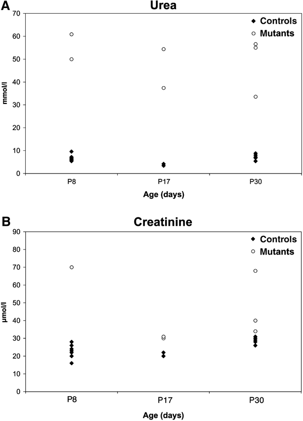
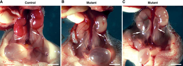
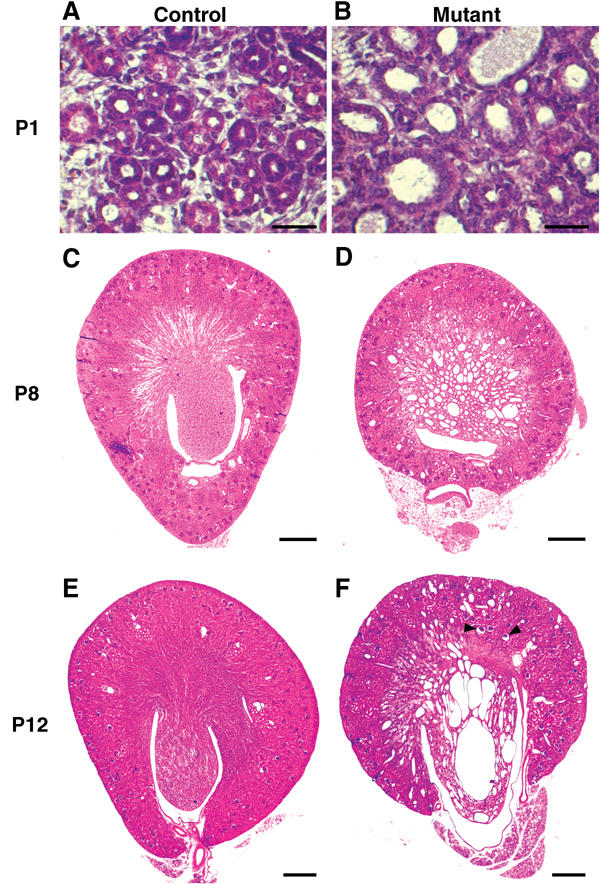
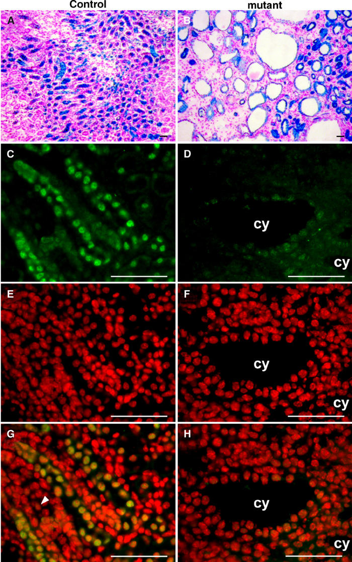
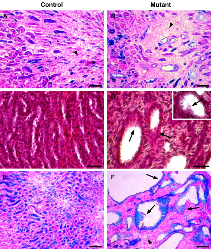
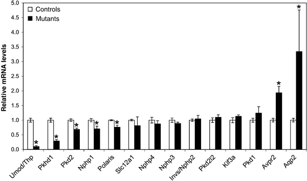
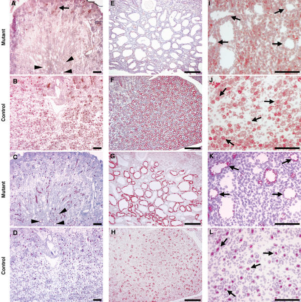
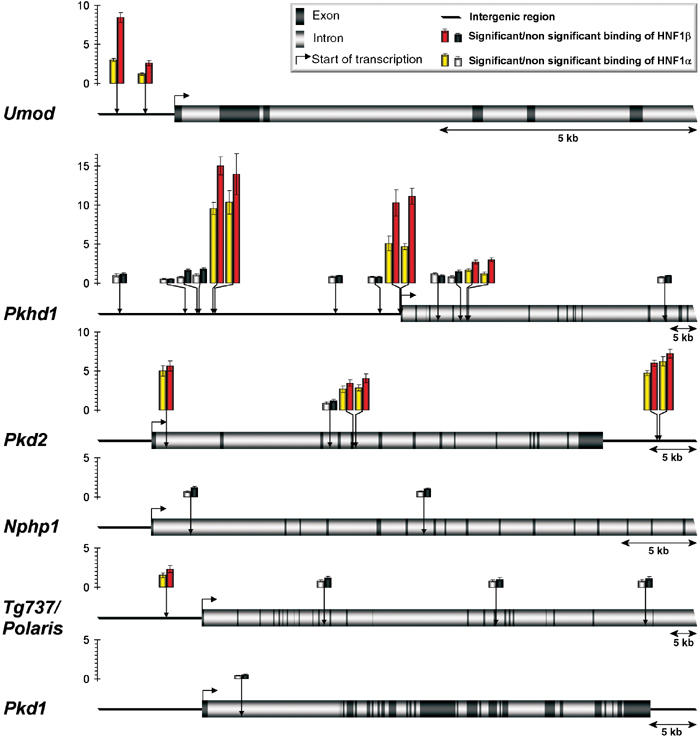
Mutations in cystic kidney disease genes represent a major genetic cause of end-stage renal disease. However, the molecular cascades controlling the expression of these genes are still poorly understood. Hepatocyte Nuclear Factor 1beta (HNF1beta) is a homeoprotein predominantly expressed in renal, pancreatic and hepatic epithelia. We report here that mice with renal-specific inactivation of HNF1beta develop polycystic kidney disease. We show that renal cyst formation is accompanied by a drastic defect in the transcriptional activation of Umod, Pkhd1 and Pkd2 genes, whose mutations are responsible for distinct cystic kidney syndromes. In vivo chromatin immunoprecipitation experiments demonstrated that HNF1beta binds to several DNA elements in murine Umod, Pkhd1, Pkd2 and Tg737/Polaris genomic sequences. Our results uncover a direct transcriptional hierarchy between HNF1beta and cystic disease genes. Interestingly, most of the identified HNF1beta target gene products colocalize to the primary cilium, a crucial organelle that plays an important role in controlling the proliferation of tubular cells. This may explain the increased proliferation of cystic cells in MODY5 patients carrying autosomal dominant mutations in HNF1beta.


Figures

Figure 1
Figure 1
Impaired renal function in mutant mice. Serum concentrations of urea (A) and creatinine (B) at P8, P17 and P30.
Figure 2
Figure 2
HNF1β inactivation results in bilateral ureteral dilation. Macroscopic view of urinary tract in control (A) and mutant (B, C) mice at P8. Some mutants exhibit a mild ureter dilation (arrows) (compare B versus A), while others present a more severe phenotype (compare C versus A). Mutant kidneys had a normal size, but were paler than controls. Scale bars: 0.25 cm.
Figure 3
Figure 3
Inactivation of HNF1β in kidney cells leads to polycystic kidney disease. Hematoxylin–eosin-stained kidney sections from control and mutant mice at P1 (A, B), P8 (C, D) and P12 (E, F). Tubular dilations were visible in the medullary region of mutants at P1, with an increased number of nuclei per tubule section compared to controls (B versus A). In mutants, the medulla was completely disrupted by large cysts by P8 (D). The size of cysts increased with age (F). A few glomerular cysts were observed in the deep cortex (F, arrowheads). Scale bars: A, B: 100 μm, C–F: 400 μm.
Figure 4
Figure 4
Cystic cells underwent recombination and lack HNF1β expression. (A, B) X-gal staining on kidney sections of control KspCre; HNF1βflox/+; ROSA26R (A) and mutant KspCre; HNF1βflox/flox; ROSA26R (B) at P14. β-gal activity is an indicator of Cre-driven recombination on the ROSA26R locus. (A) The medulla of control mice showed recombination in the tubular epithelium. (B) In mutants, all cysts were lined with recombined cells. (C–H) Kidney sections of control HNF1βflox/lacZ and mutant KspCre; HNF1βflox/lacZ at P8. (C, D) HNF1β staining (fluorescein/green). (E, F) Nuclear staining of the same section (DAPI/red). (G, H) Merging. (G) In controls, HNF1β is expressed in tubular but not in mesenchymal cells (arrowhead). (H) Mutants showed no expression of HNF1β in cysts. cy: cyst. Scale bars: 75 μm.
Figure 5
Figure 5
HNF1β inactivation leads to multilayered tubular epithelia. (A, B) X-gal staining on kidney sections of control HNF1βflox/lacZ (A) and mutant KspCre; HNF1βflox/lacZ (B) at P8. β-gal activity is an indicator of the endogenous HNF1β promoter activity. (A) HNF1β was expressed in all tubular cells but not in mesenchyme (arrowhead). (B) Mesenchyme is β-gal negative (arrowhead), indicating that it was not programmed to express HNF1β. (C, D) Hematoxylin–eosin-stained sections of control (C) and mutant (D) at P8. Multilayered tubular epithelial cells were observed (arrows), as well as polyps growing into the cyst lumen (D and insert). (E, F) X-gal staining on kidney sections of control KspCre; HNF1βflox/+; ROSA26R (E) and mutant KspCre; HNF1βflox/flox; ROSA26R (F) at P14. β-gal activity is an indicator of Cre-driven recombination. (E) KspCre-driven recombination was seen in a large proportion of medullary tubular cells. (F) Several cysts lack the typical monolayered epithelial structure (arrows). All epithelial cells are β-gal-positive, demonstrating that they underwent Cre recombination. The mesenchyme in mutants was not affected by recombination (arrowhead). Scale bars: 200 μm.
Figure 6
Figure 6
Defective transcriptional activation of cystic disease genes in mutant mice. Quantitative RT–PCR analysis of cystic disease genes and cell-specific markers. Umod/Thp, Pkhd1, Pkd2, Nphp1 and Tg737/Polaris were downregulated in mutant kidneys. Expression levels in mutants are indicated relative to controls. Results were normalized with β-2 microglobulin expression level (except for Umod, which was normalized to Slc12a1 expression). Significant differences between mutants and controls (P<0.05) are indicated (*). Error bars represent standard error of the mean (Ncontrol=5, Nmutant=3).
Figure 7
Figure 7
HNF1β inactivation leads to a strong decrease of Umod, Pkhd1 and Pkd2 protein levels in cystic cells. Immunostainings for uromodulin (A, B), Pkhd1 (E, F), Pkd2 (I, J), Slc12a1 (C, D, K, L), and Aqp2 (G, H) in mutant and control mice at P8. (A–D) Uromodulin expression pattern. In mutants, no uromodulin staining was detected in the medullary cysts (compare A versus B). Some cysts were clearly shown to be thick ascending limb (TAL)-derived (see the arrowheaded positive staining with α-Slc12a1 antibody in C). Arrowheads in A indicate the position of the corresponding cysts in the adjacent section. In controls, both uromodulin and Slc12a1 proteins colocalized (compare adjacent sections B and D, respectively). In mutants, only few cortical nondilated TAL segments expressed uromodulin (see arrow in A). (EH) Pkhd1 expression pattern. In mutants, Pkhd1 expression was strongly decreased in the vast majority of medullary cystic cells (compare E versus F). Pkhd1 downregulation is not due to the absence of collecting duct cells, as most cysts still expressed aquaporin2 (compare G versus E). (IL) Pkd2 expression pattern. Pkd2 expression is absent in cystic epithelia (compare I versus J). Again, some cysts were clearly shown to be TAL-derived (see the arrowed positive staining with α-Slc12a1 antibody in K). Arrows in I indicate the position of the corresponding cysts, in the adjacent section. In control animals, both Pkd2 and Slc12a1 proteins colocalized (compare arrows in serial sections B and D, respectively). Scale bars: 300 μm.
Figure 8
Figure 8
In vivo binding of HNF1 proteins to their chromatin target sites in cystic kidney disease genes. Predicted in silico HNF1 binding sites (vertical arrows) in Umod, Pkhd1, Pkd2, Nphp1, Tg737/Polaris and Pkd1 genes were tested in ChIP experiments for in vivo HNF1α and β binding. The relative enrichment for each DNA fragment upon immunoprecipitation of HNF1α or β is illustrated as histograms. Colored bars represent HNF1 binding sites with enrichments significantly higher than background (gray bars). PCR experiments were performed in triplicate and the standard errors of these quantifications are shown as error bars.

References

    1. Bachmann S, Metzger R, Bunnemann B (1990) Tamm-Horsfall protein-mRNA synthesis is localized to the thick ascending limb of Henle's loop in rat kidney. Histochemistry 94: 517–523 - PubMed
    1. Barbacci E, Reber M, Ott MO, Breillat C, Huetz F, Cereghini S (1999) Variant hepatocyte nuclear factor 1 is required for visceral endoderm specification. Development 126: 4795–4805 - PubMed
    1. Bernstein J, Evan AP, Gardner KD Jr. (1987) Epithelial hyperplasia in human polycystic kidney diseases. Its role in pathogenesis and risk of neoplasia. Am J Pathol 129: 92–101 - PMC - PubMed
    1. Bhunia AK, Piontek K, Boletta A, Liu L, Qian F, Xu PN, Germino FJ, Germino GG (2002) PKD1 induces p21(waf1) and regulation of the cell cycle via direct activation of the JAK-STAT signaling pathway in a process requiring PKD2. Cell 109: 157–168 - PubMed
    1. Bingham C, Bulman MP, Ellard S, Allen LI, Lipkin GW, Hoff WG, Woolf AS, Rizzoni G, Novelli G, Nicholls AJ, Hattersley AT (2001) Mutations in the hepatocyte nuclear factor-1beta gene are associated with familial hypoplastic glomerulocystic kidney disease. Am J Hum Genet 68: 219–224 - PMC - PubMed

Publication types

MeSH terms