

Early deficit of lymphocytes in Wiskott-Aldrich syndrome: possible role of WASP in human lymphocyte maturation
- PMID: 15030520
- PMCID: PMC1809006
- DOI: 10.1111/j.1365-2249.2004.02409.x
Early deficit of lymphocytes in Wiskott-Aldrich syndrome: possible role of WASP in human lymphocyte maturation
Erratum in
- Clin Exp Immunol. 2004 Jul;137(1):223
Abstract
Wiskott-Aldrich syndrome (WAS) is an X-linked platelet/immunodeficiency disease. The affected gene encodes WASP, a multidomain protein that regulates cytoskeletal assembly in blood cells. Patients have recurring infections, and their lymphocytes exhibit deficient proliferative responses in vitro. We report an evaluation of peripheral blood lymphocytes of 27 WAS patients, aged one month to 55 years. Whereas NK cells were normal, a significant deficit of T and B lymphocytes was observed. The number of lymphocytes was already decreased in infant patients, suggesting deficient output. Both CD4 and CD8 T lymphocytes were affected; the decrease was most pronounced for naïve T cells. Naïve CD4 lymphocytes of patients showed normal expression of Bcl-2, and Ki-67, and normal survival in vitro, suggesting that their in vivo survival and proliferation are normal. The collective data suggest that the patients' lymphocyte deficit results from deficient output, likely due to abnormal lymphocyte maturation in the thymus and bone marrow. We propose that WASP plays an important role not only in the function of mature T lymphocytes, but also in the maturation of human T and B lymphocytes and that impaired lymphocyte maturation is central to the aetiology of WAS immunodeficiency.
Figures
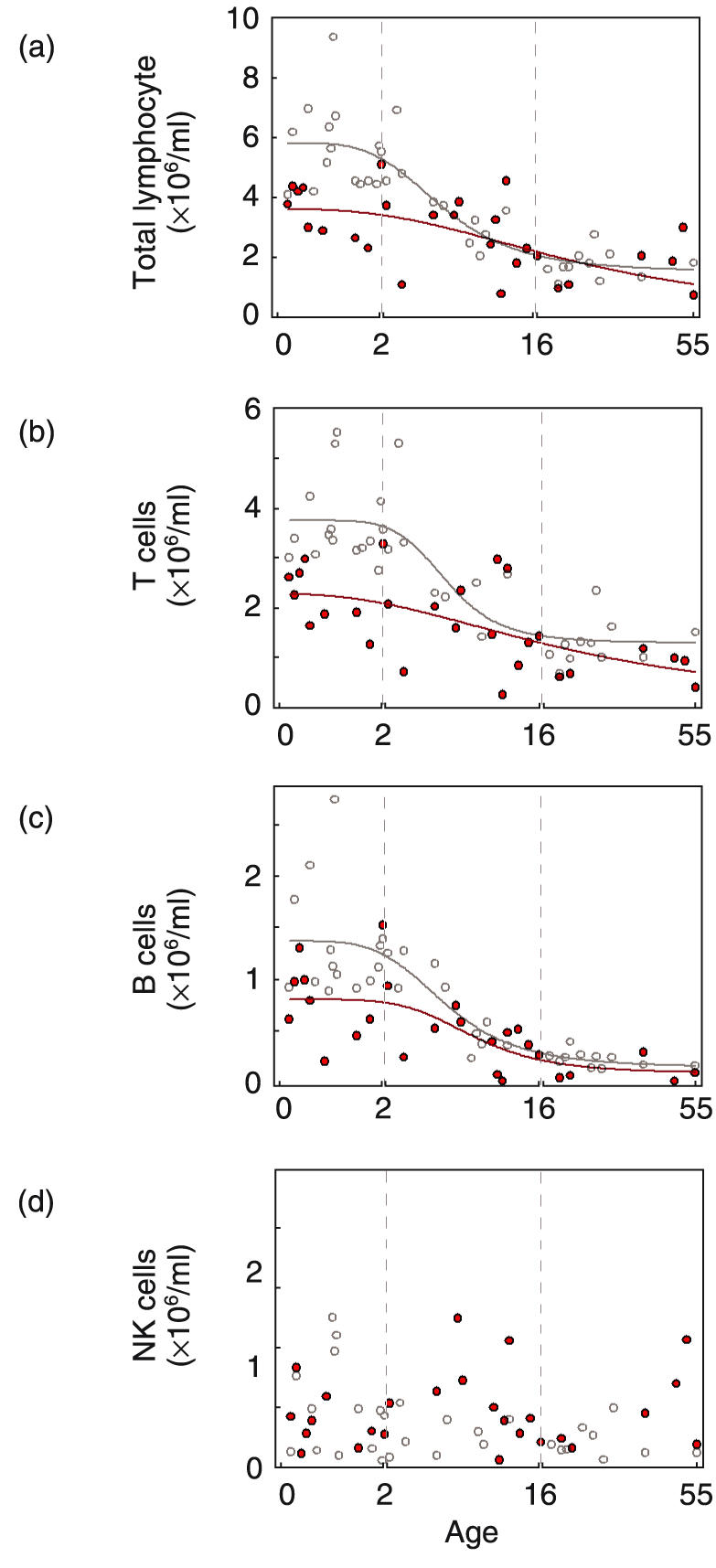
 ). Patient age is shown on the abscissa. Note that different linear scales of increasing compression were used for infants (0–2 years), children (2–16 years) and adults (16–55 years). (a) total lymphocytes (b) T lymphocytes (CD45+CD3+) (c) B lymphocytes (CD45+CD19+) (d) NK cells (CD45+CD3–CD16+CD56+).
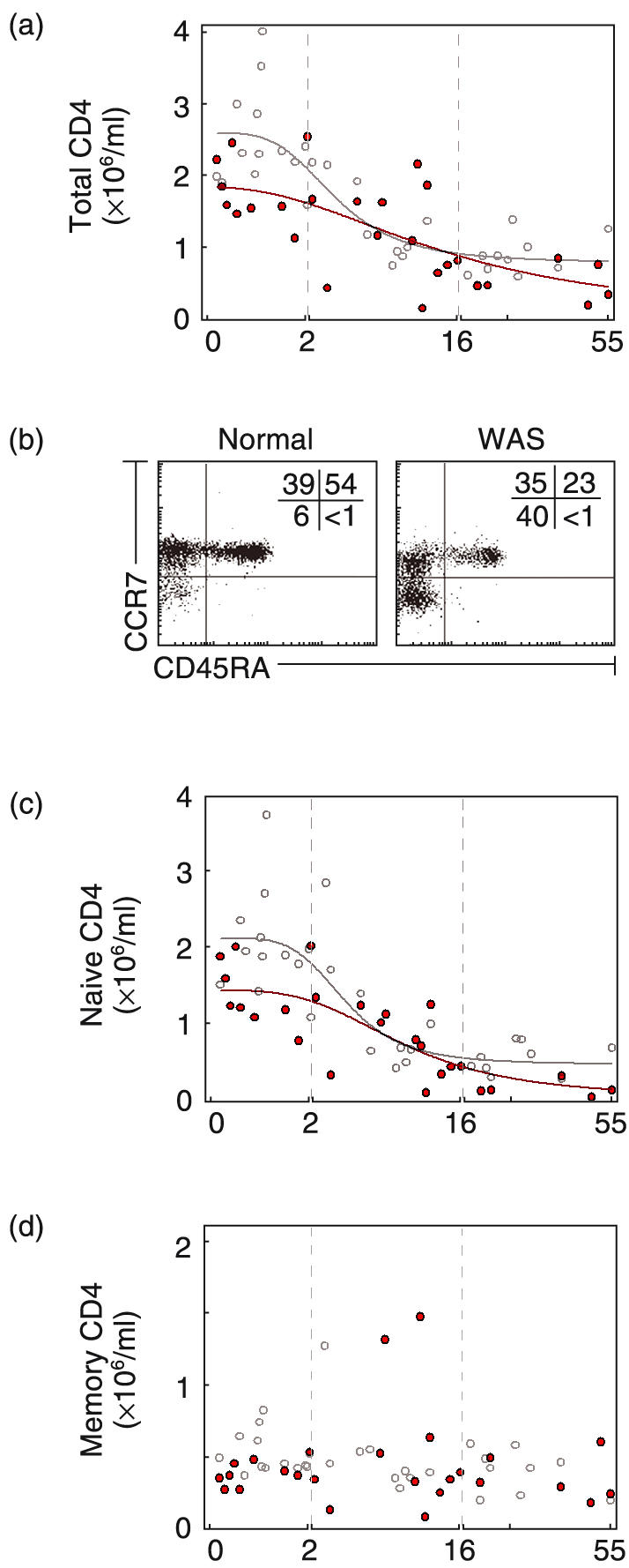
 ). (b) Dot plot of CD45RA and CCR7 expression for a normal adult and an adult patient. Shown are naïve (CD45RA+), central memory (CD45RA–CCR7+) and effector memory (CD45RA–CCR7–) CD4 cells (c) Number of naïve CD4 cells (CD45RA+). (d) Number of CD4 memory cells (CD45RA–).
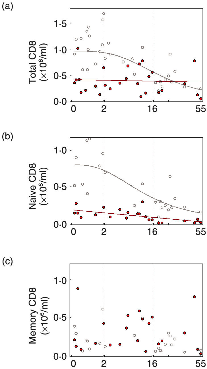
 ).
).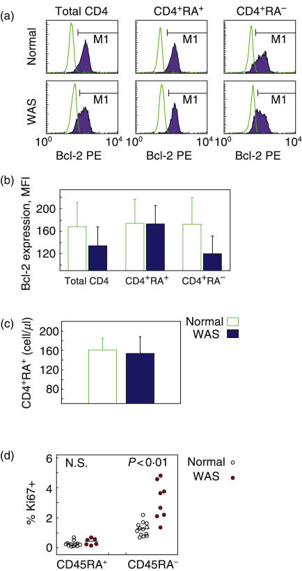
 ) and a WAS patient (
 ). (b) Mean fluorescence intensity of Bcl-2 staining ± SEM for 4 normal individuals and 3 WAS patients. (c) Survival in vitro of isolated naïve CD4 cells. CD4 cells (CD4+CD45RA+) isolated by negative selection were cultured for 3 days without exogenous agent, and the number of surviving cells was determined by flow cytometry. Shown are mean data for cells of normal individuals (
 ) and WAS patients (
 ). (n = 4). (d) Ki-67 expression of naïve and memory CD4 cells. Cells labelled with marker antibodies were stained intracellularly for Ki-67, and the percent positive cells for normal individuals (^) and WAS patients (
 ) was quantified by flow cytometry. The bars represent mean values.
References
-
- Wiskott A. Familiarer, angeobren Morbus Werlhofi? Monatschrift Kinderheil. 1937;68:212–6.
-
- Aldrich R, Steinberg A, Campbell D. Pedigree demonstrating a sex-linked recessive condition characterized by draining ears, eczematoid dermatitis and blood diarrhea. Pediatrics. 1954;13:133–8. - PubMed
-
- Molina IJ, Sancho J, Terhorst C, Rosen FS, Remold-O’Donnell E. T cells of patients with the Wiskott–Aldrich syndrome have a restricted defect in proliferative responses. J Immunol. 1993;151:4383–90. - PubMed
-
- Thrasher AJ. WASp in immune-system organization and function. Nat Rev Immunol. 2002;2:635–46. - PubMed
-
- Kenney D, Cairns L, Remold-O’Donnell E, Peterson J, Rosen FS, Parkman R. Morphological abnormalities in the lymphocytes of patients with the Wiskott–Aldrich syndrome. Blood. 1986;68:1329–32. - PubMed
Publication types
MeSH terms
Substances
Grants and funding
LinkOut - more resources
Full Text Sources
Other Literature Sources
Research Materials
