. 2004 Feb;10(2):320-6.
doi: 10.3201/eid1002.030913.

Ultrastructural characterization of SARS coronavirus

Affiliations

Ultrastructural characterization of SARS coronavirus

Cynthia S Goldsmith et al. Emerg Infect Dis. 2004 Feb.

Abstract

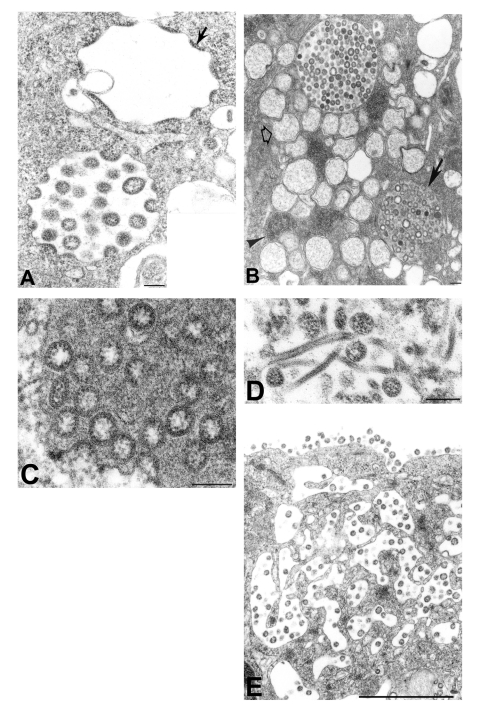
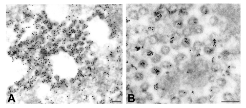
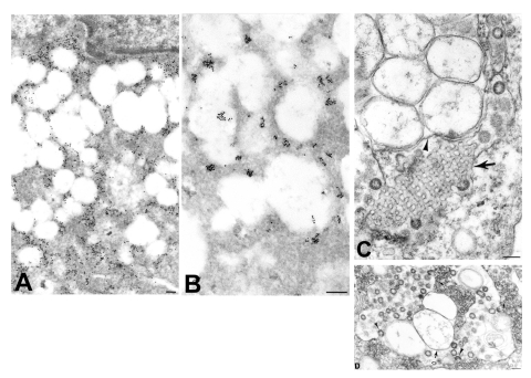
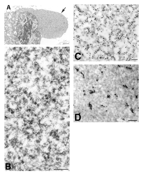
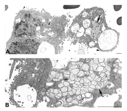
Severe acute respiratory syndrome (SARS) was first described during a 2002-2003 global outbreak of severe pneumonia associated with human deaths and person-to-person disease transmission. The etiologic agent was initially identified as a coronavirus by thin-section electron microscopic examination of a virus isolate. Virions were spherical, 78 nm in mean diameter, and composed of a helical nucleocapsid within an envelope with surface projections. We show that infection with the SARS-associated coronavirus resulted in distinct ultrastructural features: double-membrane vesicles, nucleocapsid inclusions, and large granular areas of cytoplasm. These three structures and the coronavirus particles were shown to be positive for viral proteins and RNA by using ultrastructural immunogold and in situ hybridization assays. In addition, ultrastructural examination of a bronchiolar lavage specimen from a SARS patient showed numerous coronavirus-infected cells with features similar to those in infected culture cells. Electron microscopic studies were critical in identifying the etiologic agent of the SARS outbreak and in guiding subsequent laboratory and epidemiologic investigations.


Figures

Figure 1
Figure 1
Assembly of severe acute respiratory syndrome–associated coronavirus (SARS-CoV) particles in infected Vero E6 cells. A) Apposition of nucleocapsids (arrow) along membranes of the budding compartment as particles developed and budded. Nucleocapsids measured 6 nm in diameter and were mostly seen in cross-section. Some virions had an electron-lucent center, with the nucleocapsid juxtaposed to the envelope, while others were relatively dark when the nucleocapsid was present throughout the particle. Tannic acid pre-treatment enhanced the visibility of the club-shaped viral projections (inset), which averaged 14 nm in length. B) SARS-CoV–infected cell with virus-containing vesicles, double-membrane vesicles (open arrow), and nucleocapsid inclusions (arrowhead). Note the vesicle with granular material interspersed among the virions (arrow). C) Higher magnification of a virus-containing vesicle with dark granular material. D) Tubular structures in a virus-containing vesicle. E) Virions in vesicles, which appeared to migrate toward and fuse with the plasma membrane. The characteristic lining of particles along the cell surface is seen. Bars: A, inset; B–D, 100 nm; E, 1 μm.
Figure 2
Figure 2
Detection of viral proteins and viral RNA associated with intracytoplasmic virions. A) Immunogold labeling of viral proteins by using hyperimmune mouse ascitic fluid directed against severe acute respiratory syndrome–associated coronavirus (12 nm gold). B) Ultrastructural in situ hybridization detection of viral RNA by using a pool of polymerase and nucleocapsid riboprobes (6 nm gold). Bars, 100 nm.
Figure 3
Figure 3
Ultrastructural characteristics of double-membrane vesicles. A) Immunogold labeling of viral proteins by using hyperimmune mouse ascitic fluid (12 nm gold) in areas of cytoplasm in close proximity to the double-membrane vesicles. B) Ultrastructural in situ hybridization detection of viral mRNA, genRNA, or both (6 nm gold) in the same areas and also at times associated with diffuse granular material within the double-membrane vesicles. C) Double-membrane vesicles showing several single-membrane vesicles enclosed within an outer membrane (arrowhead). Also present is a tubuloreticular structure (arrow) with virus particles budding from the membranes. D) Double-membrane vesicles with a large space between the inner (arrow) and outer (open arrow) membranes of the vesicles. Virions are seen budding into (arrowheads) and accumulating within the dilated inter-membrane space. At the periphery of the double-membrane vesicles are nucleocapsid inclusions; arrows point to discernable nucleocapsids (small arrows). Bars, 100 nm.
Figure 4
Figure 4
Immunogold and in situ hybridization (ISH) labeling of severe acute respiratory syndrome–associated coronavirus–infected cells. A) Cytoplasmic area that is relatively free of organelles (arrow). B) At higher magnification, these regions are shown to consist of ribosomelike and filamentous structures. Within these regions, C) viral proteins are detected by immunolabeling, using hyperimmune mouse ascitic fluid (12 nm gold), and D) ultrastructural ISH detects viral mRNA, genRNA, or both, by using a pool of riboprobes (6 nm gold). Bars, A,1 μm; B–D, 100 nm.
Figure 5
Figure 5
Ultrastructural characteristics of a bronchial alveolar lavage (BAL) from a patient with severe acute respiratory syndrome. A) Infected cells from a BAL specimen, showing numerous cytoplasmic and extracellular virions (arrowheads). Note the region of double-membrane vesicles (arrow), a common feature of coronavirus-infected cells. B) At higher magnification, double-membrane vesicles (arrow) are shown to contain diffuse, granular material. Bars, 1 μm.

References

    1. Centers for Disease Control and Prevention. Outbreak of severe acute respiratory syndrome—worldwide, 2003. [Erratum in: MMWR Morb Mortal Wkly Rep 2003;52:284]. MMWR Morb Mortal Wkly Rep. 2003;52:226–8. - PubMed
    1. World Health Organization. Severe acute respiratory syndrome (SARS) multi-country outbreak—update 4. Outbreak reported March 19, 2003. Available from: URL: http://www.who.int/csr/don/2003_03_19/en/
    1. World Health Organization. Summary of probable SARS cases with onset of illness from 1 November 2002 to 31 July 2003. Revised September 26, 2003. Available from: URL: http://www.who.int/csr/sars/country/table2003_09_23/en/
    1. World Health Organization. A multicentre collaboration to investigate the cause of severe acute respiratory syndrome. Lancet. 2003;361:1730–3. 10.1016/S0140-6736(03)13376-4 - DOI - PMC - PubMed
    1. Drosten C, Gunther S, Preiser W, van der Werf S, Brodt HR, Becker S, et al. Identification of a novel coronavirus in patients with severe acute respiratory syndrome. N Engl J Med. 2003;348:1967–76. 10.1056/NEJMoa030747 - DOI - PubMed

MeSH terms

LinkOut - more resources