

A new form of retinopathy associated with myocardial infarction treated with percutaneous coronary intervention
- PMID: 15031163
- PMCID: PMC1772107
- DOI: 10.1136/bjo.2003.027136
A new form of retinopathy associated with myocardial infarction treated with percutaneous coronary intervention
Abstract
Aim: To report a new form of retinopathy that was observed in patients who had undergone percutaneous coronary intervention (PCI) following acute myocardial infarction (AMI).
Methods: Serial ophthalmological examinations were conducted in 40 patients who underwent PCI. Thirty patients were diagnosed with AMI, and another 10 had stable angina pectoris.
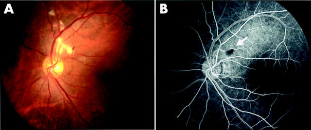
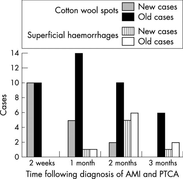
Results: Cotton wool spots developed in 17 (57%) patients from the group with AMI undergoing PCI (n = 30) within 2 months. Of these, 41% (seven patients) also developed superficial haemorrhages. Retinopathy was most prominent 1-2 months after AMI and then tended to become quiescent afterwards, without treatment.
Conclusion: We have identified a new form of retinopathy in patients with AMI that spontaneously subsides without treatment.
Figures
References
-
- Destro M, Gragoudas ES. Arterial occlusions. In: Albert DM, Jakobiec FA, eds. Principle and practice of ophthalmology. Philadelphia: WB Saunders Company, 1994:727–8.
-
- Brown GC. Retinal arterial obstructive disease. In: Ryan SJ, ed. Retina. 2nd ed. St Louis, MO: Mosby-Year Book, Inc, 1994:1373–5.
-
- Brown GC, Brown MM, Hiller T, et al. Cotton wool spots. Retina 1985;5:206–14. - PubMed
-
- Teitelbaum BA. Asymptomatic unilateral microembolic retinopathy secondary to percutaneous transluminal coronary angioplasty. J Am Optom Assoc 1999;70:587–92. - PubMed
MeSH terms
LinkOut - more resources
Full Text Sources
Medical
Miscellaneous