. 2004 Apr;16(4):977-92.
doi: 10.1105/tpc.020156. Epub 2004 Mar 18.

Formation of monoterpenes in Antirrhinum majus and Clarkia breweri flowers involves heterodimeric geranyl diphosphate synthases

Affiliations

Formation of monoterpenes in Antirrhinum majus and Clarkia breweri flowers involves heterodimeric geranyl diphosphate synthases

Dorothea Tholl et al. Plant Cell. 2004 Apr.

Abstract

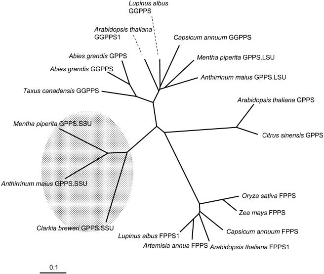
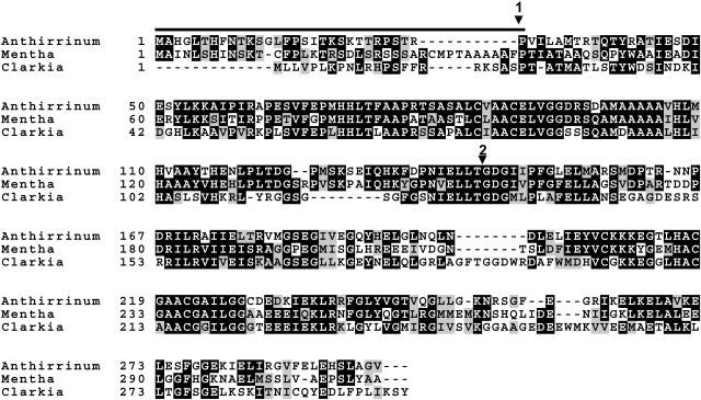
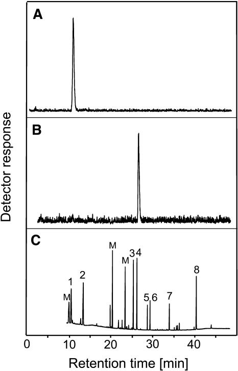
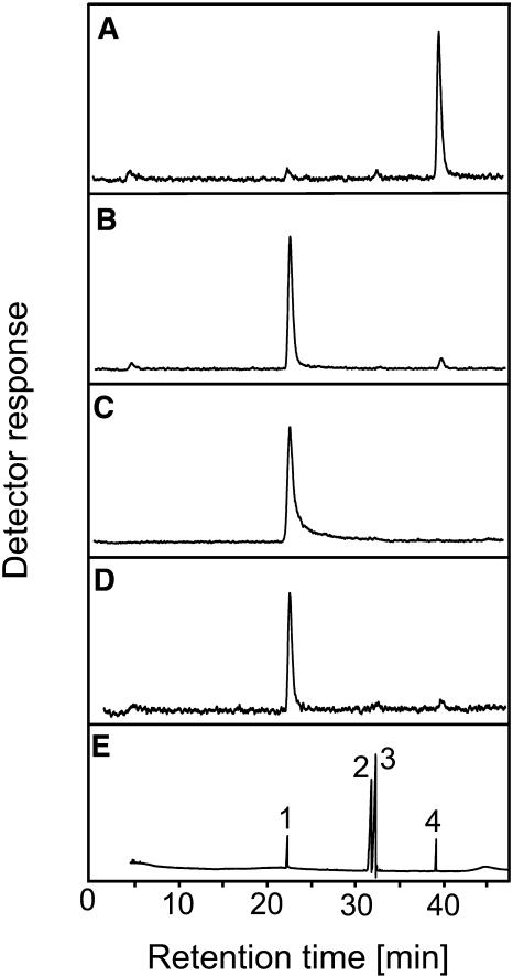
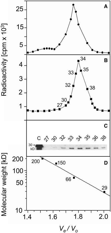
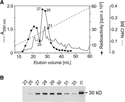
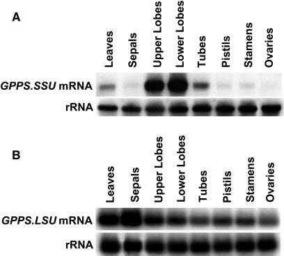
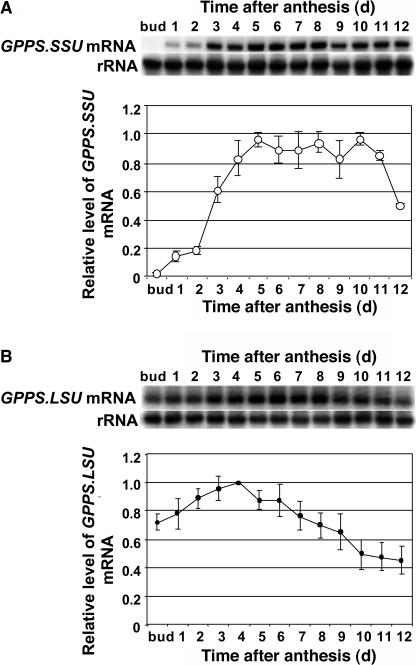
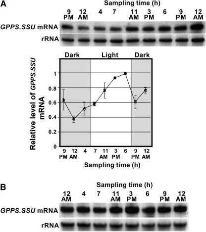
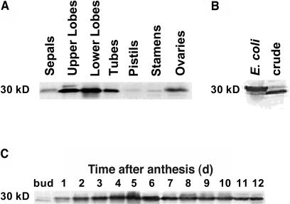
The precursor of all monoterpenes is the C10 acyclic intermediate geranyl diphosphate (GPP), which is formed from the C5 compounds isopentenyl diphosphate and dimethylallyl diphosphate by GPP synthase (GPPS). We have discovered that Antirrhinum majus (snapdragon) and Clarkia breweri, two species whose floral scent is rich in monoterpenes, both possess a heterodimeric GPPS like that previously reported from Mentha piperita (peppermint). The A. majus and C. breweri cDNAs encode proteins with 53% and 45% amino acid sequence identity, respectively, to the M. piperita GPPS small subunit (GPPS.SSU). Expression of these cDNAs in Escherichia coli yielded no detectable prenyltransferase activity. However, when each of these cDNAs was coexpressed with the M. piperita GPPS large subunit (GPPS.LSU), which shares functional motifs and a high level of amino acid sequence identity with geranylgeranyl diphosphate synthases (GGPPS), active GPPS was obtained. Using a homology-based cloning strategy, a GPPS.LSU cDNA also was isolated from A. majus. Its coexpression in E. coli with A. majus GPPS.SSU yielded a functional heterodimer that catalyzed the synthesis of GPP as a main product. The expression in E. coli of A. majus GPPS.LSU by itself yielded active GGPPS, indicating that in contrast with M. piperita GPPS.LSU, A. majus GPPS.LSU is a functional GGPPS on its own. Analyses of tissue-specific, developmental, and rhythmic changes in the mRNA and protein levels of GPPS.SSU in A. majus flowers revealed that these levels correlate closely with monoterpene emission, whereas GPPS.LSU mRNA levels did not, indicating that the levels of GPPS.SSU, but not GPPS.LSU, might play a key role in regulating the formation of GPPS and, thus, monoterpene biosynthesis.


Figures

Figure 1.
Figure 1.
A Neighbor-Joining Tree Based on Degree of Sequence Similarity between Plant GPPS, FPPS, and GGPPS. Sequence analysis was performed using ClustalX, and the nearest neighbor-joining method was applied to create trees. TreeView was used to visualize the resulting trees. Only some of the known plant FPPS and GGPPS are shown.
Figure 2.
Figure 2.
Alignment of Deduced Amino Acid Sequences of A. majus and C. breweri GPPS.SSUs. A. majus GPPS.SSU, C. breweri GPPS.SSU, and GPPS.SSU from M. piperita are included. Residues shaded in black indicate conserved residues (identical in at least two out of three sequences shown), and residues shaded in gray are similar in at least two out of three sequences shown. Dashes indicate gaps that have been inserted for optimal alignment. The horizontal line indicates the putative N-terminal transit peptide region. Arrowhead 1 indicates the truncation site, and arrowhead 2 indicates the intron position in A. majus GPPS.SSU.
Figure 3.
Figure 3.
Identification by Radio Gas Chromatograms of Labeled Dephosphorylated Reaction Products Generated by A. majus GPPS.SSU. (A) and (B) The radioactivity detector responses to the dephosphorylated compounds present in the reaction solution after incubation of [14C]-IPP and DMAPP with recombinant A. majus GPPS.SSU (A) and with recombinant A. majus GPPS.SSU coexpressed with GPPS.LSU from M. piperita (B). (C) The detector response to coinjected authentic standards: isopentenol (peak 1), dimethylallyl alcohol (peak 2), nerol (peak 3), geraniol (peak 4), cis- and trans-nerolidol (peaks 5 and 6, respectively), trans-farnesol (peak 7), geranylgeraniol (peak 8), and monoterpenes (M).
Figure 4.
Figure 4.
Comparison of the Predicted Amino Acid Sequences of A. majus GPPS.LSU and Related Proteins. A. majus GPPS.LSU sequence was aligned with GPPS.LSU from M. piperita and GGPPS from Arabidopsis (At4g36810), A. grandis, and T. canadensis. Residues shaded in black indicate conserved residues (identical in at least three out of six sequences shown), and residues shaded in gray are similar in at least three out of six sequences shown. Dashes indicate gaps that have been inserted for optimal alignment.
Figure 5.
Figure 5.
Radio Gas Chromatograms of the Labeled Dephosphorylated Reaction Products Generated by A. majus GPPS.LSU and Recombinant and Native GPPS. (A) to (D) The radioactivity detector responses to the dephosphorylated compounds present in the reaction solution after incubation of [14C]-IPP and DMAPP with recombinant A. majus GPPS.LSU (A), with recombinant A. majus GPPS.SSU coexpressed with A. majus GPPS.LSU (B), with recombinant A. majus GPPS.SSU coexpressed with A. majus GPPS.LSU at [1-14C]-IPP to DMAPP ratio of 1:3 (C), and with GPPS isolated from A. majus upper and lower lobes (D). (E) The detector response to coinjected authentic standards: geraniol (peak 1), cis-farnesol (peaks 2), trans-farnesol (peak 3), and geranylgeraniol (peak 4).
Figure 6.
Figure 6.
Determination of the Molecular Size of A. majus Recombinant and Native GPPS. (A) and (B) Gel filtration chromatography of A. majus recombinant GPPS (A) and partially purified native GPPS (B) on Superdex 200-HR. (C) Immunodetection of GPPS.SSU in corresponding gel filtration chromatography fractions with native GPPS activity using polyclonal anti-GPPS.SSU antibodies. Crude petal protein extract was loaded in the first (left) lane and used as a control (C). (D) The elution behavior of the standards: β-amylase (200 kD), alcohol dehydrogenase (150 kD), BSA (66 kD), and carbonic anhydrase (29 kD). Ve/Vo is the ratio of elution volume to void volume.
Figure 7.
Figure 7.
Purification of Native GPPS from A. majus Upper and Lower Petal Lobes. (A) Anion exchange chromatography (Mono Q) of GPPS activity containing fractions after initial DEAE-Sepharose chromatography. Absorbance at 280 nm, prenyltransferase activities (in cpm), and the NaCl gradient are indicated. Radio GC analysis of labeled dephospholylated reaction products revealed FPPS activity in peak area 1 (fractions 15 to 25) and GPPS activity in peak area 2 (fractions 26 to 31) (see Figure 5D). (B) Immunodetection of GPPS.SSU in the corresponding fractions separated by anion exchange chromatography (A) using the polyclonal antibodies raised against the native GPPS.SSU protein. Crude extract was loaded in the last (right) lane and used as a control (C).
Figure 8.
Figure 8.
Intracellular Localization of GPPS.SSU in A. majus. (A) Transmission electron microscopy (TEM) image of conical cells of 1-d-old A. majus lower petal lobe labeled with anti-GPPS.SSU antibodies and gold-conjugated goat anti-rabbit antibodies. p, plastid. (B) TEM image of conical cells of 7-d-old A. majus lower petal lobe labeled with anti-GPPS.SSU antibodies and gold-conjugated goat anti-rabbit antibodies. m, mitochondria; p, plastid. (C) TEM image of conical cells of 7-d-old A. majus lower petal lobe treated with preimmune serum and gold-conjugated goat anti-rabbit antibodies. p, plastid.
Figure 9.
Figure 9.
Tissue Specificity of A. majus GPPS.SSU and GPPS.LSU Gene Expression. (A) RNA gel blot of total RNA (5 μg per lane) isolated from leaves, upper and lower petal lobes, tubes, pistils, stamens, and ovaries of 3-d-old A. majus flowers. The top gel represents the results of hybridization with a coding region of the GPPS.SSU gene as a probe. Autoradiography was performed overnight. The blot was rehybridized with an 18S rDNA probe (bottom gel) to standardize samples. (B) RT-PCR with GPPS.LSU gene-specific primers was performed on RNA isolated from leaves, upper and lower petal lobes, tubes, pistils, stamens, and ovaries of 3-d-old A. majus flowers. The amplified products were run on 1.2% agarose gel, blotted onto nitrocellulose membrane, and hybridized with the GPPS.LSU gene as a probe. The RT-PCR products for rRNA are shown at the bottom.
Figure 10.
Figure 10.
Developmental Changes in Steady State GPPS.SSU and GPPS.LSU mRNA Levels in Upper and Lower Lobes of A. majus Petals. (A) Representative RNA gel blot hybridization with mRNA isolated from upper and lower petal lobes at different stages of development, from mature flower buds to day 12 after anthesis. Each lane contained 5 μg of total RNA. A coding region of the GPPS.SSU gene was used as a probe. Autoradiography was performed overnight. The blots were rehybridized with an 18S rDNA probe (bottom gel) to standardize samples. RNA gel blots were scanned with a Storm 860 PhosphorImager, and the values were corrected by standardizing for the amounts of 18S rRNA measured in the same runs. The maximum transcript level was taken as 1. Each point is the average of six different experiments (including the one shown in [A]). Standard error values are indicated by vertical bars. (B) Representative RT-PCR with GPPS.LSU gene-specific primers on RNA isolated from upper and lower petal lobes at different stages of development, from mature flower buds to day 12 after anthesis. The amplified products were run on 1.2% agarose gel, blotted onto nitrocellulose membrane, and hybridized with the GPPS.LSU gene as a probe. The RT-PCR products for rRNA are shown at the bottom. Hybridization signals were analyzed using a Storm 860 PhosphorImager and ImageQuant software (Molecular Dynamics). The maximum transcript level was taken as 1. Each point is the average of four different experiments (including the one shown in [B]). Standard error values are indicated by vertical bars.
Figure 11.
Figure 11.
GPPS.SSU mRNA Expression in Petal Tissue (Upper and Lower Lobes) of 3-d-old A. majus Flowers during a Normal Light/Dark Cycle and in Continuous Dark. (A) RNA gel blot analysis of steady state GPPS.SSU mRNA levels in A. majus petals during a normal light/dark cycle. Total RNA was isolated from upper and lower petal lobes of 3-d-old flowers at time points indicated at the top of the figure, and 5 μg of total RNA was loaded in each lane. The top gel represents the results of hybridization with a GPPS.SSU probe. Autoradiography was performed overnight. The blot was rehybridized with an 18S rDNA probe (bottom gel) to standardize samples. RNA gel blots were scanned with a PhosphorImager and corrected by standardizing for the amounts of 18S rRNA measured in the same runs. The maximum transcript level was taken as 1. Each point is the average of four different experiments (including the one shown in [A]). Standard error values are indicated by vertical bars. Shaded and light areas correspond to dark and light, respectively. (B) RNA gel blot analysis of steady state GPPS.SSU mRNA levels in A. majus flowers exposed to continuous dark. Total RNA was isolated from upper and lower petal lobes of 5-d-old flowers exposed to the second constant dark cycle at time points indicated at the top of the figure, and 5 μg of total RNA was loaded in each lane. The blot was rehybridized with an 18S rDNA probe (bottom gel) to standardize samples.
Figure 12.
Figure 12.
GPPS.SSU Protein Levels in Different Floral Tissues and in Petals during Flower Development. (A) Expression of GPPS.SSU protein in sepals, upper and lower petal lobes, tubes, pistils, stamens, and ovaries of 4-d-old A. majus flowers. Representative protein gel blot shows the 30-kD protein recognized by anti-GPPS.SSU antibodes. Proteins were extracted from different floral tissues, and 20 μg of protein was loaded in each lane. The blot shown represents a typical result of two independent experiments. (B) Immunodetection of GPPS.SSU in the recombinant GPPS and in crude petal extract. (C) Expression of the GPPS.SSU protein in upper and lower petal lobes at different stages of development. Proteins were extracted from upper and lower petal lobes at different stages of development, and 20 μg of protein was loaded in each lane. The blot shown represents a typical result of six independent experiments.

References

    1. Bouvier, F., Suire, C., d'Harlingue, A., Backhaus, R.A., and Camara, B. (2000). Molecular cloning of geranyl diphosphate synthase and compartmentation of monoterpene synthesis in plant cells. Plant J. 24, 241–252. - PubMed
    1. Bradford, M.M. (1976). A rapid and sensitive method for the quantitation of microgram quantities of protein utilizing the principle of protein-dye binding. Anal. Biochem. 72, 248–254. - PubMed
    1. Burke, C.C., and Croteau, R. (2002. a). Interactions with the small subunit of geranyl diphosphate synthase modifies the chain length specificity of geranylgeranyl diphosphate synthase to produce geranyl diphosphate. J. Biol. Chem. 277, 3141–3149. - PubMed
    1. Burke, C.C., and Croteau, R. (2002. b). Geranyl diphosphate synthase from Abies grandis: cDNA isolation, functional expression, and characterization. Arch. Biochem. Biophys. 405, 130–136. - PubMed
    1. Burke, C.C., Wildung, M.R., and Croteau, R. (1999). Geranyl diphosphate synthase: Cloning, expression, and characterization of this prenyltransferase as a heterodimer. Proc. Natl. Acad. Sci. USA 96, 13062–13067. - PMC - PubMed

Publication types

MeSH terms

LinkOut - more resources