. 2004 Apr;16(4):1008-20.
doi: 10.1105/tpc.020701. Epub 2004 Mar 18.

The novel gene HOMOLOGOUS PAIRING ABERRATION IN RICE MEIOSIS1 of rice encodes a putative coiled-coil protein required for homologous chromosome pairing in meiosis

Affiliations

The novel gene HOMOLOGOUS PAIRING ABERRATION IN RICE MEIOSIS1 of rice encodes a putative coiled-coil protein required for homologous chromosome pairing in meiosis

Ken-Ichi Nonomura et al. Plant Cell. 2004 Apr.

Abstract

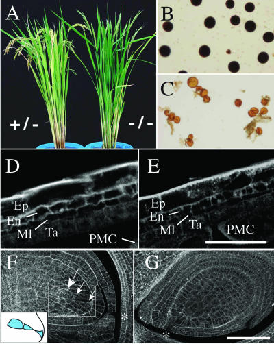
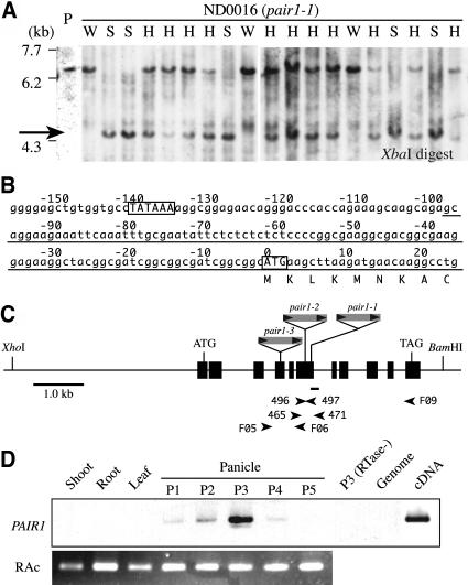
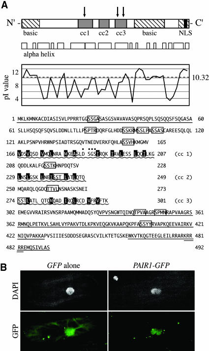
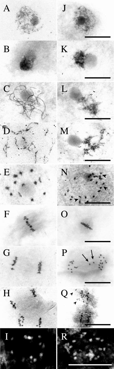
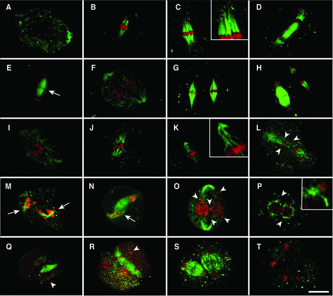
We have identified and characterized a novel gene, PAIR1 (HOMOLOGOUS PAIRING ABERRATION IN RICE MEIOSIS1), required for homologous chromosome pairing and cytokinesis in male and female meiocytes of rice (Oryza sativa). The pair1 mutation, tagged by the endogenous retrotransposon Tos17, exhibited meiosis-specific defects and resulted in complete sterility in male and female gametes. The PAIR1 gene encodes a 492-amino acid protein, which contains putative coiled-coil motifs in the middle, two basic regions at both termini, and a potential nuclear localization signal at the C terminus. Expression of the PAIR1 gene was detected in the early stages of flower development, in which the majority of the sporocytes had not entered meiosis. During prophase I of the pair1 meiocyte, all the chromosomes became entangled to form a compact sphere adhered to a nucleolus, and homologous pairing failed. At anaphase I and telophase I, chromosome nondisjunction and degenerated spindle formation resulted in multiple uneven spore production. However, chromosomal fragmentation frequent in plant meiotic mutants was never observed in all of the pair1 meiocytes. These observations clarify that the PAIR1 protein plays an essential role in establishment of homologous chromosome pairing in rice meiosis.


Figures

Figure 1.
Figure 1.
Defects of the pair1 Mutation Observed Only in Male and Female Sporocytes. (A) The plant morphology of the homozygous pair1-1 mutant (−/−) was normal, whereas complete sterility resulted in erected panicles and prolonged greening of leaves compared with the heterozygous wild-type sibling (+/−). (B) Fertile pollens in a heterozygous sibling, stained by iodium potassium iodide solution. (C) Completely sterile pollen in the homozygous pair1-1 mutant. (D) and (E) In anther wall development, no discernible difference was observed between the wild type (D) and the pair1-1 mutant (E), respectively. Ep, epidermis; En, endothecium; Ml, middle layer; Ta, tapetum cells; PMC, pollen mother cell. Bar = 20 μm. (F) and (G) In the wild-type ovule, three of four female spores were degenerated (arrows and bottom left illustration, smaller arrows for degenerated spores; [F]), whereas no obvious structure of spores was observed in the mutant ovules (G). Bar = 20 μm.
Figure 2.
Figure 2.
Identification and Characterization of the PAIR1 Gene. (A) DNA gel blot analysis of a sample of the 317 progenies of the pair1-1 heterozygote probed with the PAIR1 cDNA. A Tos17 insertion induced a 4.5-kb polymorphic band (arrow), whereas the original Nipponbare showed a 6.5-kb band (lane P). Presence of the 4.5-kb bands was completely linked with the sterile phenotype (S). Homozygosity (W) or heterozygosity (H) of fertile plants was determined in their progeny. (B) Genomic sequence 150 bp upstream of the PAIR1 coding region. Underlining indicates the 5′ end predicted by three independent reactions of RACE. The predicted TATA box 35 bp upstream of the 5′ end and the translation initial codon ATG are boxed. (C) PAIR1 coding region and Tos17-inserted sites. Shaded bars indicate allelic Tos17 insertions, in which arrowheads represent long terminal repeats and their orientation. A small bar under the gene structure indicates the position of the DNA probe used in (A) and (D). Arrowheads indicate the position and orientation of the PCR primers. (D) RT-PCR for the PAIR1 mRNA. The panicles were classified into five groups (P1 to P5) according to their development stage (see the text). In lane 9, the P3 total RNA was templated without RTase as a negative control. In lanes 10 and 11, total DNA from Nipponbare and the PAIR1 cDNA clone were templated as negative and positive controls, respectively.
Figure 3.
Figure 3.
Primary and Secondary Structure Prediction of PAIR1. (A) The vertical arrows indicate the position of Tos17 insertion in the respective three alleles. The α-helical regions were predicted according to Chou and Fasman (1978). The pI values of 20 amino acids were estimated in each 10–amino acid interval along the PAIR1 peptide and plotted on the graph. The coiled-coil structure with three heptad clusters (cc1, cc2, and cc3), separated by two nonhelical sequences, was predicted at the middle of the peptide (shaded bars and wavy lines). The coiled-coil regions contained 13 heptad repeats in total, whose first and fourth residues were frequently hydrophobic (highlighted). The cc1 sequence was predicted to make a helix-turn-helix structure (turned at the residues with dots). Three basic amino acid clusters were detected (hatched bars and underlines). The C-terminal basic region including a KRRRR sequence was predicted to act as a nuclear localization signal (NLS; closed box and double underline). The S/T-P-X-X or S/T-S/T-X-X motifs, characteristic of DNA binding proteins (Suzuki 1989), are boxed. (B) Nuclear localization of GFP-PAIR1 fusion protein during transient expression in A. cepa epidermal cells.
Figure 4.
Figure 4.
Homologous Chromosome Pairing Is Defective in Male and Female Meiocytes of the Wild Type and pair1-1 Mutant. (A) to (H) Chromosomal spreads from wild-type male meiocytes in various stages: leptotene (A), zygotene (B), pachytene (C), diplotene (D), diakinesis (E), metaphase I (F), metaphase II (G), and anaphase II (H). (I) Homologous chromosomes at diakinesis in wild-type female meiocytes. (J) to (Q) Chromosomal spreads from pair1-1 male meiocytes in various stages. No noticeable aberration was observed at leptotene (J) and synizetic zygotene (K). However, synizetic conformation still remained at pachytene and diplotene in the mutant ([L] and [M]). At late diakinesis, 24 complete univalents were observed in the mutant (N), in which several univalents were frequently connected by a thin-chromatin thread (arrowheads). Though all univalents aligned on the division plane (O), nondisjunction and delayed univalents (arrows) were observed in the mutant (P). Most of the chromatids were synchronously divided to the opposite poles, whereas several chromatids independently moved (arrowheads; [Q]). (R) Approximately 24 univalents were observed in the mutant female meiocytes at diakinesis. Bar = 20 μm.
Figure 5.
Figure 5.
Frequency of Each Meiotic Stage Observed in Anthers of the Wild Type and the pair1-1 Mutant. Staging was performed using anthers of 0.6, 0.7, 0.8, and 0.9 mm in length. Meiotic stages were classified into leptotene (red), zygotene with synizetic conformation (light blue), pachytene (yellow), diplotene (green), diakinesis (orange), and metaphase I or later stages (dark blue). In the mutant anthers, typical features of pachytene and diplotene chromosomes were seldom observed. Then, the meiocytes were classified according to the extent of chromosome condensation.
Figure 6.
Figure 6.
Chromosome Pairing in Male Meiocytes of the 0.8-mm Anthers. The pictures, a part of the experiments shown in Table 2, are superimposed images of DAPI-stained chromatin (blue) and FISH (green) with a probe for the 25S rDNA. Most of the wild-type meiocytes showed a single focus (left), whereas most of the mutant meiocytes showed two unpaired foci (right).
Figure 7.
Figure 7.
Abnormal Male Sporulation in the pair1-1 Mutant. (A) to (D) DAPI-stained male sporocytes in dyad stage. All wild-type anthers carried normal dyads and tetrads. The mutant carried abnormal dyads that had one or more of the following: nuclei of different sizes (A), multiple nuclei (B), unequal cytoplasm (C), and micronuclei (D). (E) to (I) DAPI-stained male spores in the tetrad stage. The mutant carried abnormal tetrads with an unequal volume or misdivided nuclei ([F] and [G]) with several micronuclei. Abnormal pentayads (H) and hexayads (I) were also observed.
Figure 8.
Figure 8.
Immunostaining of Spindles in Wild-Type and pair1-1 Male Meiocytes. (A) to (H) Male meiocytes of the wild type. (I) to (T) Male meiocytes of pair1-1 mutant. (A) Tubulin fibers were present throughout the cytoplasm at diakinesis. Chromosomes were stained by PI (red), and microtubules were immunologically stained by Alexa488 (green). (B) Bipolar-oriented microtubules had developed at early metaphase I. (C) Mature spindles were formed at metaphase I. (D) Bivalents were separated by the spindle into two sets of 12 univalents at anaphase I. (E) The phragmoplast had begun to form between daughter pronuclei at telophase I, in which diminished staining in the midzone indicated the cytokinetic plane (arrow). (F) A division plate with a phragmoplast ring had grown to separate the daughter cells. (G) Two sets of univalents aligned at metaphase-II plates. (H) Two sets of univalents were separated to the opposite poles by spindles. (I) and (J) No obvious abnormality was observed in the microtubule array in pair1-1 mutant at diakinesis (I) and early metaphase I (J). (K) The mutant spindle is composed of wavy, thin bundles of microtubules. (L) Several delayed univalents (arrowheads) were always observed at anaphase I. (M) Extra microtubule fibers (arrows) extended to a different direction from the poleward axis at telophase I. (N) The ectopic extension of microtubule fibers (arrow) also appeared at late telophase I. (O) Several micronuclei (arrowheads) were observed in the mutant dyad. (P) Daughter nuclei were enclosed by the microtubule corona, in which micronuclei were clearly detected outside the corona at the dyad stage. (Q) to (T) During meiosis II, the spindles exhibited normal appearance but not around the micronuclei (arrowheads). Bar = 10 μm.

Similar articles

Cited by

References

    1. Altschul, S.F., Madden, T.L., Schaffer, A.A., Zhang, J., Zhang, Z., Miller, W., and Lipman, D.J. (1997). Gapped BLAST and PSI-BLAST: A new generation of protein database search programs. Nucleic Acids Res. 25, 3389–3402. - PMC - PubMed
    1. Armstrong, S.J., Caryl, A.P., Jones, G.H., and Franklin, F.C. (2002). Asy1, a protein required for meiotic chromosome synapsis, localizes to axis-associated chromatin in Arabidopsis and Brassica. J. Cell Sci. 115, 3645–3655. - PubMed
    1. Armstrong, S.J., Franklin, F.C., and Jones, G.H. (2001). Nucleolus-associated telomere clustering and pairing precede meiotic chromosome synapsis in Arabidopsis thaliana. J. Cell Sci. 114, 4207–4217. - PubMed
    1. Bai, X., Peirson, B.N., Dong, F., Xue, C., and Makaroff, C.A. (1999). Isolation and characterization of SYN1, a RAD21-like gene essential for meiosis in Arabidopsis. Plant Cell 11, 417–430. - PMC - PubMed
    1. Bass, H.W., Marshall, W.F., Sedat, J.W., Agard, D.A., and Cande, W.Z. (1997). Telomeres cluster de novo before the initiation of synapsis: A three-dimensional spatial analysis of telomere positions before and during meiotic prophase. J. Cell Biol. 137, 5–18. - PMC - PubMed

Publication types

Associated data