. 2004 Apr 13;101(15):5577-82.
doi: 10.1073/pnas.0307917101. Epub 2004 Mar 18.

Suppression of nuclear oscillations in Saccharomyces cerevisiae expressing Glu tubulin

Affiliations

Suppression of nuclear oscillations in Saccharomyces cerevisiae expressing Glu tubulin

A C Badin-Larçon et al. Proc Natl Acad Sci U S A. .

Abstract

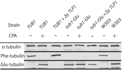
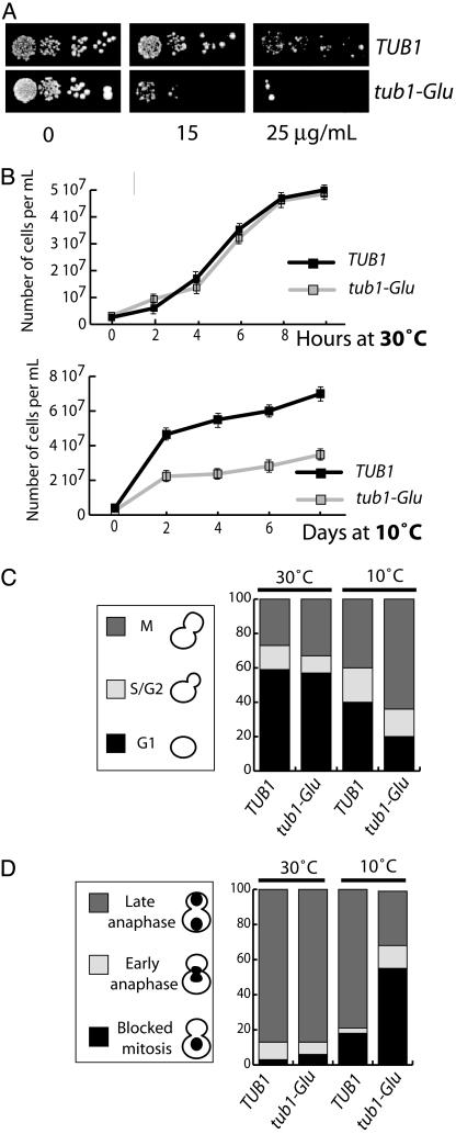
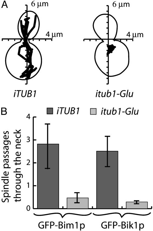
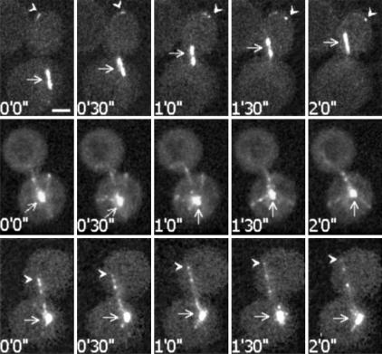
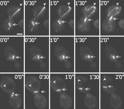
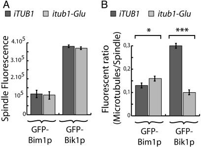
In most eukaryotic cells, the C-terminal amino acid of alpha-tubulin is aromatic (Tyr in mammals and Phe in Saccharomyces cerevisiae) and is preceded by two glutamate residues. In mammals, the C-terminal Tyr of alpha-tubulin is subject to cyclic removal from the peptide chain by a carboxypeptidase and readdition to the chain by a tubulin-Tyr ligase. There is evidence that tubulin-Tyr ligase suppression and the resulting accumulation of detyrosinated (Glu) tubulin favor tumor growth, both in animal models and in human cancers. However, the molecular basis for this apparent stimulatory effect of Glu tubulin accumulation on tumor progression is unknown. Here we have developed S. cerevisiae strains expressing only Glu tubulin and used them as a model to assess the consequences of Glu tubulin accumulation in cells. We find that Glu tubulin strains show defects in nuclear oscillations. These defects are linked to a markedly decreased association of the yeast ortholog of CLIP170, Bik1p, with microtubule plus-ends. These results indicate that the accumulation of Glu tubulin in cells affects microtubule tip complexes that are important for microtubule interactions with the cell cortex.


Figures

Fig. 1.
Fig. 1.
Analysis of the α-tubulin C terminus in control and mutant strains. Western blotting of whole-cell extracts from TUB1 and tub1-Glu, expressing or not the Tlp1p protein (2μ TLP1) and W303 strains. Extracts were exposed or not to carboxypeptidase A (CPA) before immunoblotting. Primary antibodies were directed against total α-tubulin (YOL34), Phe tubulin (YL1/2), or Glu tubulin (NS8) as indicated.
Fig. 2.
Fig. 2.
Benomyl supersensitivity and mitotic block in tub1-Glu strains. (A) Benomyl sensitivity. Yeast cells of the indicated genotype were spotted on solid rich media containing the indicated benomyl concentration. (B) Cell growth. At each time point a minimum of 100 cells were counted. Four independent experiments were run. Growth rates were similar at 30°C in both strains. At 10°C, the tub1-Glu strain showed impaired growth. (C) Mitotic block in tub1-Glu cells. The percent of unbudded cells (G1), cells containing small buds (S), and cells with large buds (M) were scored in TUB1 and tub1-Glu strain (n = 150) at both warm and cold temperatures as indicated. (D) Inhibition of nuclear division and the position of DNA in control and mutant cells. Cells grown at 30°C and 10°C were stained with Hoescht, and the nucleus number and position were determined by fluorescence microscopy (n = 150).
Fig. 3.
Fig. 3.
Inhibition of nuclear oscillations in Glu tubulin strains. (A) Tracking of spindle motions over a 30-min time duration was performed, and a representative example is shown in both control and mutated strains. (B) Quantitative analysis of spindle passage through the neck in control or mutated strains expressing GFP-Bim1p (number of films: control, n = 5; mutated, n = 11) or GFP-Bik1p (number of films: control, n = 16; mutated, n = 21) as indicated. The ordinate represent the absolute number of spindle passages through the neck during the 30 min of the observation.
Fig. 4.
Fig. 4.
Video microscopy analysis of spindle and microtubule behavior in Phe tubulin or Glu tubulin strains expressing GFP-Bim1p. (Top) Phe tubulin GFP-Bim1p strain during a nuclear oscillation event. Arrows indicate the spindle. Arrowheads indicate microtubules plus-ends. Microtubule ends were apparently captured at the cell cortex with subsequent microtubule sliding and spindle pulling. (Middle) One aspect observed in Glu tubulin GFP-Bim1p strains. Arrows indicate the spindle. In Glu tubulin strains, microtubules often seemed to lack directional cues and showed little net spindle motion. (Bottom) Another aspect observed in Glu tubulin GFP-Bim1p strains. Arrows indicate the spindle. Arrowheads indicate microtubules plus-ends. In some cases, microtubules had apparent directional cues. However, microtubule ends were not clustered, and microtubule capture and sliding at the cell cortex were rarely observed. Time points corresponding to image capture are indicated. (Bar, 2 μm.)
Fig. 5.
Fig. 5.
Video microscopy analysis of spindle and microtubule behavior in Phe tubulin and Glu tubulin strains expressing GFP-Bik1p. (Top) Phe tubulin GFP-Bik1p strain during a nuclear oscillation. Arrows indicate spindle. Arrowheads indicate microtubules plus-ends. With GFP-Bik1p, long comets were observed on the bundled microtubules. Such comets remained conspicuous during microtubule capture and sliding at the cell cortex. (Middle) One aspect observed in Glu microtubule GFP-Bik1p strains. Arrows indicate the spindle. Microtubules apparently lack directional cues. Microtubule plus-ends are poorly labeled with GFP-Bik1p, and no comet is visible. (Bottom) Glu tubulin GFP-Bik1p strain during a nuclear oscillation. Arrows indicate spindle with a translocation event. Arrowheads indicate microtubules plus-ends. Nuclear oscillations were rarely observed in Glu tubulin strains (Fig. 3B). During such oscillations, the GFP-Bik1p labeling remained conspicuously different from controls, with barely apparent and evanescent GFP-Bik1p comets and poorly labeled microtubule ends. Times when images are captured are indicated. (Bar, 2 μm.)
Fig. 6.
Fig. 6.
Quantitative analysis of GFP-Bim1p and GFP-Bik1p labeling in Phe tubulin and Glu tubulin strains. (A) Spindle fluorescence measured in arbitrary units by using metamorph software. Spindle labeling was averaged by using the first 10 images of each filmed cell. Labeling was stronger with GFP-Bik1p compared with GFP-Bim1p in both Phe strains and Glu strains. No statistically significant differences were observed between the two types of strains. (B) Ratio of microtubule/spindle fluorescence labeling in Phe tubulin and Glu tubulin strains. With GFP-Bim1p, the ration of microtubule/spindle fluorescence was slightly (but significantly) higher in Glu tubulin cells compared with Phe tubulin cells. In contrast, with GFP-Bik1p, the same ratio was strongly reduced in Glu tubulin cells compared with Phe tubulin cells, indicating impaired association of GFP-Bik1p with microtubule plus-ends in the Glu tubulin cells. Results were analyses with ANOVA. *, P < 0.05; ***, P < 0.001.

Comment in

References

    1. Valiron, O., Caudron, N. & Job, D. (2001) Cell Mol. Life Sci. 58, 2069–2084. - PMC - PubMed
    1. Lafanechere, L., Courtay-Cahen, C., Kawakami, T., Jacrot, M., Rudiger, M., Wehland, J., Job, D. & Margolis, R. L. (1998) J. Cell Sci. 111, 171–181. - PubMed
    1. Mialhe, A., Lafanechere, L., Treilleux, I., Peloux, N., Dumontet, C., Bremond, A., Panh, M. H., Payan, R., Wehland, J., Margolis, R. L. & Job, D. (2001) Cancer Res. 61, 5024–5027. - PubMed
    1. Schatz, P. J., Solomon, F. & Botstein, D. (1986) Mol. Cell. Biol. 6, 3722–3733. - PMC - PubMed
    1. Schatz, P. J., Solomon, F. & Botstein, D. (1988) Genetics 120, 681–695. - PMC - PubMed

Publication types

MeSH terms