Review
. 2004 Apr;2(4):301-14.
doi: 10.1038/nrmicro865.

Cellular autophagy: surrender, avoidance and subversion by microorganisms

Affiliations
Review

Cellular autophagy: surrender, avoidance and subversion by microorganisms

Karla Kirkegaard et al. Nat Rev Microbiol. 2004 Apr.

Abstract

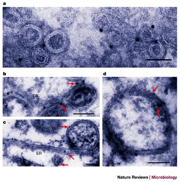
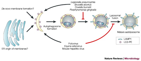
Intracellular bacteria and viruses must survive the vigorous antimicrobial responses of their hosts to replicate successfully. The cellular process of autophagy — in which compartments bound by double membranes engulf portions of the cytosol and then mature to degrade their cytoplasmic contents — is likely to be one such host-cell response. Several lines of evidence show that both bacteria and viruses are vulnerable to autophagic destruction and that successful pathogens have evolved strategies to avoid autophagy, or to actively subvert its components, to promote their own replication. The molecular mechanisms of the avoidance and subversion of autophagy by microorganisms will be the subject of much future research, not only to study their roles in the replication of these microorganisms, but also because they will provide — as bacteria and viruses so often have — unique tools to study the cellular process itself.


Conflict of interest statement

The authors declare no competing financial interests.

Figures

Figure 1
Figure 1. Immunoelectron microscopy of GFP–Atg5-expressing human cells undergoing autophagy.
Atg5−/− mouse embryonic stem cells were stably transfected with yeast Atg5 tagged with green fluorescent protein (GFP–Atg5) and cultured in Hanks' solution for 2 h to induce autophagy. The localization of GFP–Atg5 was examined by silver-enhanced immuno-gold electron microscopy using an anti-GFP antibody. ad | A series of images showing the presumed progression of membrane extension and cytosolic sequestration during autophagosome formation. e | An example of an autophagosome. f | An example of an autolysosome. Scale bar, 1 μm. Reproduced with permission from Ref. © (2001) Rockerfeller University Press.
Figure 2
Figure 2. The autophagic pathway.
The known steps of induction, execution and maturation of autophagosomes and autolysosomes. Green lines and arrows indicate activation or inhibition events, respectively, that induce autophagy; red lines indicate events that inhibit autophagy. The markers that are present at each morphological step are indicated in the key, as are several known inhibitors and the steps at which they are thought to act (red boxes). Caution must be used in interpreting the results obtained using all of these inhibitors, due to their pleiotropic effects. 3-MA, 3-methyladenine; DAMP, N-(3-[2,4-dinitrophenyl]-amino) propyl-l-N-(3-aminopropyl-methylamine) dihydrochloride); LAMP, lysosome-associated membrane protein; LC3, microtubule-associated-protein light-chain 3; MDC, monodansylcadaverine; PE, phosphatidylethanolamine; PI3K, phosphatidylinositol-3-kinase; TOR, target of rapamycin.
Figure 3
Figure 3. Autophagic sequestration of bacteria.
a | Electron micrograph of Rickettsia conorii infecting mouse endothelial cells 36 h after inoculation and addition of cytokines. The arrow indicates a rickettsial cell that is contained within a double-membrane-bound compartment. Arrowheads indicate intact R. conorii in the cytosol. b | A situation in which bacterial infection stimulates autophagosome function (red arrow) and the bacteria avoid a phagosomal fate, as has been proposed for Porphyromonas and Brucella infection. c | A situation in which bacterial infection delays autophagosome formation (red blunt-ended arrow), as has been proposed for Legionella and Rickettsia infection. a reproduced with permission from Ref. © (1997) Nature Publishing Group.
Figure 4
Figure 4. Formation of double-membrane-bound vesicles during poliovirus infection.
| a | Electron micrograph of a chemically fixed, poliovirus-infected HeLa cell 7 h post-infection. 'B' indicates the apparently cytosolic lumens of double-membrane-bound vesicular bodies. 'Va' indicates a vacuole that does not seem to be multilamellar. 'M' indicates a mitochondrion. Poliovirus particles, either single or in groups, can be seen in the cytoplasm and, occasionally, within the lumen of the double-membrane-bound vesicles (arrows). The inset shows the region outlined by dashed white lines in the main image at a higher magnification. Scale bar in main image, 0.6 μm; scale bar in inset, 0.25 μm. b | Electron micrograph of poliovirus-infected HeLa cells 2.5 h post-infection preserved by high-pressure cryopreservation and freeze substitution. Arrows indicate double-membrane-bound structures with apparently cytosolic lumens that are similar to those seen at later times after infection. Membranes that have distributions and morphologies that are characteristic of the endoplasmic reticulum are indicated (ER). 'N' indicates the nucleus. Scale bar, 0.2 μm. c | Immunolocalization of the late endosomal/lysosomal protein LAMP1 in poliovirus-infected HeLa cells 4.5 h post-infection. Arrows indicate selected gold particles coupled to a secondary antibody. 'L' indicates a lysosome. Scale bar, 0.2 μm. a reproduced with permission from Ref. © (1965) Elsevier Science; b and c reproduced with permission from Ref. © (1996) ASM.
Figure 5
Figure 5. Formation of double-membrane-bound vesicles during EAV infection and on expression of EAV proteins nsp2 and nsp3.
a | RK-13 cells were infected with equine arterivirus (EAV) for 8 h and cryosections were labelled with anti-nsp2 followed by protein-A–gold detection. Scale bar, 0.1 μm. bd | Cryoimmunoelecron microscopy of RK-13 cells in which EAV proteins nsp2 and nsp3 were expressed in precursor forms using a Sindbis virus vector. Gold labelling detects nsp2 (a,c) and nsp3 (d); arrows indicate selected gold particles. Cells were harvested 8–12 h after infection. Scale bars, 0.1 μm. ER, endoplasmic reticulum. a Reproduced with permission from Ref. © (1999) ASM. bd Reproduced with permission from Ref. © (2001) Society for General Microbiology.
Figure 6
Figure 6. Potential subversion of the autophagic pathway or its components by bacteria and viruses.
Proposed stages at which intracellular bacteria and viruses might induce or interfere with autophagosome development. The viruses and bacteria listed induce the formation of double-membrane-bound compartments that bear markers from the autophagic pathway. The persistence of the double-membrane-bound morphology of these structures indicates that, if they are similar to autophagosomes, their maturation into autolysosomes is arrested. In Legionella, membranes show delayed acquisition of lysosome-associated membrane protein 1 (LAMP1), whereas the poliovirus-induced membranes contain LAMP1; therefore, Legionella and poliovirus are proposed to block autophagic maturation at different steps. ER, endoplasmic reticulum; LC3, microtubule-associated-protein light-chain 3; PE, phosphatidylethanolamine.

References

    1. Stromhaug PE, Klionsky DJ. Approaching the molecular mechanism of autophagy. Traffic. 2001;2:524–531. doi: 10.1034/j.1600-0854.2001.20802.x. - DOI - PubMed
    1. Klionsky DJ, et al. A unified nomenclature for yeast autophagy-related genes. Dev. Cell. 2003;5:539–545. doi: 10.1016/S1534-5807(03)00296-X. - DOI - PubMed
    1. Liang XH, et al. Induction of autophagy and inhibition of tumorigenesis by beclin 1. Nature. 1999;402:672–676. doi: 10.1038/45257. - DOI - PubMed
    1. Melendez A, et al. Autophagy genes are essential for dauer development and life-span extension in C. elegans. Science. 2003;301:1387–1391. doi: 10.1126/science.1087782. - DOI - PubMed
    1. Otto GP, Wu MY, Kazgan N, Anderson OR, Kessin RH. Macroautophagy is required for multicellular development of the social amoeba Dictyostelium discoideum. J. Biol. Chem. 2003;278:17636–17645. doi: 10.1074/jbc.M212467200. - DOI - PubMed

Publication types