

Histological organization and its relationship to function in the femur of Alligator mississippiensis
- PMID: 15032909
- PMCID: PMC1571257
- DOI: 10.1111/j.0021-8782.2004.00275.x
Histological organization and its relationship to function in the femur of Alligator mississippiensis
Abstract
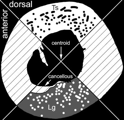
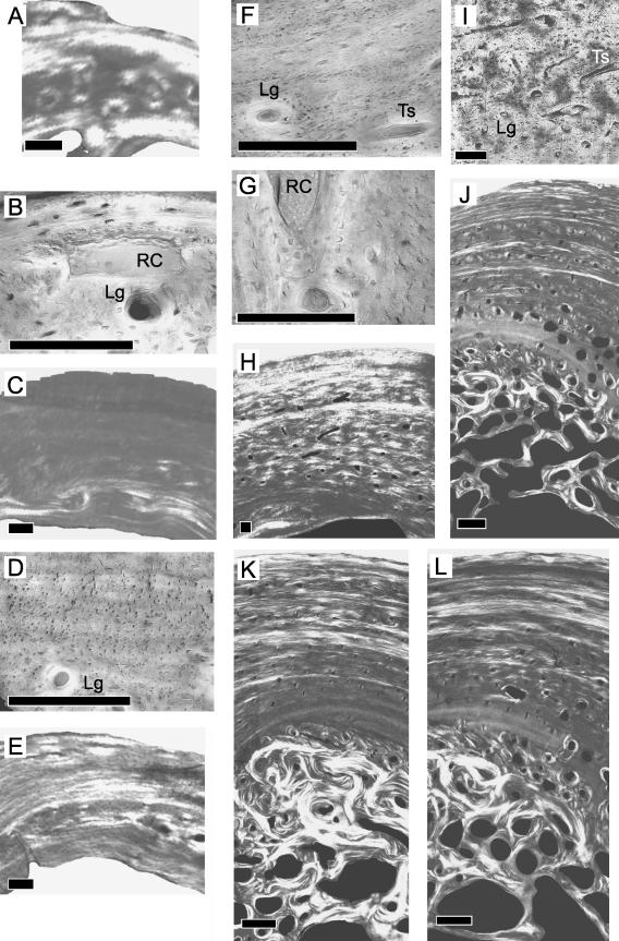
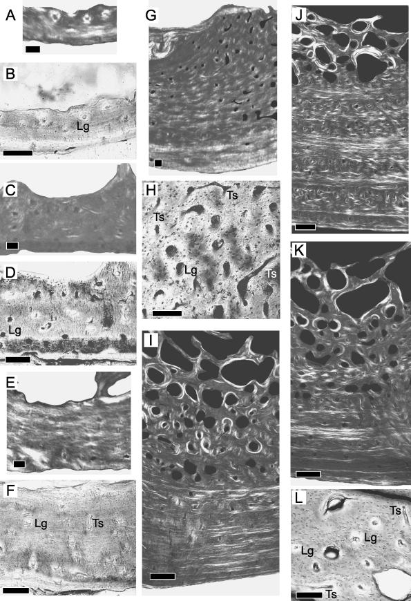
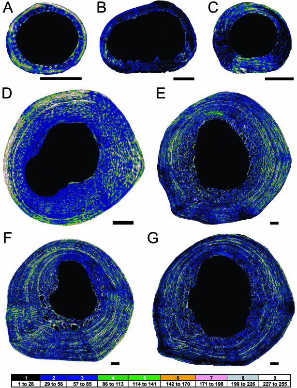
Histological analysis of a growth series of alligator femora tests the correlation between strain milieu and microstructure. From mid-diaphyseal cross-sections of these femora (n = 7), vascular canal orientation and density as well as collagen fibre organization were recorded. Throughout ontogeny, the proportion of transverse-spiral (TS) collagen in the dorsal cortex is significantly greater than it is in the ventral cortex (P = 0.008). This regional difference in the proportion of TS collagen is correlated with a regional difference in the state of peak principal strain (compressive or tensile). Nevertheless, the predominant orientation of collagen fibres is longitudinal, which is inconsistent with biomechanical hypotheses that involve peak principal or shear strains. Although the density and orientation of vascular canals do not show significant regional differences (P = 0.26 and P = 0.26, respectively), as with collagen orientation, the vascular canal orientation is predominantly longitudinal. The longitudinal organization of both the vascular canals and the collagen fibres is probably a consequence of longitudinal shifting of subperiosteal osteoid during femoral lengthening. When taken together, these data suggest that growth dynamics is the dominant influence on the histological organization of primary bony tissues in alligator femora.
Figures
Similar articles
-
Contrast-enhanced XROMM reveals in vivo soft tissue interactions in the hip of Alligator mississippiensis.J Anat. 2020 Feb;236(2):288-304. doi: 10.1111/joa.13101. Epub 2019 Nov 6. J Anat. 2020. PMID: 31691966 Free PMC article.
-
Structural design and mechanical behavior of alligator (Alligator mississippiensis) osteoderms.Acta Biomater. 2013 Nov;9(11):9049-64. doi: 10.1016/j.actbio.2013.07.016. Epub 2013 Jul 24. Acta Biomater. 2013. PMID: 23891812
-
Mechanics of limb bone loading during terrestrial locomotion in the green iguana (Iguana iguana) and American alligator (Alligator mississippiensis).J Exp Biol. 2001 Mar;204(Pt 6):1099-122. doi: 10.1242/jeb.204.6.1099. J Exp Biol. 2001. PMID: 11222128
-
The consequences of calcium: investigating intracortical reproductive signals in the American alligator for sex determination.Anat Rec (Hoboken). 2025 Feb;308(2):629-635. doi: 10.1002/ar.25533. Epub 2024 Jul 3. Anat Rec (Hoboken). 2025. PMID: 38958219
-
Motor control of locomotor hindlimb posture in the American alligator (Alligator mississippiensis).J Exp Biol. 2003 Dec;206(Pt 23):4327-40. doi: 10.1242/jeb.00688. J Exp Biol. 2003. PMID: 14581602
Cited by
-
Vertebrae-Based Body Length Estimation in Crocodylians and Its Implication for Sexual Maturity and the Maximum Sizes.Integr Org Biol. 2020 Nov 24;2(1):obaa042. doi: 10.1093/iob/obaa042. eCollection 2020. Integr Org Biol. 2020. PMID: 33791579 Free PMC article.
-
Ontogenetic relationships between in vivo strain environment, bone histomorphometry and growth in the goat radius.J Anat. 2007 Mar;210(3):272-93. doi: 10.1111/j.1469-7580.2007.00696.x. J Anat. 2007. PMID: 17331177 Free PMC article.
-
Bone histology in Dysalotosaurus lettowvorbecki (Ornithischia: Iguanodontia)--variation, growth, and implications.PLoS One. 2012;7(1):e29958. doi: 10.1371/journal.pone.0029958. Epub 2012 Jan 6. PLoS One. 2012. PMID: 22238683 Free PMC article.
-
Osteohistological insight into the growth dynamics of early dinosaurs and their contemporaries.PLoS One. 2024 Apr 3;19(4):e0298242. doi: 10.1371/journal.pone.0298242. eCollection 2024. PLoS One. 2024. PMID: 38568908 Free PMC article.
-
The effect of growth rate on the three-dimensional orientation of vascular canals in the cortical bone of broiler chickens.J Anat. 2018 Oct;233(4):531-541. doi: 10.1111/joa.12847. Epub 2018 Jul 18. J Anat. 2018. PMID: 30022496 Free PMC article.
References
-
- Bassett CAL. Electrical effects in bone. Sci. Am. 1965;213:18–25. 10.1111/j.0021-8782.2004.00275.x. - DOI - PubMed
-
- Blob RW, Biewener AA. In vivo locomotor strain in the hindlimb bones of Alligator mississippiensis and Iguana iguana: implications for the evolution of limb bone safety factor and non-sprawling limb posture. J. Exp. Biol. 1999;202:1023–1046. 10.1111/j.0021-8782.2004.00275.x. - DOI - PubMed
-
- Boyde A, Riggs CM. The quantitative study of the orientation of collagen in compact bone slices. Bone. 1990;11:35–40. 10.1111/j.0021-8782.2004.00275.x. - DOI - PubMed
-
- Carando S, Portigliatti-Barbos M, Ascenzi A, Riggs CM, Boyde A. Macroscopic shape of, and lamellar distribution within, the upper limb shafts, allowing inferences about mechanical properties. Bone. 1991;12:265–270. 10.1111/j.0021-8782.2004.00275.x. - DOI - PubMed
-
- Carter DR, Orr TE, Fyhrie DP, Schurman DJ. Influences of mechanical stress on prenatal and postnatal skeletal development. Clin. Orthop. 1987;219:237–250. 10.1111/j.0021-8782.2004.00275.x. - DOI - PubMed
Publication types
MeSH terms
Substances
LinkOut - more resources
Full Text Sources
