. 2004 Apr 1;172(7):4520-6.
doi: 10.4049/jimmunol.172.7.4520.

Aerobic exercise attenuates airway inflammatory responses in a mouse model of atopic asthma

Affiliations

Aerobic exercise attenuates airway inflammatory responses in a mouse model of atopic asthma

Amy Pastva et al. J Immunol. .

Abstract

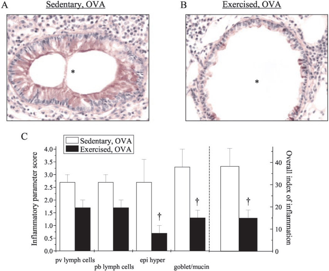
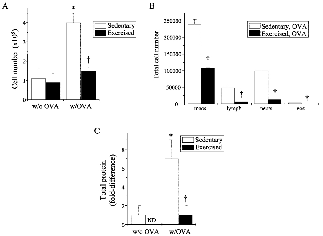
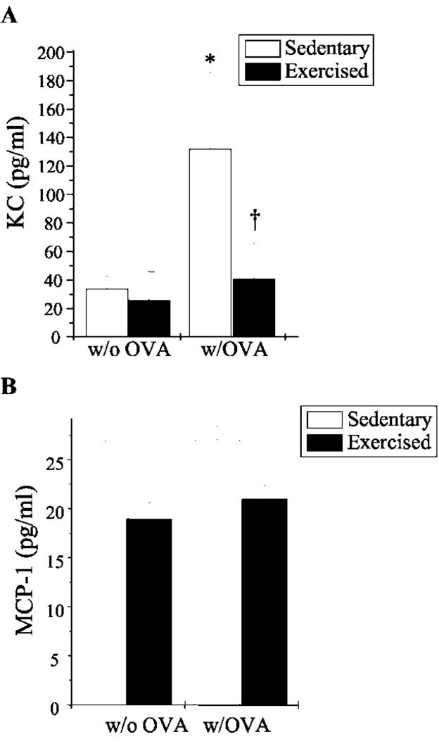
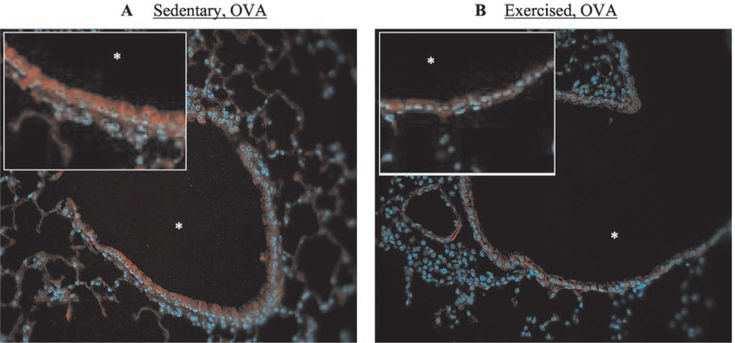
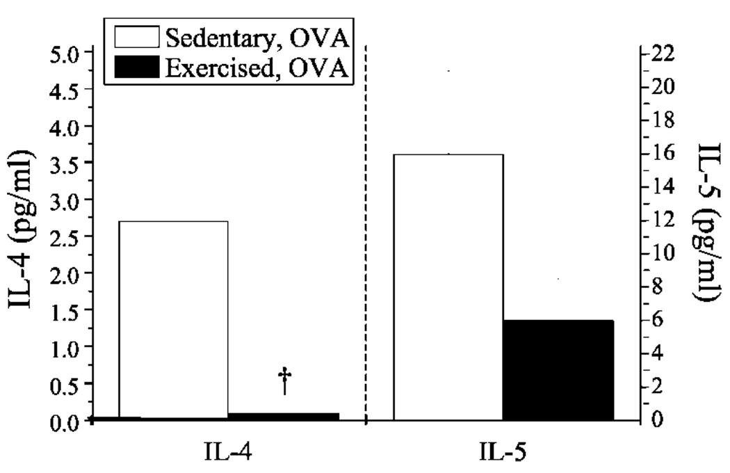
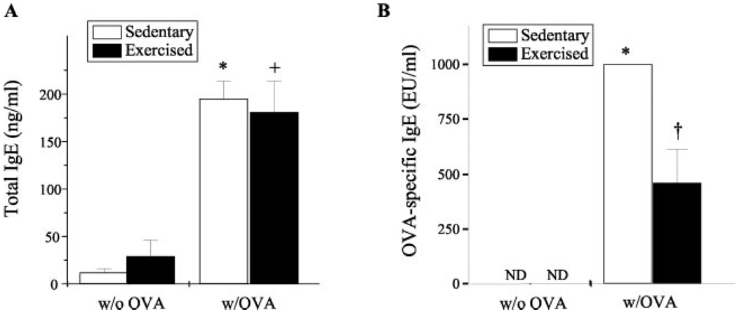
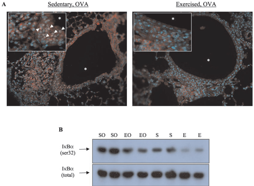
Recent reports indicate that aerobic exercise improves the overall physical fitness and health of asthmatic patients. The specific exercise-induced improvements in the pathology of asthma and the mechanisms by which these improvements occur, however, are ill-defined; thus, the therapeutic potential of exercise in the treatment of asthma remains unappreciated. Using an OVA-driven mouse model, we examined the role of aerobic exercise in modulating inflammatory responses associated with atopic asthma. Data demonstrate that moderate intensity aerobic exercise training decreased leukocyte infiltration, cytokine production, adhesion molecule expression, and structural remodeling within the lungs of OVA-sensitized mice (n = 6-10; p < 0.05). Because the transcription factor NF-kappaB regulates the expression of a variety of genes that encode inflammatory mediators, we monitored changes in NF-kappaB activation in the lungs of exercised/sensitized mice. Results show that exercise decreased NF-kappaB nuclear translocation and IkappaBalpha phosphorylation, indicating that exercise decreased NF-kappaB activation in the lungs of sensitized mice (n = 6). Taken together, these results suggest that aerobic exercise attenuates airway inflammation in a mouse model of atopic asthma via modulation of NF-kappaB activation. Potential exists, therefore, for the amelioration of asthma-associated chronic airway inflammation through the use of aerobic exercise training as a non-drug therapeutic modality.


Figures

FIGURE 1
FIGURE 1
Exercise lessened sensitization-induced changes in leukocyte infiltration and lung architecture. A and B, Mice were sensitized/exercised and lungs were prepared for analysis of leukocyte infiltrate, epithelial hypertrophy, and mucus production as described in Materials and Methods. Representative results of five to six separate experiments are shown (*, airway lumen; magnification, ×40). C, Tissue sections were coded randomly and scored subjectively for the inflammatory parameters of: 1) perivascular (pv) and peribronchial (pb) lymphoid accumulation; 2) hypertrophy/hyperplasia of the mucosal epithelium (epi hyper); 3) goblet cell and mucin production; and 4) overall index of inflammation. Grading scale: 0, none; 1, <25% 2, 25–50% 3, 50–75%; 4, >75%. Scores are represented as average scores per group (n = 5–6/group; †, p < 0.05 as compared with SO). Each average score per category was multiplied by the extent of appearance in the lungs (also rated 0–4) to devise an overall index of inflammation. Scores from nonsensitized groups totaled zero (data not shown).
FIGURE 2
FIGURE 2
Exercise decreased cellular infiltrate and total lavage protein in sensitized mice. A, Cells collected from BALF of mice were analyzed for differences in total cell count as described in Materials and Methods. Results are reported as cell number (×105) (n = 5–6 for each group; *, p < 0.05 as compared with S; †, p < 0.05 as compared with SO). B, Differential BALF cell counts were performed using standard hematological criteria. Results are reported as percentage of total cell number (n = 3–4 for each group; †, p < 0.05 as compared with SO). C, BALF was monitored for changes in total protein concentrations via detergent-compatible assay. Results are reported as fold differences in total protein, as compared with nonsensitized mice (n = 3–5; *, p < 0.05 as compared with S; †, p < 0.05 as compared with SO; ND, none detected).
FIGURE 3
FIGURE 3
Exercise decreased KC- and MCP-1-secreted protein levels in the lungs of OVA-sensitized mice. BALF was analyzed for KC (A)- and MCP-1-secreted protein levels (B) via ELISA. Results are reported as picograms per milliliter of respective protein (n = 5–6; *, p < 0.05 as compared with S; †, p < 0.05 as compared with SO).
FIGURE 4
FIGURE 4
Exercise decreased VCAM-1 surface expression in the lungs of OVA-sensitized mice. Formalin-fixed, paraffin-embedded sections from SO (A) and EO (B) mice were analyzed for VCAM-1 expression as described in Materials and Methods. Tissue was counterstained with Hoescht to detect nuclei (blue). Representative results from three independent experiments are shown (*, airway lumen; magnification, ×25; inset, ×50).
FIGURE 5
FIGURE 5
Exercise decreased IL-4-and IL-5-secreted protein levels in the lungs of OVA-sensitized mice. BALF samples were analyzed for differences in secreted IL-4 and IL-5 protein via ELISA. Results are reported as picograms per milliliter of respective protein (n = 5–6; †, p < 0.05 as compared with SO; IL-5 results approached significance with p = 0.06).
FIGURE 6
FIGURE 6
Exercise decreased OVA-specific IgE levels but not total IgE levels in the serum of OVA-sensitized mice. Serum samples were monitored for changes in total IgE (A) and OVA-specific IgE (B) levels via ELISA as described in Materials and Methods. Results for total IgE levels are reported as nanograms per milliliter protein; results for OVA-specific IgE totals are reported as EU/ml, with OVA-induced IgE arbitrarily assigned 1000 EU/ml (n = 6–9; *, p < 0.05 as compared with S; +, p < 0.05 as compared with E; †, p < 0.05 as compared with SO; ND, none detected).
FIGURE 7
FIGURE 7
Exercise decreased NF-κB nuclear translocation and IκBα in OVA-sensitized mice. A, Translocation of the NF-κB subunit p65 (denoted by arrows) was monitored in SO (left) and EO (right) mice via immunohistochemical analysis as described in Materials and Methods. Representative photomicrographs of six separate experiments are shown (*, airway lumen; magnification, ×25; inset, ×50). B, Lung IκBα phosphorylation was analyzed via immunoblotting as described in Materials and Methods. Representative results from six independent experiments are shown.

References

    1. O’Byrne PM. Airway inflammation and asthma. Aliment. Pharmacol. Ther. 1996;10:18. - PubMed
    1. Christman JW, Sadikot RT, Blackwell TR. The role of nuclear factor-κB in pulmonary diseases. Chest. 2000;117:1482. - PubMed
    1. Mannino DM, Homa DM, Pertowski CA, Ashizawa A, Nixon LL, Johnson CA, Ball LB, Jack E, Kang DS. Surveillance for asthma—United States, 1960–1995. MMWR. 1998;47:1. - PubMed
    1. Pedersen BK, Hoffman-Goetz L. Exercise and the immune system: regulation, integration, and adaptation. Physiol. Rev. 2000;80:1055. - PubMed
    1. Woods JA, Lu Q, Ceddia MA, Lowder T. Exercise-induced modulation of macrophage function. Immunol. Cell Biol. 2000;78:545. - PubMed

MeSH terms