. 2004 May 15;557(Pt 1):133-46.
doi: 10.1113/jphysiol.2003.059014. Epub 2004 Mar 19.

Excitability of the T-tubular system in rat skeletal muscle: roles of K+ and Na+ gradients and Na+-K+ pump activity

Affiliations

Excitability of the T-tubular system in rat skeletal muscle: roles of K+ and Na+ gradients and Na+-K+ pump activity

O B Nielsen et al. J Physiol. .

Abstract

Strenuous exercise causes an increase in extracellular [K(+)] and intracellular Na(+) ([Na(+)](i)) of working muscles, which may reduce sarcolemma excitability. The excitability of the sarcolemma is, however, to some extent protected by a concomitant increase in the activity of muscle Na(+)-K(+) pumps. The exercise-induced build-up of extracellular K(+) is most likely larger in the T-tubules than in the interstitium but the significance of the cation shifts and Na(+)-K(+) pump for the excitability of the T-tubular membrane and the voltage sensors is largely unknown. Using mechanically skinned fibres, we here study the role of the Na(+)-K(+) pump in maintaining T-tubular function in fibres with reduced chemical K(+) gradient. The Na(+)-K(+) pump activity was manipulated by changing [Na(+)](i). The responsiveness of the T-tubules was evaluated from the excitation-induced force production of the fibres. Compared to control twitch force in fibres with a close to normal intracellular [K(+)] ([K(+)](i)), a reduction in [K(+)](i) to below 60 mM significantly reduced twitch force. Between 10 and 50 mM Na(+), the reduction in force depended on [Na(+)](i), the twitch force at 40 mM K(+) being 22 +/- 4 and 54 +/- 9% (of control force) at a [Na(+)](i) of 10 and 20 mM, respectively (n= 4). Double pulse stimulation of fibres at low [K(+)](i) showed that although elevated [Na(+)](i) increased the responsiveness to single action potentials, it reduced the capacity of the T-tubules to respond to high frequency stimulation. It is concluded that a reduction in the chemical gradient for K(+), as takes place during intensive exercise, may depress T-tubular function, but that a concomitant exercise-induced increase in [Na(+)](i) protects T-tubular function by stimulating the Na(+)-K(+) pump.


Figures

Figure 1
Figure 1. Effect of different intracellular solutions on the force—pCa relationship of a rat fibre
The relationship between force and [Ca2+] in a skinned fibre from rat EDL muscle was ascertained in various intracellular solutions which differed in [Na+], [K+] and [NH4+]. In a given solution, the fibre was exposed to progressively higher free [Ca2+] (i.e. pCa from 9 to 4.5) and corresponding forces were fitted by a Hill curve. A solution set with 127 mm K+ and 36 mm Na+ was examined both before (▪) and after (□) other sets containing (mm) 10 Na+, 40 K+, 113 NH4+ (▾) and 50 Na+, 40 K+, 73 NH4+ (○). The force—pCa relationships in the different conditions were virtually indistinguishable.
Figure 2
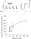
Figure 2. Effect of intracellular [K+] on excitation-induced contractions in skinned muscle fibres
A, traces of force recordings from a fibre segment bathed in control intracellular solution (113 mm K+ and 30 mm Na+) or in a solution with 40 mm K+ and 20 mm Na+, as indicated. Times show minutes after start of the experiment. Traces a: twitch stimulation. Trace b: force elicited by release of SR Ca2+ when exposing the fibre to the low Mg2+-solution (see Methods). Trace c: maximal Ca2+-activated force induced by exposure to the Ca-EGTA solution at pCa 4.5. B, twitch force when the bathing solution contained 20–113 mm K+ and either 20 (▵, ▴) or 50 (○, •) mM Na+ expressed relative to the average of the force response in the bracketing determinations of twitch force in control solution (113 mm K+ and 30 mm Na+). Open symbols show the mean (±s.e.m.) force response at the indicated combination of K+ and Na+ for cases where 3 or more fibres were examined (3 and 4 fibres at 80 and 113 mm K+, respectively, and 5–8 fibres in other cases). Filled symbols show individual values in cases where only two fibres were examined.
Figure 6
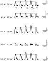
Figure 6. Contractions in a skinned muscle fibre in response to single and double pulse stimulation
In each intracellular solution, the force response was tested using single and double pulses with a pulse configuration of single (s) - single (s) - double (d8) - double (d20) - single (s). The interpulse interval in the two double pulses was 8 (d8) and 20 ms (d20). Traces show force responses during consecutive exposure to control and test solutions with the indicated concentrations of Na+ and K+. Other details as in Fig. 3. Note the expanded force scale for contractions in test solutions.
Figure 3
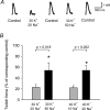
Figure 3. Significance of the intracellular [Na+] for twitch force in skinned fibres exposed to low intracellular [K+]
For determinations of control force, the fibre segment was bathed for 2 min in control solution (113 mm K+, 30 mm Na+). Force responses in solutions with low [K+] (30 or 40 mm K+) were determined after 30 s exposure to the solution. Determinations of force in test conditions were bracketed by determinations of control force. A, recordings of twitch force from an experiment in which the fibre was exposed to intracellular solutions with 30 mm K+ combined with 20 or 50 mm Na+, as indicated. B, effect of varying the Na+ concentration on the maximal twitch force in fibres exposed to 30 mm or 40 mm K+. Each fibre was only exposed to one of the two test K+ concentrations, and the tests of force at the two Na+ concentrations were done in a semi-random order. For each test condition, force is expressed relative to the average of the two bracketing control contractions. Columns show mean +s.e.m. from experiments on 4 and 5 fibres.
Figure 4
Figure 4. Effect of removal of intracellular Na+ on the excitation-induced twitch force
Skinned fibre segments were incubated in control solution (113 mm K+, 30 mm Na+) and control twitch was determined. At time zero, the fibres were exposed to a solution having the same constituents as the control solution but with all Na+ replaced by NH4+. The fibres were stimulated at the indicated time points using single pulses. Force responses are expressed relative to the initial control force. Curves show individual data from 3 fibres.
Figure 5
Figure 5. Effect of interpulse interval on the force of fused contractions elicited by electrical stimulation with double pulses in a skinned muscle fibre
The fibre segment was bathed in control solution (113 mm K+ and 30 mm Na+) and contractions were stimulated with single pulses or with double pulses with interpulse intervals from 4 to 20 ms. Contractions were evoked every 10 s and the interpulse interval was progressively increased from 4 to 20 ms and then decreased again, with the force response at a given interpulse interval being similar on both occasions. Maximal force elicited is expressed relative to the twitch force elicited by single pulse stimulation. The figure shows data from one fibre; very similar results were obtained in two other fibres.
Figure 7
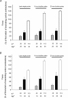
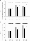
Figure 7. Effect of changes in intracellular [K+] and [Na+] on the force response to double pulse stimulation
Experimental conditions as in Fig. 6. For each condition the force responses were tested using both single and double pulses with a pulse configuration of one or two single pulses followed by two double pulses and a single pulse. The interpulse interval in the two double pulses was 8 and 20 ms. The figure shows means +s.e.m. for the two double pulses and the last single pulse in the pulse configuration. The forces are expressed relative to the force elicited by the first single pulse in the bracketing determinations of control contractions. A, intracellular solutions with 30 mm K+ and 20 or 50 mm Na+, n = 5. B, solutions with 40 mm K+ and 10 or 20 mm Na+, n = 4.
Figure 8
Figure 8. Effect of interpulse interval on force production at different combinations of intracellular [K+] and [Na+]
Data from Fig. 7. For each condition the force responses were tested using both single and double pulses with a set pulse configuration of one or two single pulses followed by two double pulses and a single pulse. The interpulse interval in the two double pulses was 8 and 20 ms. The figure shows means +s.e.m. for the two double pulses and the last single pulse in the pulse configuration. The forces are expressed relative to the force elicited by the last single pulse in the set pulse configuration. A, intracellular solutions with 30 mm K+ and 20 or 50 mm Na+, n = 5. B, solutions with 40 mm K+ and 10 or 20 mm Na+, n = 4.

References

    1. Cairns SP, Buler SJ, Loiselle DS, Renaud JM. Changes of action potentials and force at lowered [Na+]o in mouse skeletal muscle: implications for fatigue. Am J Physiol. 2003;285:C1131–C1141. - PubMed
    1. Cairns SP, Flatman JA, Clausen T. Relation between extracellular [K+], membrane potential and contraction in rat soleus muscle: modulation by the Na+–K+ pump. Pflugers Arch. 1995;430:909–915. - PubMed
    1. Cairns SP, Hing WA, Slack JR, Mills RG, Loiselle DS. Different effects of raised [K+]o on membrane potential and contraction in mouse fast- and slow-twitch muscle. Am J Physiol. 1997;273:C598–C611. - PubMed
    1. Caputo C, Bezanilla F, Horowicz P. Depolarization-contraction coupling in short frog muscle fibres. J General Physiol. 1984;84:133–154. - PMC - PubMed
    1. Chua M, Dulhunty AF. Inactivation of excitation-contraction coupling in rat extensor digitorum longus and soleus muscles. J General Physiol. 1988;91:737–757. - PMC - PubMed

Publication types