. 2004 May 15;557(Pt 1):147-57.
doi: 10.1113/jphysiol.2004.062661. Epub 2004 Mar 19.

BK potassium channels control transmitter release at CA3-CA3 synapses in the rat hippocampus

Affiliations

BK potassium channels control transmitter release at CA3-CA3 synapses in the rat hippocampus

Giacomo Raffaelli et al. J Physiol. .

Abstract

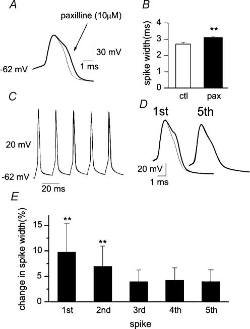
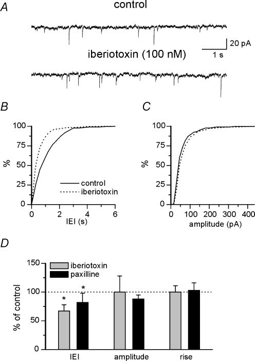
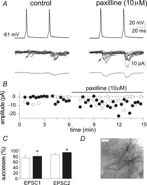
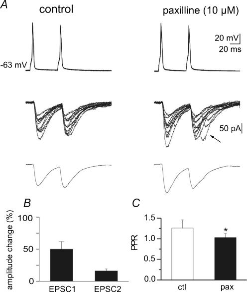
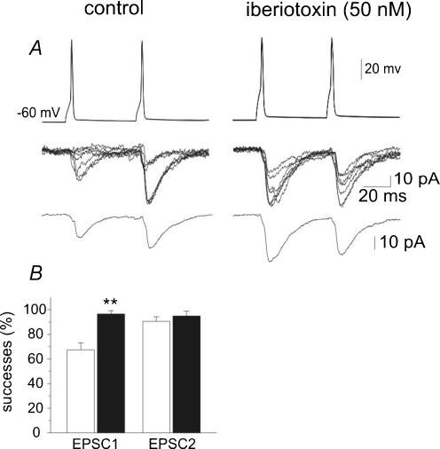
Large conductance calcium- and voltage-activated potassium channels (BK channels) activate in response to calcium influx during action potentials and contribute to the spike repolarization and fast afterhyperpolarization. BK channels targeted to active zones in presynaptic nerve terminals have been shown to limit calcium entry and transmitter release by reducing the duration of the presynaptic spike at neurosecretory nerve terminals and at the frog neuromuscular junction. However, their functional role in central synapses is still uncertain. In the hippocampus, BK channels have been proposed to act as an 'emergency brake' that would control transmitter release only under conditions of excessive depolarization and accumulation of intracellular calcium. Here we demonstrate that in the CA3 region of hippocampal slice cultures, under basal experimental conditions, the selective BK channel blockers paxilline (10 microM) and iberiotoxin (100 nM) increase the frequency, but not the amplitude, of spontaneously occurring action potential-dependent EPSCs. These drugs did not affect miniature currents recorded in the presence of tetrodotoxin, suggesting that their action was dependent on action potential firing. Moreover, in double patch-clamp recordings from monosynaptically interconnected CA3 pyramidal neurones, blockade of BK channels enhanced the probability of transmitter release, as revealed by the increase in success rate, EPSC amplitude and the concomitant decrease in paired-pulse ratio in response to pairs of presynaptic action potentials delivered at a frequency of 0.05 Hz. BK channel blockers also enhanced the appearance of delayed responses, particularly following the second action potential in the paired-pulse protocol. These results are consistent with the hypothesis that BK channels are powerful modulators of transmitter release and synaptic efficacy in central neurones.


Figures

Figure 1
Figure 1. BK channels are involved in action potential repolarization
A, action potentials generated under control conditions (thin line) and in the presence of paxilline (thick line) are aligned at threshold and superimposed. B, mean changes in spike width before (open column) and during paxilline application (filled column; n = 10). C, two bursts of five consecutive action potentials generated by brief depolarizing current pulses (5 ms duration, each delivered at 50 Hz) under control conditions and in the presence of paxilline (10 μm) are superimposed. D, the first and the fifth spikes in the train before (thin line) and during paxilline (thick line) are superimposed. E, mean changes in spike duration (as percentage of controls) obtained in the presence of paxilline (10 μm) during repetitive firing (n = 3). Note that paxilline clearly broadened only the first two action potentials. P < 0.001.
Figure 2
Figure 2. BK channels increase the frequency but not the amplitude of spontaneous action potential-dependent EPSCs
A, traces showing spontaneous EPSCs recorded from a CA3 pyramidal neurone at the holding potential of −60 mV under control conditions and in the presence of iberiotoxin (100 nm). B and C, cumulative interevent-interval (B) and amplitude distribution (C) of spontaneous EPSCs (same neurone shown in A) under control conditions (continuous line) and during iberiotoxin application (dotted line). Bin size was 0.3 s in B and 15 pA in C. D, mean changes of interevent interval (IEI), amplitude and rise time, compared to control (dotted line) during application of iberiotoxin (n = 7) or paxilline (n = 7). P <0.05.
Figure 3
Figure 3. Blocking BK channels with paxilline increases synaptic efficacy at low probability CA3−CA3 connections
A, pairs of action potentials are generated (50 ms intervals, 0.05 Hz) in the presynaptic cell (upper traces) while EPSCs are recorded from the postsynaptic cell in control (left) and in the presence of paxilline (10 μm, right). Eight traces are superimposed and shown in the middle, while the average of all responses (successes plus failures) is shown at the bottom. Note reduced failure rate and increased amplitude of successes after paxilline. B, time course of the peak amplitude of the first (○) and second (•) EPSCs recorded from the cell shown in A. C, mean success rate of EPSC1 and EPSC2 obtained in 7 cells under control conditions (open columns) and during paxilline application (filled columns). P <0.05. D, a pair of interconnected cells labelled with biocytin (the bar is 50 μm).
Figure 4
Figure 4. In high probability CA3−CA3 connections paxilline increases the amplitude of evoked EPSCs and decreases the PPR
A, the upper traces represent pairs of 10 superimposed action potentials generated at 0.05 Hz in the presynaptic cell. Pairs of 10 EPSCs recorded from the postsynaptic cell are superimposed in the middle. Average EPSCs (in this particular neurone no failures were detected) are recorded at the bottom. Note that paxilline (right, 10 μm) increased EPSC amplitude and induced the appearance of delayed responses. B, summary data from 5 cells showing mean amplitude of EPSC1 and EPSC2 in paxilline, normalized to control values. C, mean PPR obtained in 5 neurones under control conditions (open column) and during application of paxilline (filled column). P < 0.05
Figure 5
Figure 5. Blocking BK channels with iberiotoxin increases synaptic efficacy at CA3−CA3 connections
Pairs of action potentials are generated in the presynaptic cell (upper traces) while EPSCs are recorded from the postsynaptic cell in control (left) and in the presence of iberiotoxin (50 nm, right). Seven superimposed traces are shown in the middle while average responses are shown at the bottom (average of 15 responses including failures). Note changes in PPR after iberiotoxin. B, mean success rate of EPSC1 and EPSC2 obtained in 4 cells under control conditions (open columns) and during paxilline application (filled columns). P <0.01.
Figure 6
Figure 6. At high probability synapses, iberiotoxin increases the amplitude of EPSC1 and EPSC2
A, pairs of seven EPSCs recorded from the postsynaptic cell in control (left) and in the presence of iberiotoxin (right) are superimposed in the upper traces. Average EPSCs are represented in the lower traces. B, time course of the peak amplitude of the first (○) and second (•) EPSCs recorded from the cell shown in A. C, summary data from 6 cells showing mean amplitude of EPSC1 and EPSC2 in control (open column) and in the presence of iberiotoxin (filled column). D, mean PPR obtained in 6 neurones under control conditions (open column) and during application of iberiotoxin (filled column). P < 0.05; P < 0.005.

References

    1. Adams PR, Constanti A, Brown DA, Clark RB. Intracellular Ca2+ activates a fast voltage-sensitive K+ current in vertebrate sympathetic neurones. Nature. 1982;296:746–749. - PubMed
    1. Blundon JA, Wright SN, Brodwick MS, Bittner GD. Presynaptic calcium-activated potassium channels and calcium channels at a crayfish neuromuscular junction. J Neurophysiol. 1995;73:178–189. - PubMed
    1. Candia S, Garcia ML, Latorre R. Mode of action of iberiotoxin, a potent blocker of the large conductance Ca2+-activated K+ channel. Biophys J. 1992;63:583–590. - PMC - PubMed
    1. Clements JD, Bekkers JM. Detection of spontaneous synaptic events with an optimally scaled template. Biophys J. 1997;73:220–229. - PMC - PubMed
    1. De Simoni A, Griesinger CB, Edwards FA. Development of rat CA1 neurones in acute versus organotypic slices: role of experience in synaptic morphology and activity. J Physiol. 2003;550:135–147. - PMC - PubMed

Publication types

MeSH terms

LinkOut - more resources