. 2004 Jun;15(6):2568-79.
doi: 10.1091/mbc.e03-11-0836. Epub 2004 Mar 19.

A point mutation in the motor domain of nonmuscle myosin II-B impairs migration of distinct groups of neurons

Affiliations

A point mutation in the motor domain of nonmuscle myosin II-B impairs migration of distinct groups of neurons

Xuefei Ma et al. Mol Biol Cell. 2004 Jun.

Abstract

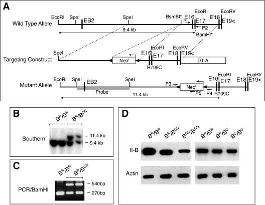
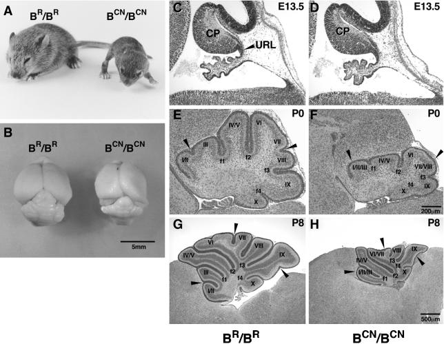
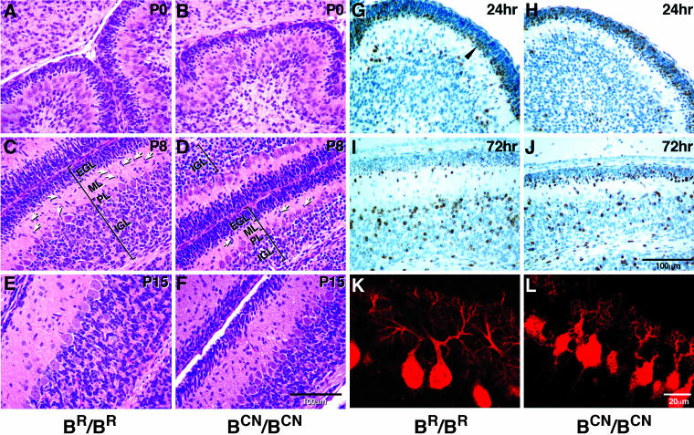
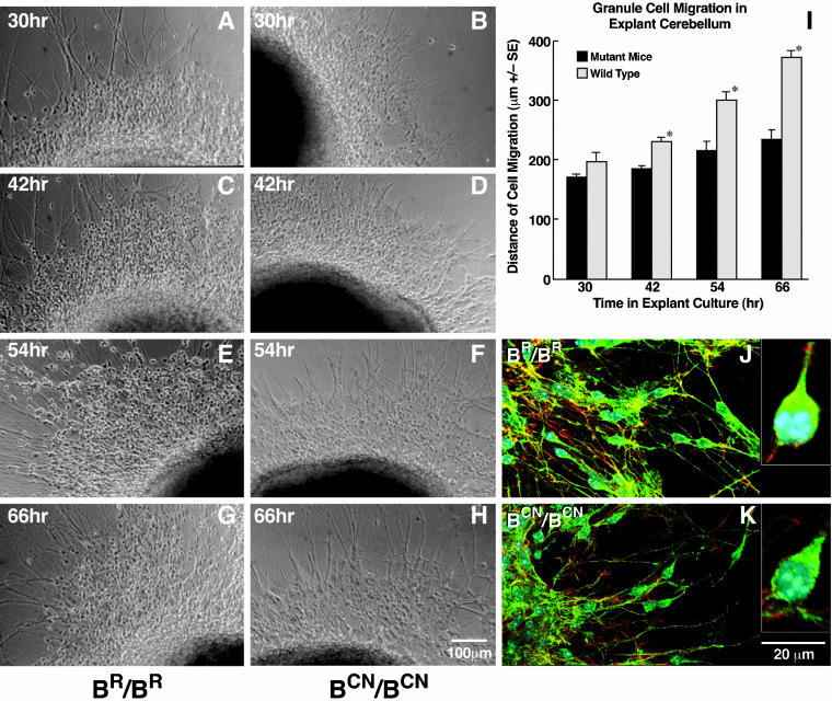
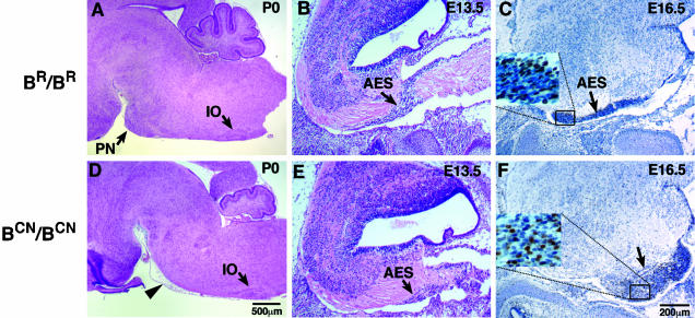
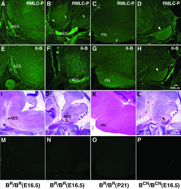
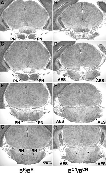
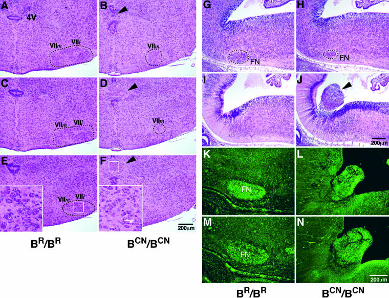
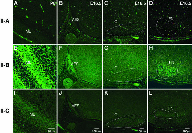
We generated mice harboring a single amino acid mutation in the motor domain of nonmuscle myosin heavy chain II-B (NMHC II-B). Homozygous mutant mice had an abnormal gait and difficulties in maintaining balance. Consistent with their motor defects, the mutant mice displayed an abnormal pattern of cerebellar foliation. Analysis of the brains of homozygous mutant mice showed significant defects in neuronal migration involving granule cells in the cerebellum, the facial neurons, and the anterior extramural precerebellar migratory stream, including the pontine neurons. A high level of NMHC II-B expression in these neurons suggests an important role for this particular isoform during neuronal migration in the developing brain. Increased phosphorylation of the myosin II regulatory light chain in migrating, compared with stationary pontine neurons, supports an active role for myosin II in regulating their migration. These studies demonstrate that NMHC II-B is particularly important for normal migration of distinct groups of neurons during mouse brain development.


Figures

Figure 1.
Figure 1.
Generation of hypomorphic NMHC II-B R709C mutant mice. (A) Diagram of the wild-type and R709C mutant NMHC II-B allele and targeting construct. The R709C mutation was generated by changing the coding sequence CGG (Arg) to TGC (Cys) in exon 17 (E17). The mutation destroyed a BamHI recognition site in the wild-type allele in E17 (BamHI*). A floxed Neor cassette was inserted at the BsmBI* site upstream to the mutation for positive selection. The Diphtheria Toxin-A cassette (DT-A) was placed at the 3′ end of the construct for negative selection. (B) Southern blot analysis of genomic DNA obtained from ES cell clones. Genomic DNA was digested with EcoRI and probed with the indicated SpeI/SpeI fragment (A). Two bands at 11.4 and 9.4 kb represent the targeted (with Neocassette) and wild-type allele, respectively. (C) PCR screen of Southern positive ES cell clones. PCR products of genomic DNA amplified by primers p1 and p2 (A) were digested by BamHI to yield fragments of 270 bp (wild-type) and 540 bp (mutant). (D) Western blot analysis of the protein extract obtained from hypomorphic and nonhypomorphic mutant and wild-type mouse brains. The presence of Neor in BCN/BCN mice resulted in a 73% reduction in the expression of mutant NMHC II-B compared with wild-type NHMC II-B in BR/BR mice (left). Removal of Neor restored the expression of the mutant II-B in BC/BC mice to the same level as the wild-type II-B in BR/BR mice (right). Actin staining is included as a loading control.
Figure 2.
Figure 2.
Motor dysfunction and abnormal cerebellar foliation in mutant mice. (A) Picture of a P15 R709C homozygous mutant (BCN/BCN) mouse and wild-type littermate (BR/BR). The BCN/BCN mouse shows severe growth retardation, progressive hydrocephalus and a broad-based gait compared with wild-type littermate. (B) Gross view of the brains of BCN/BCN and BR/BR mice. The BCN/BCN mouse brain shows a distorted cerebral cortex due to hydrocephalus and an underdeveloped cerebellum. (C–H) Midsagittal cerebellar sections of mouse brains at the ages indicated stained with H&E. No obvious differences in the formation of the cerebellar primordium (CP) are found in the mutant mice (BCN/BCN) compared with the wild-type littermate (BR/BR) at E13.5 (C and D). E–H demonstrate the impaired cerebellar foliation pattern with missing fissures in the mutant mice (cf. arrowheads in F with E at P0, and H with G at P8) compared with the wild-type littermates. URL, upper rhombic lip.
Figure 3.
Figure 3.
Abnormal layer formation in the mutant cerebellum. (A–F) Midsagittal cerebellar sections of mouse brains at ages indicated stained with H&E. At P0, a small decrease in the thickness of the external germinal layer (EGL) of the cerebellum is evident in BCN/BCN mice (B) compared with that in BR/BR mice (A). At P8, the mutant EGL is similar in thickness to the wild-type, but the IGL is only about one-half the size of the wild-type (cf. the mutant D with wild-type C). A decreased number of migrating granule cells is also found in the ML in mutant mice compared with wild-type mice (arrows). At P15, the EGL of the mutant cerebellum is thicker than the wild-type and the mutant IGL remains thinner than the wild-type (cf. the mutant F with wild-type E). (G–J) Sagittal sections of mouse cerebellum immunostained for BrdU and counterstained with hematoxylin. BrdU was injected at P6. BrdU-positive postmitotic granule cells (brown staining) are localized to the premigratory zone of the EGL 24 h after injection (G, arrowhead), and most of them have migrated to the IGL by 72 h in wild-type (BR/BR) mice (I). However, in the mutant (BCN/BCN) mice, many BrdU-positive cells are located throughout the EGL 24 h after injection (H), and many of these cells are still found in the EGL at 72 h, although most have now accumulated in the premigratory zone (J). (K and L) Confocal immunofluorescent microscopic images of the sagittal sections of P8 mouse cerebellum stained with antibody against calbindin showing dendritic trees and cell bodies of the Purkinje cells. The BCN/BCN mice show underdeveloped Purkinje cells with less arborization and stunted dendritic trees (L) compared with BR/BR mice. PL, Purkinje cell layer.
Figure 4.
Figure 4.
Slowed migration of mutant cerebellar granule cells in microexplant cultures. (A–H) Examples of phase contrast images of the migrating granule cells at indicated times in microexplant cultures from BR/BR (A, C, E, and G) and BCN/BCN mice (B, D, F, and H). (I) Bar plot comparing the distance traversed by the front wave of the migrating cell bodies between explants from wild-type and mutant mice, showing a significantly slowed progression of the granule cells from mutant mice (*p < 0.0001, n = 8). (J and K) Immunofluorescence confocal images. Green indicates NMHC II-B stained with anti-NMHC II-B, red indicates actin stained with rhodamine-phalloidin, blue indicates nuclei stained with 4,6-diamidino-2-phenylindole. Accumulation of NMHC II-B in the perinuclear area of migrating cerebellar granule cells is seen in wild-type mice (J), but it is not consistently seen in the mutant cells (K). Insets in J and K show asymmetric distribution of NMHC II-B in migrating granule cells at high magnification for wild-type and mutant cells.
Figure 5.
Figure 5.
Abnormal migration of the mutant AES. Sagittal sections of mouse brains at indicated ages stained with H&E (A, B, D, and E) or immunostained for BrdU and counterstained with hematoxylin (C and F). Pontine nuclei (PN) are absent at their normal destination in BCN/BCN mice at P0 (D, arrowhead); however, no obvious abnormalities in the formation of the inferior olive (D, IO) are found in these mice. The AES is initiated in mutant mice (E, arrow) similar to wild-type mice (B, arrow) at E13.5. However, defects in the progression of the AES are evident in BCN/BCN mice at E16.5 (F, arrow). The insets in C and F demonstrate the presence of BrdU-labeled neurons (brown staining) in the AES and are enlarged from the indicated areas.
Figure 9.
Figure 9.
Increased phosphorylation of the RMLC in the AES. (A–D) Immunofluorescence confocal images stained for RMLC-P in the AES, containing migrating pontine neurons (A and B), and stationary pontine neurons (C, PN) in BR/BR mice and the abnormally accumulated neurons of the putative AES in BCN/BCN mice (D, arrowhead). At E16.5, the migrating pontine neurons in both medial (A) and lateral (B) sections and the vasculature (B, arrows) show a marked increase in phosphorylation of the RMLC. This increase is not present in stationary pontine neurons at P21, where strong staining remains in the vasculature (C, arrows). In BCN/BCN mice, the migration of the AES is slowed (Figure 5), but the increase in RMLC-P is still observed (D, arrowhead). (E–H) Corresponding sections stained for NMHC II-B. Both the migrating (E and F) and stationary pontine neurons (G) show increased amounts of NMHC II-B compared with the surrounding areas. In BCN/BCN mice, this increase in staining is seen in the mutant putative AES (H, arrowhead). (I–L) H&E staining of the corresponding sections at low magnification to show identification of neurons. (M–P) Negative control for immunofluorescence staining by using normal rabbit IgG as primary antibody.
Figure 6.
Figure 6.
Coronal sections of the abnormal migration of the AES in mutant mice. Serial coronal sections (anterior to posterior) of a P0 mouse brain stained with H&E, showing absence of pontine nuclei (PN) in a BCN/BCN mouse (cf. normal A, C, and E with mutant B, D, and F) and an abnormal accumulation of the AES at the lateral posterior region (D, F, and H, arrows), which is not seen in the wild-type (BR/BR) mouse (C, E, and G). Note also that the reticular nucleus of the pons is seen in its normal location in the wild-type mouse (G, RN), but not in the mutant mouse (H, arrows).
Figure 7.
Figure 7.
Diminished facial neurons and their abnormal protrusion into the fourth ventricle in mutant mice. (A–F) Serial coronal sections (anterior to posterior) of P0 mouse brains stained with H&E. Diminished facial neurons are evident in BCN/BCN mice (B, D, and F) compared with BR/BR littermates (A, C, and E). Only some medial facial neurons (VIIm) are observed in the mutant mice, but the lateral facial neurons (VIIl) are not seen (cf. Band D with A and C). In addition, the fourth ventricle (4V) in BCN/BCN mice is disrupted by an ectopic group of large-sized neurons (B, D, and F, arrowhead) similar to facial neurons of the wild-type mice (cf. inserts in F with E). The white box outlines the region enlarged in the insert. (G–J) Serial sagittal sections (lateral to medial) of mouse brains at E13.5 stained with H&E. At E13.5, facial neurons have just completed their migration. A diminished number of facial neurons (FN) is seen in BCN/BCN mice (H) compared with BR/BR littermates (G). Protrusion of a group of neurons into the fourth ventricle near the region where the facial neurons originate is observed in the mutant mice (J, arrowhead) but not in the wild-type mice (I). (K–N) Sagittal sections of mouse brains at E16.5 immunostained for two different marker proteins showing positive staining of the protruded neurons (L and N) and normal facial neurons (K and M) for both Hoxb1 (K and L) and Phox2b (M and N).
Figure 8.
Figure 8.
Differential distribution of NMHC II-B in wild-type mouse brain. Immunofluorescence confocal images stained for NMHC II-A (A–D), II-B (E–H), and II-C (I–L) in developing cerebellum (A, E, and I), areas around the AES (B, F, and J), inferior olive (C, G, and K), and facial neurons (D, H, and L). Strong staining of the vasculature is obvious for NMHC II-A (A–D). NMHC II-B is detected throughout the brain, but more intense staining is present in the ML of the developing cerebellum (E), the AES (F), and facial neurons (H, FN). In contrast, neurons of the inferior olive (G, IO) do not show any increased staining with II-B. NMHC II-C is uniformly stained throughout all the brain sections. Negative controls using normal rabbit IgG as the primary antibody did not show fluorescence.

References

    1. Altman, J., and Bayer, S.A. (1987). Development of the precerebellar nuclei in the rat. J. Comp. Neurol. 257, 477-552. - PubMed
    1. Altman, J., and Bayer, S.A. (1997). Development of the cerebellar system in relation to its evolution, structure, and functions, Boca Raton, FL: CRC Press.
    1. Bloch-Gallego, E., Ezan, F., Tessier-Lavigne, M., and Sotelo, C. (1999). Floor plate and netrin-1 are involved in the migration and survival of inferior olivary neurons. J. Neurosci. 19, 4407-4420. - PMC - PubMed
    1. Brown, M.E., and Bridgman, P.C. (2003). Retrograde flow is increased in growth cones from myosin IIB knockout mice. J. Cell Sci. 116, 1087-1094. - PubMed
    1. Buxton, D.B., Golomb, E., and Adelstein, R.S. (2003). Induction of nonmuscle myosin heavy chain II-C by butyrate in RAW 264.7 mouse macrophages. J. Biol. Chem. 278, 15449-15455. - PubMed

MeSH terms

Substances