. 2004 Jun;15(6):2549-57.
doi: 10.1091/mbc.e03-11-0857. Epub 2004 Mar 19.

RalA activation at nascent lamellipodia of epidermal growth factor-stimulated Cos7 cells and migrating Madin-Darby canine kidney cells

Affiliations

RalA activation at nascent lamellipodia of epidermal growth factor-stimulated Cos7 cells and migrating Madin-Darby canine kidney cells

Akiyuki Takaya et al. Mol Biol Cell. 2004 Jun.

Abstract

RalA, a member of the Ras-family GTPases, regulates various cellular functions such as filopodia formation, endocytosis, and exocytosis. On epidermal growth factor (EGF) stimulation, activated Ras recruits guanine nucleotide exchange factors (GEFs) for RalA, followed by RalA activation. By using fluorescence resonance energy transfer-based probes for RalA activity, we found that the EGF-induced RalA activation in Cos7 cells was restricted at the EGF-induced nascent lamellipodia, whereas under a similar condition both Ras activation and Ras-dependent translocation of Ral GEFs occurred more diffusely at the plasma membrane. This EGF-induced RalA activation was not observed when lamellipodial protrusion was suppressed by a dominant negative mutant of Rac1, a GTPase-activating protein for Cdc42, inhibitors of phosphatidylinositol 3-kinase, or inhibitors of actin polymerization. On the other hand, EGF-induced lamellipodial protrusion was inhibited by microinjection of the RalA-binding domains of RalBP1 and Sec5. Furthermore, we found that RalA activity was high at the lamellipodia of migrating Madin-Darby canine kidney cells and that the migration of Madin-Darby canine kidney cells was perturbed by the microinjection of RalBP1-RalA-binding domain. Thus, RalA activation is required for the induction of lamellipodia, and conversely, lamellipodial protrusion seems to be required for the RalA activation, suggesting the presence of a positive feedback loop between RalA activation and lamellipodial protrusion. Our observation also demonstrates that the spatial regulation of RalA is conducted by a mechanism distinct from the temporal regulation conducted by Ras-dependent plasma membrane recruitment of Ral guanine nucleotide exchange factors.


Figures
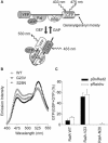
Figure 1.
Figure 1.
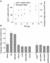
Basic properties of Raichu-RalA. (A) Schematic representation of Raichu-RalA bound to GDP or GTP. Ral, RBD, GEF, and GAP indicate RalA, the Ral-binding domain of RalBP1, the guanine nucleotide exchange factor for Ral, and the Ral GTPase-activating protein, respectively. YFP and CFP denote yellow-emitting and cyan-emitting mutants of GFP, respectively. (B) Emission spectra of Raichu-RalA expressed in 293T cells. Cells were lysed and analyzed with a fluorescence spectrometer at an excitation wavelength of 433 nm. WT, V23, and N28 denote the wild-type, constitutively active mutant, and dominant negative mutant, respectively. (C) GTP/GDP loading of Raichu-RalA and DsRed2-tagged RalA. 293T cells expressing Raichu-RalA or DsRed2-RalA were labeled with 32Pi. Raichu-RalA and DsRed2-RalA were precipitated with anti-GFP and anti-RFP rabbit sera, respectively, followed by TLC analysis. Separated GTP and GDP were quantitated with a BAS-1000 image analyzer and GTP/(GTP + GDP) (%) values were plotted with SD.
Figure 2.
Figure 2.
Sensitivity of Raichu-RalA to GEFs and GAPs. (A) Correlation of GTP-loading and FRET efficiency of Raichu-RalA. pRaichu-RalA and varying quantities of pcDNA3-Rlf were transfected into pairs of 293T cells. One set of cells was labeled with 32Pi and determined for GTP/(GTP + GDP) (%) on Raichu-RalA by TLC. The other set of cells was lysed and examined for the emission ratio (intensity at 475 vs. 530 nm) with an excitation wavelength of 433 nm with a fluorescence spectrometer. Error bars are shown either for the upward or the downward for the brevity. (B) FRET efficiency of Raichu-RalA in the presence of various GEFs and GAPs. pRaichu-RalA was transfected into 293T cells with expression vectors encoding GEFs or GAPs as indicated at the bottom of the figure. FRET efficiency was measured as in A. Data from at least two independent experiments are shown with SD.
Figure 3.
Figure 3.
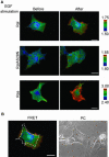
RalA activity in EGF-stimulated Cos7 cells. Cos7 cells were transfected with pRaichu-RalA or pRaichu-Ras. Cells were imaged for YFP, CFP, and phase contrast (PC) every 1 min with a time-lapse epifluorescence microscope equipped with a cooled CCD camera. The ratio images (YFP/CFP) were generated with MetaMorph software. In the IMD mode shown here, eight colors from red to blue are used to represent the YFP/CFP ratio, with the intensity of each color indicating the mean intensity of YFP and CFP. The upper and lower limits of the ratio range are shown on the right. (A) Representative ratio images of Raichu-RalA and RaidhuRas at the indicated time points. EGF (50 ng/ml) was inoculated at 0 min. Cell imaging was repeated >20 times, and the most representative images are shown. (B) Ratio and phase contrast images of cells expressing Raichu-RalA at 10 min after EGF stimulation. White broken lines show the contours of the cells before EGF stimulation. Bars, 25 μm.
Figure 4.
Figure 4.
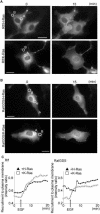
Translocation of RalGEFs to the plasma membrane upon EGF stimulation. (A and B) pCAGGS-EGFP-Rlf or -RalGDS was cotransfected with pIRM-Flag-H-Ras or pIRM-Flag-K-Ras into Cos7 cells. After 24 h, cells were serum starved for 4 h and stimulated with 50 ng/ml EGF. Expression of the GTPases was confirmed by the fluorescence of dsFP593, which was translated from the internal ribosomal entry site of pIRM. Images of EGFP-Rlf and -RalGDS were obtained every 1 min, and EGF was added 5 min after the start of recording. The images of H-Ras and K-Ras were taken are at 0 and 15 min. (C) Intensity ratios of the regions denoted by circles A and B in A and B were plotted against time. The intensity ratio was defined as IB/IA, where IA is the intensity of perinuclear region A, and IB is the intensity of peripheral region B. Bars, 25 μm.
Figure 5.
Figure 5.
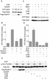
Inhibition of EGF-induced RalA activation by reagents that perturb lamellipodia. (A) Cos7 cells transfected with or without an expression vector for a dominant negative mutant of Rac, Rac1-N17, or GAP for Cdc42, CdGAP were serum starved for 8 h and stimulated with EGF. Cells were analyzed by Bos' pull-down method. (B) Levels of GTP-RalA were quantitated with an LAS-1000 image analyzer. Data from two independent experiments are shown with SD. (C and D) Cos7 cells left untreated or treated with wortmannin (Wort), LY294002 (LY), latrunculin B (LatB), or cytochalasin D (CytD) for 30 min were stimulated with EGF and analyzed as described in A. Data from three independent experiments are shown with SD. (E) Cells treated as in A and C were analyzed for the phosphorylation of Akt with anti-phospho-Akt (Thr308) antibody.
Figure 6.
Figure 6.
Inhibition of EGF-induced lamellipodial protrusion by RalBP1-RBD and Sec5-RBD. GST, GST-RalBP1-RBD, Sec5-RBD, or GST-PAK-CRIB were microinjected into Cos7 cells. Beginning at 30 min, phase contrast images of these cells were obtained every 30 s, followed by the stimulation with EGF (50 ng/ml). Cells images were further collected for 30 min. (A) Outline of the quantification of lamellipodial induction. The maximum length of nascent lamellipodia (X) and the breadth of the nucleus (Y) were measured on the video image of each microinjected cell. When the value of X/Y was >0.3, the cell was scored as lamellipodia positive. (B) More than 60 cells were analyzed for each protein in at least six independent experiments, and the percentage of lamellipodia-positive cells is shown.
Figure 7.
Figure 7.
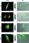
Imaging of activities of low-molecular-weight GTPases in migrating MDCK cells. Confluent MDCK cells transfected with an expression plasmid for Raichu-RalA, Raichu-Ras, Raichu-Rac1, or Raichu-Cdc42 were wounded 1 h before FRET imaging. Images of CFP, YFP, and DIC were obtained every 2 min for 4 h. Shown here are representative pseudocolor images of FRET efficiency (YFP/CFP) and overlay images of DIC and YFP at 2 h from the start of FRET imaging. Ratio ranges are shown on the right. Each cell imaging was repeated at least three times with similar results. Bars, 25 μm.
Figure 8.
Figure 8.
Effect of RalA inhibition on wound closure. MDCK monolayers were wounded and, 1 h later, the cells at the wound edge were microinjected with FITC-dextran and GST, GST-RalBP1-RBD, or GST-PAK-CRIB. Cells were imaged for FITC and DIC every 2 min for 4 h. (A) Overlay image of FITC and phase contrast at 4 h after microinjection and outlines the method for the quantification. The white dotted line labeled as I indicates the initial position of the wound edge. The ratio of migration distance is defined as Dn/Di, where Dn and Di are the migration distances of control cells and microinjected cells, respectively. Data from four independent experiments are averaged and shown with SD.

References

    1. Anderson, K.E., Coadwell, J., Stephens, L.R., and Hawkins, P.T. (1998). Translocation of PDK-1 to the plasma membrane is important in allowing PDK-1 to activate protein kinase B. Curr. Biol. 8, 684-691. - PubMed
    1. Aoki, K., Nakamura, T., and Matsuda, M. (2004). Spatio-temporal regulation of Rac1 and Cdc42 activity during nerve growth factor-induced neurite outgrowth in PC12 cells. J. Biol. Chem. 279, 713-719. - PubMed
    1. Bhattacharya, M., Anborgh, P.H., Babwah, A.V., Dale, L.B., Dobransky, T., Benovic, J.L., Feldman, R.D., Verdi, J.M., Rylett, R.J., and Ferguson, S.S. (2002). beta-Arrestins regulate a Ral-GDS Ral effector pathway that mediates cytoskeletal reorganization. Nat. Cell Biol. 4, 547-555. - PubMed
    1. Bhullar, R.P., and Seneviratne, H.D. (1996). Characterization of human platelet GTPase activating protein for the Ral GTP-binding protein. Biochim. Biophys. Acta 1311, 181-188. - PubMed
    1. Bos, J.L. (1998). All in the family? New insights and questions regarding interconnectivity of Ras, Rap1 and Ral. EMBO J. 17, 6776-6782. - PMC - PubMed

Publication types

MeSH terms